The surfaces and borders of the heart
Orientation and surfaces:
Apex of the heart points in an anteroinferior direction
In the anatomical position, the heart has 5 surfaces formed by different chambers of the heart:
Anterior (sternocostal) - right ventricle
Posterior (base of pyramid) - left atrium
Inferior (diaphragmatic) - left & right ventricles
Right pulmonary - right atrium
Left pulmonary - left ventricle
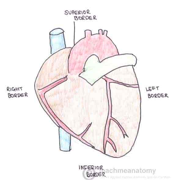
Borders:
4 main borders separating the surfaces of the heart:
Right border - right atrium
Inferior border - left & right ventricle
Left border - left ventricle (and some of left atrium)
Superior border - right & left atrium and the great vessels
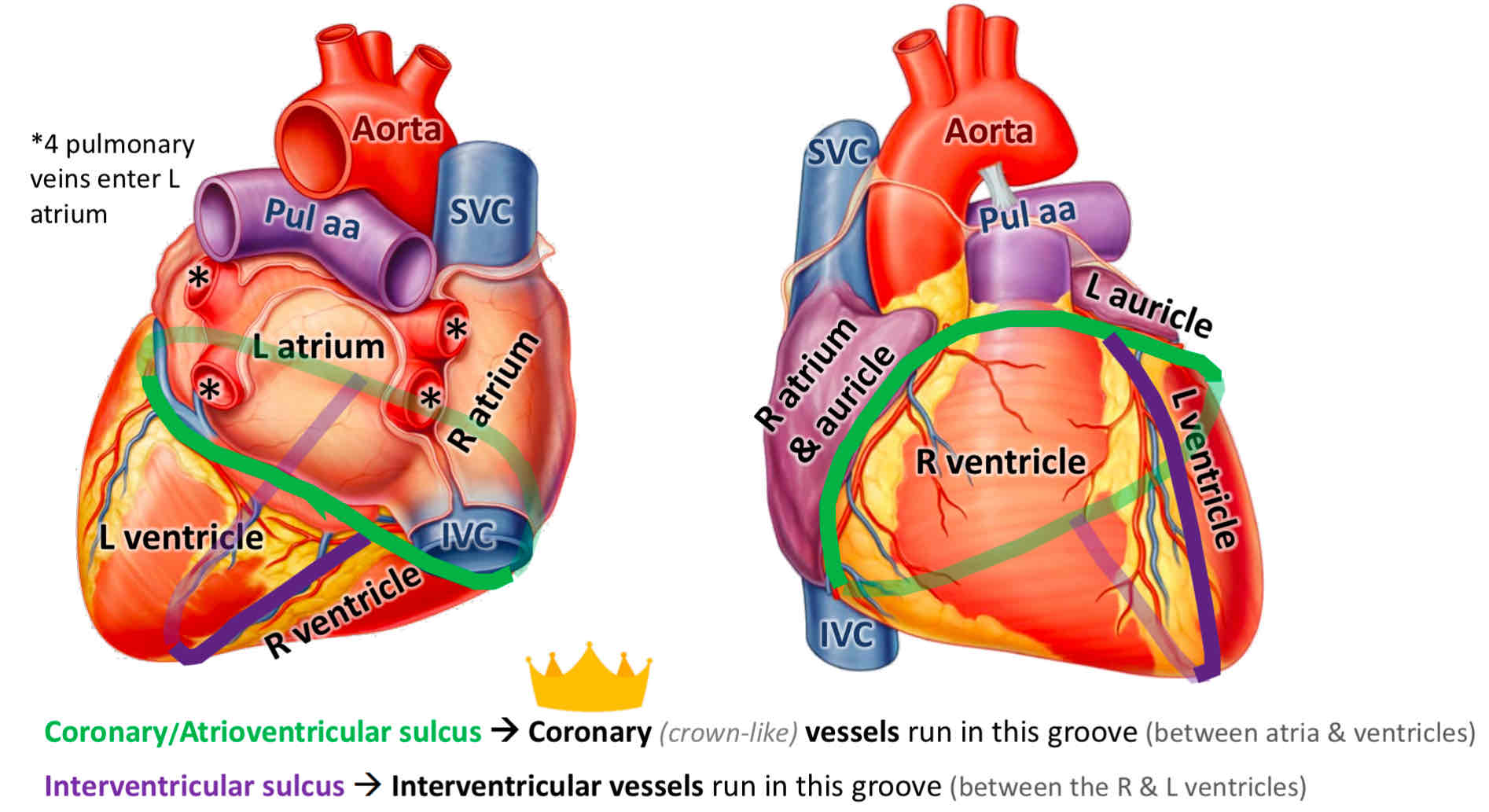
Sulci of the heart:
Heart is divided internally into 4 chambers - these divisions create grooves on the external surface of the heart known as sulci
3 main sulci:
Coronary sulcus (atrioventricular groove) - circles around the heart and represents the separation of the atria from the ventricles
Contains the right coronary artery, circumflex branch of the left CA, small cardiac vein & coronary sinus
Anterior interventricular sulcus - located on the anterior surface of the heart and represents the separation of the left and right ventricle
Contains the anterior interventricular artery (aka the left anterior descending artery) and great cardiac vein
Posterior interventricular sulcus - located on the posterior surface of the heart and represents the separation of the left and right ventricle
Contains the posterior interventricular artery and middle cardiac vein
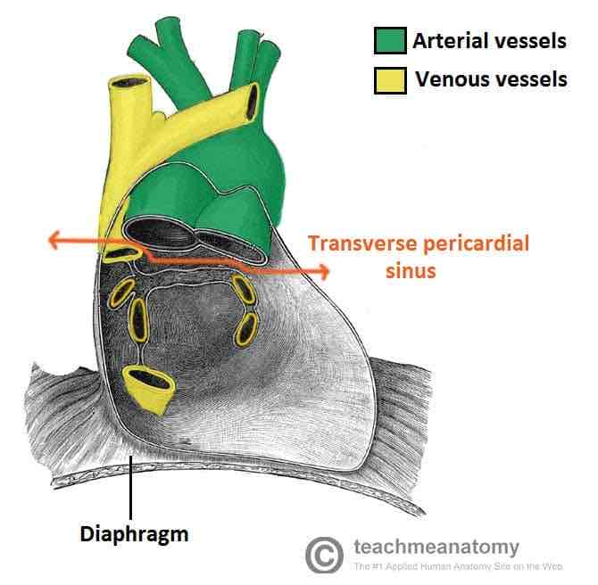
Pericardial sinuses:
Passageways formed when the pericardium folds around the great vessels
oblique pericardial sinus: blind ending passageway located on the posterior surface of the heart
Transverse pericardial sinus: found superiorly on the heart, can be used in coronary artery bypass grafting
Posterior to the ascending aorta and pulmonary trunk, anterior to superior VC, superior to the LA
Separate the arterial and venous vessels of the heart
Can be used to identify and subsequently litigate (tie off) the arteries of the heart during coronary artery bypass grafting