1b: Central regulation and signal transduction
Learning Outcomes
Contrast the afferent (sensory) and efferent (motor) branches of the somatic and autonomic nervous systems (voluntary/conscious control vs involuntary regulation of internal organs).
Understand Afferent:
Prefix "A" (Latin Ad) implies towards the CNS (central nervous system).
Understand Efferent:
Prefix "E" (Latin Ex) implies away from the CNS.
Discuss the relative contributions of the sympathetic and parasympathetic divisions of the autonomic nervous system in the regulation of physiology.
Appreciate the link between autonomic control centers in the brainstem and unconscious functions of the ANS.
Describe the parasympathetic functions of the vagus nerve.
Appreciate the range of sensory receptor types.
Explain why the sympathetic nervous system is commonly referred to as the “fight or flight” response while the parasympathetic nervous system is referred to as “rest and digest.”
Describe the role of the adrenal gland in sympathetic mass discharge.
Explain the concept of autonomic tone and the relative contribution of the two divisions of the autonomic nervous system in maintaining the basal activity of an organ at the resting state.
Appreciate the example of autonomic tone being the cardiovascular system.
Describe the major anatomical and physiological differences of the sympathetic and parasympathetic neurons.
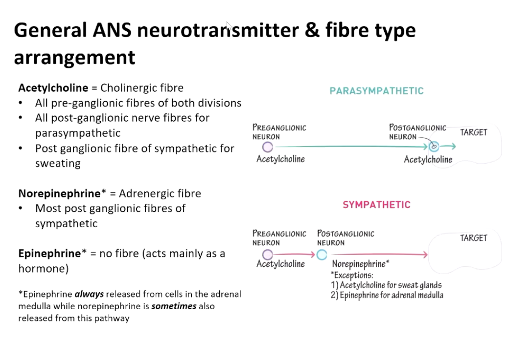
Define adrenergic and cholinergic neurons.
Describe the three types of signal transducing membrane receptors and their mechanisms.
Define the role of second messengers in signal transduction and identify
The major components (enzymes and second messengers) within G protein signal transduction pathways involving:
Gs
Gi
Gq
Explain the concepts of amplification and termination in signal transduction.
Overview of the Nervous System
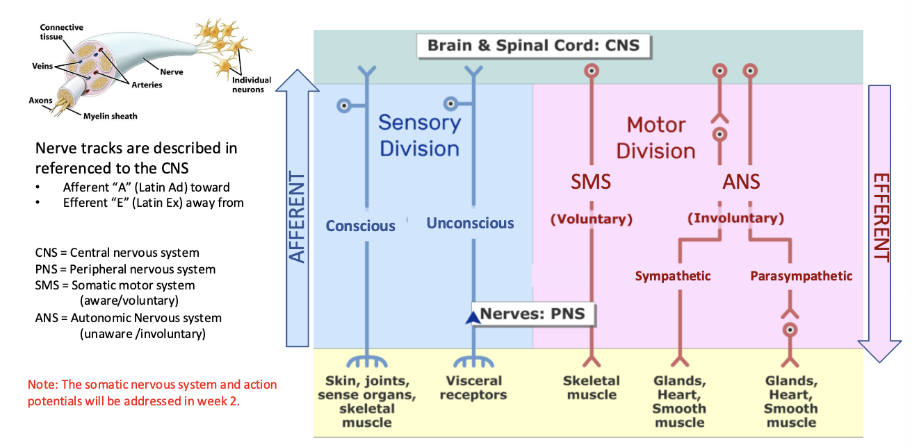
The nervous system comprises two major parts:
Central Nervous System (CNS)
Peripheral Nervous System (PNS)
The autonomic nervous system (ANS) can be divided into two branches:
Sympathetic Nervous System (SNS) - fight or flight, turbo
Parasympathetic Nervous System (PNS) - rest and digest, recharge
Note: The somatic nervous system and action potentials will be addressed in week 2.
Somatic vs. Autonomic Systems
Somatic Nervous System - conscious control, voluntary
Controls voluntary movements via conscious awareness.
Neurotransmitter: Acetylcholine
Effector organs: Skeletal muscle
Autonomic Nervous System - automatic regulation of internal organs, involuntary
Regulates involuntary functions and homeostasis.
Divided into sympathetic and parasympathetic divisions.
Neurotransmitters:
Sympathetic: Norepinephrine (NE)
Parasympathetic: Acetylcholine (ACh)
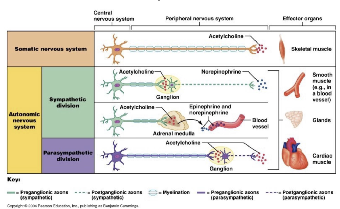
Includes the adrenal medulla, which releases epinephrine and norepinephrine.
Effector Organs: Smooth muscle, cardiac muscle, glands.
Autonomic Nervous System Functions
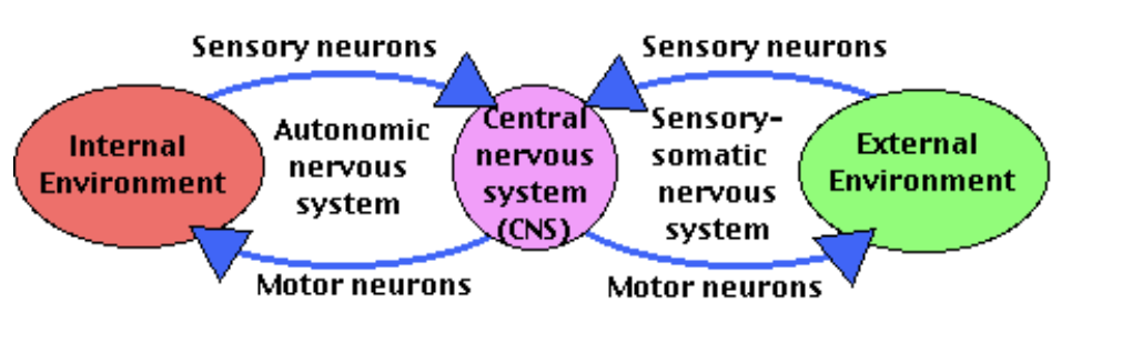
Evaluates the internal environment and maintains homeostasis.
Sensory neurons connect sensory receptors to the CNS (hypothalamus and medulla oblongata).
Motor neurons connect the CNS to effectors (organs or tissues).
Autonomic functions often operate as reflexes (e.g., enteric nervous system).
Significant role in cardiovascular and respiratory system functioning, important pharmacological targets.
Sensory Receptors
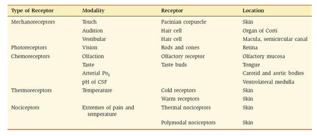
Different types of receptors exist, each responsible for detecting different environmental stimuli:
Mechanoreceptors (e.g., Pacinian corpuscle for touch)
Photoreceptors (e.g., rods and cones for vision)
Chemoreceptors (e.g., for taste and olfaction)
Thermoreceptors (e.g., for temperature)
Nociceptors (e.g., for pain)
Each receptor type has specific modalities, locations, and functions.
Autonomic Control Centers in the Brain Stem
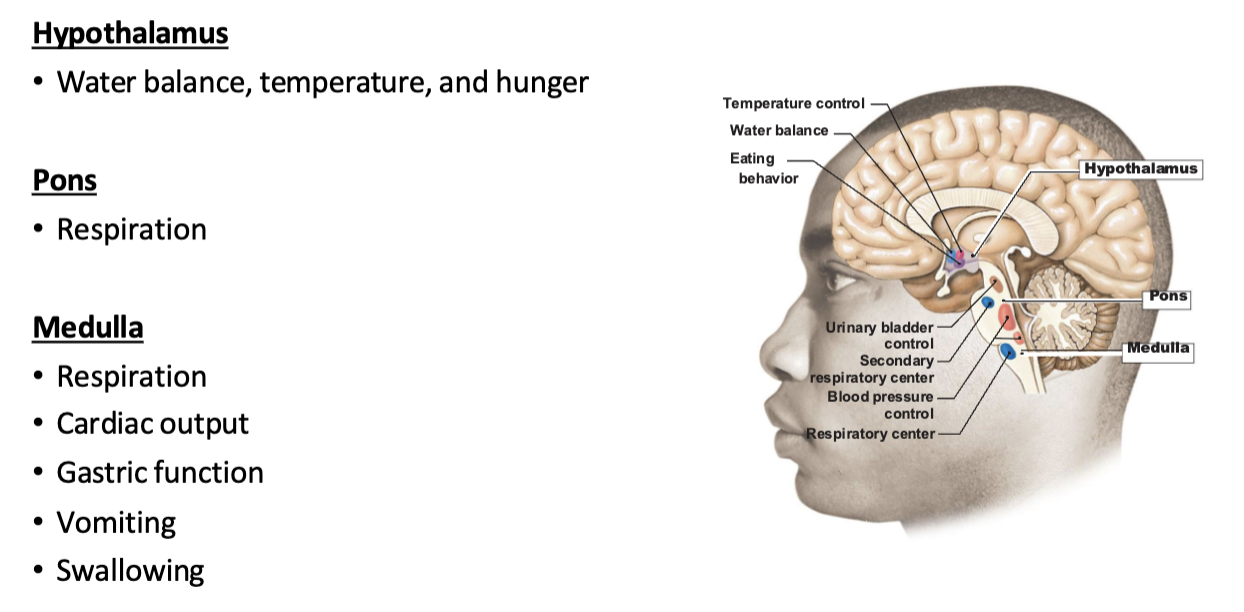
Control centers in the brainstem regulate bodily functions governed by the ANS.
Structures Include:
Hypothalamus: regulates water balance, temperature, hunger, and eating behavior.
Pons: involved in respiration.
Medulla: regulates cardiac output, gastric function, and automatic reflexes like vomiting and swallowing.
Note: Focus on the concept of regulatory centers; specific functions of brain structures should not be memorized.
Divisions of the Autonomic Nervous System
Sympathetic Division: fight or flight
Increases alertness, energy, heart rate, blood pressure, and breathing rate.
Triggered by stressors like fear and pain.
Parasympathetic Division: rest and digest
Decreases alertness, heart rate, blood pressure, and aids in digestion and relaxation.
Heavily involved in the functions of the vagus nerve.
vagus nerve also helps with defection, urination, and sexual arousal
Opposing Effects of ANS Divisions
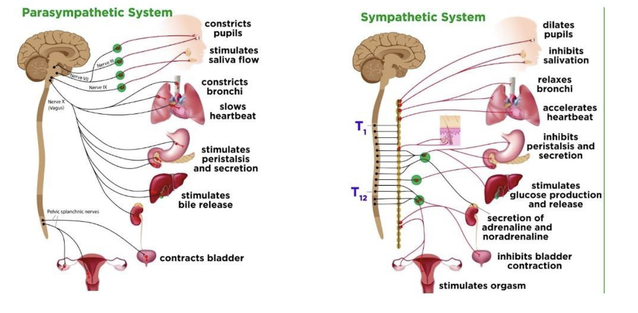
Physiological changes induced are opposite between the parasympathetic and sympathetic divisions.
Examples:
Parasympathetic: constricts pupils; stimulates salivation; slows heartbeat.
Sympathetic: dilates pupils; inhibits salivation; accelerates heartbeat.
Anatomical and Functional Differences of ANS Divisions
Spinal cord exit site:
Sympathetic: thoracolumbar region.
sympathetic output can activate many organs at once
coordinated fight or flight response
Parasympathetic: craniosacral region.
parasympathetic fibres go directly to specific organs
Neurotransmitters used in postganglionic fibers.
Receptors expressed at target tissues.
Activity of target tissues relates to specialized functions of target cells.
Ganglia and Neurons
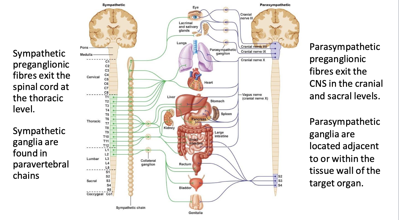
sympathetic:
Preganglionic neurons originate within the CNS.
preganglionic fibres exit at the thoracic level
ganglia - little clusters of neuron cell bodies located outside of the CNS
acts as a ‘relay station’
sympathetic ganglia are found in paravertebral chains
ganglia far from organs
preganglionic length - short
postganglionic length - long
parasympathetic:
preganglionic fibres exit the CNS in the cranial and sacral levels
ganglia are located adjacent to or within the tissue wall of the target organ
post ganglionic length - short
pre ganglionic length - long
length: how far each axon has to travel
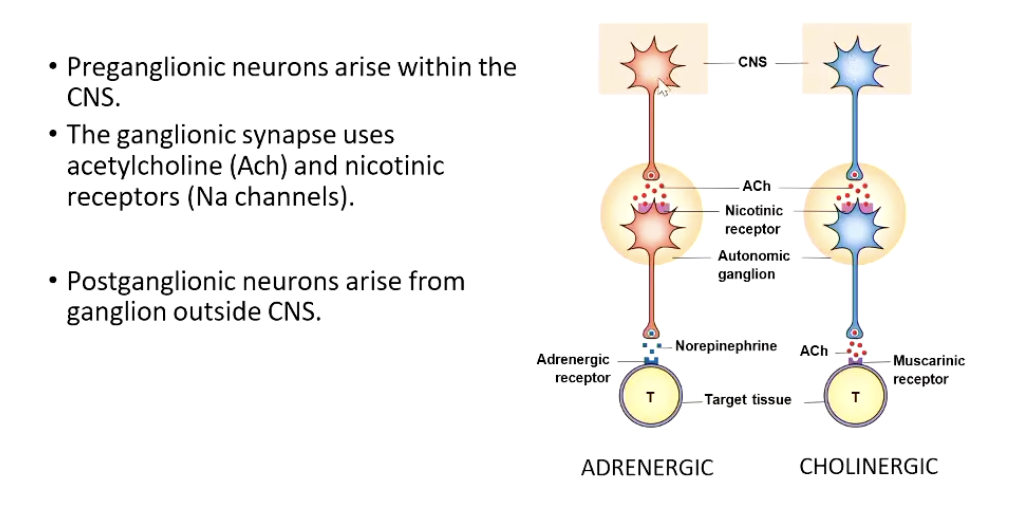
preganglionic neurons are within the CNS, their cell bodies are in the brainstem or spinal cord
postganglionic neurons, their cell bodies are outside the CNS, in the autonomic ganglia
types of neurotransmitter that postganglionic neuron releases:
adrenergic
when a neuron releases norepinephrine (NE)
most sympathetic postganglionic neurons
for fight or flight
cholinergic
when a neuron releases acetylcholine (ACh)
parasympathetic postganglionic neurons
for rest and digest
Mass Discharge and Sympathetic Activation
Definition: Coordinated discharge of sympathetic neurons in response to stress.
Known as the “fight or flight,” “alarm response,” or “stress response.”
triggered by fear and pain
hypothalamus activates sympathetic division of nervous system
heart rate, blood pressure, and respiration increase
adrenal medulla secrete epinephrine and norepinephrine
blood flow to skeletal msucles increases
stomach contractions are inhibited, contracts less
acute vs chronic activation (oral pres vs chased by bear)
homeostasis is a dynamic balance between autonomic branches
organ systems are balanced between the input from the sympathetic and parasympathetic divisions
when something upsets that balance, the homeostatic mechanisms strive to return it to its regular state
for each organ system, there may be more of a sympathetic or parasympathetic dominance to the resting state.
even when you’re not stressed and not actively digesting, one branch of the autonomic nervous system tends to be the ‘default controller’ for that organ
Autonomic Tone: single system
Refers to the baseline activity of both the sympathetic and parasympathetic systems.
Sympathetic activity regulates increases and decreases in organ activity.
Local sympathetic tone modulates blood flow, maintaining rest conditions at approximately 50% arterial diameter.
when sympathetic nervous system regulates both increases and decrease in activity of a stimulated organ
usually in the blood vessels
vascular tone: baseline level of contraction in your blood vessels
local cympathetic vascular tone regulates local blood flow into tissues
under resting conditions:
sympathetic NS maintains artery diameter at ~50% of maximum
increasing sympathetic stimulation
increasing sympathetic stimulation constricts the smooth muscles, increasing resistance in the arteries = increased blood pressure = decreased blood flow
decreasing sympathetic activity relaxes the muscle, decreasing resistance in the arteries = increased flow, decreased blood pressure
Signal Transduction
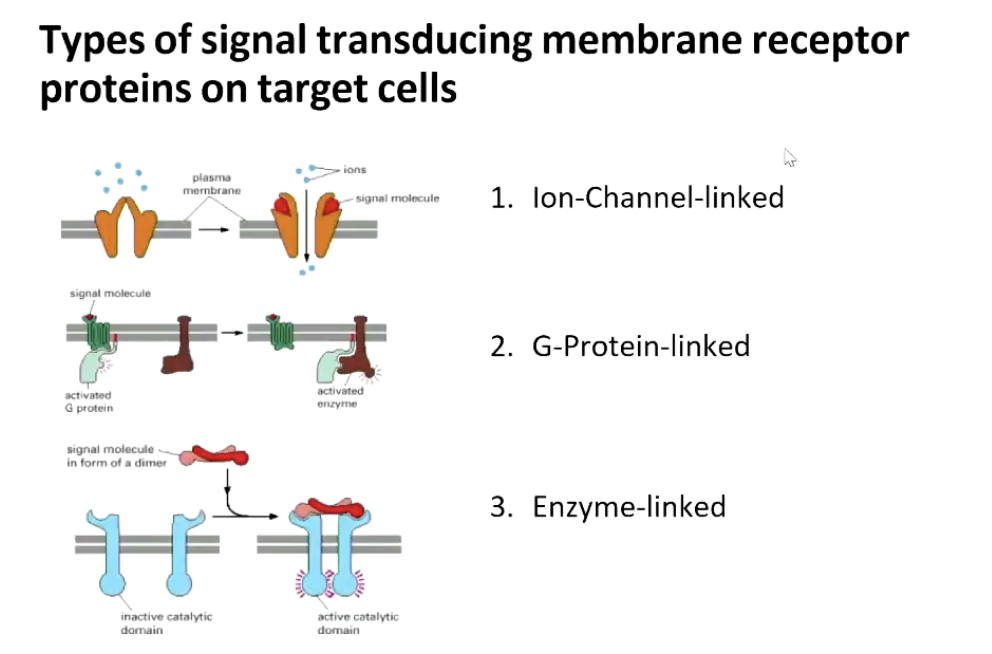
Three types of signal transducing membrane receptor proteins exist:
Ion-Channel-linked
muscle related
G-Protein-linked
regulates enzyme activity and are characterised by their self-limiting process via GTPase activity
Enzyme-linked
related to growth factors
cytokines
G Proteins
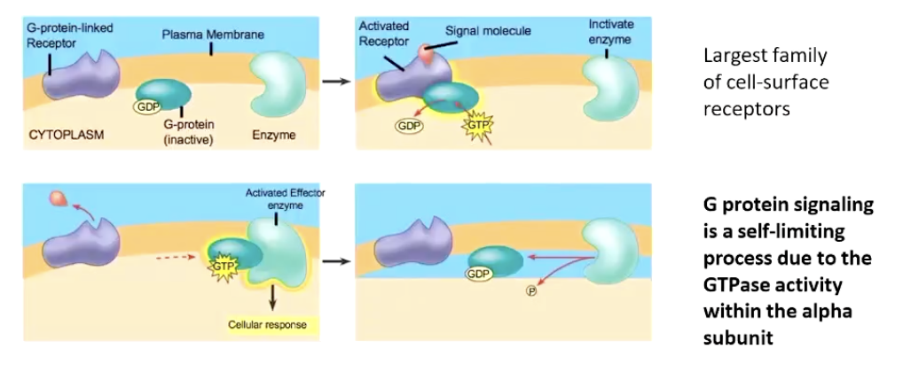
Features of G proteins:
cell-surface receptors
GTP = high energy state
GDP = low energy state
the alpha subunit of the g protein can turn itself off by breaking down GTP to GDP
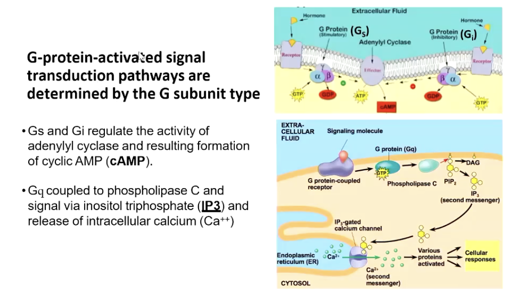
Gs - stimulatory
Gi - inhibitory
Gs + Gi regulates the activity of adenylyl cyclase and resulting formation of cyclic AMP (cAMP)
Gq coupled to phospholipase C and signal via inositol triphosphate (IP3) and release of intracellular calcium (Ca++)
Rapid amplification.
Rapid termination of enzyme activation.
GTPase activity leads to attenuation of secondary signals.
Various pathways regulated include:
Phosphodiesterase cleaving cAMP.
K+ flux restoration by Na+/K+ ATPase.
Ca2+ flux reverses from the ER/SR by Ca2+ ATPase.
Rapid degradation of IP3 by monophosphatases.
Signal Transduction Pathways
G subunit types define the pathways via which signals are transduced:
Gs and Gi regulate cyclic AMP production by adenylyl cyclase.
Gq couples with phospholipase C leading to inositol triphosphate (IP3) and intracellular calcium release.
Receptor Activity and Signaling Mechanisms
Ligand binding to GPCR activates G proteins and signal transduction pathways, including phospholipase C activation, leading to further signaling events.
Termination of signaling occurs via GTPase activity and hydrolysis of signaling molecules like IP3.
Conclusion
Understanding the complexity and interrelation between the sympathetic and parasympathetic divisions is crucial for grasping autonomic regulation in physiology.
The balance between these two systems is essential for maintaining homeostasis in various bodily functions.
Nature of the receptor on the target tissue
sympathetic = adrenergic receptors that signal via G protein complexes
alpha forms mostly activate Gq (cuts, calcium mobilising), Gi
beta forms mostly activate Gs
parasympathetic - cholinergic synapses
muscarinic receptors that mostly signal through Gi or Gq
Nicotinic receptors that activate gated ion channels