Renal Failure
1. Demonstrate an understanding of the pathophysiology and manifestations of acute kidney injury (acute renal failure) and chronic kidney disease (chronic renal failure).
Functions of the Kidney
The kidneys are vital organs with diverse functions crucial for maintaining overall body homeostasis. Main functions include:
Maintaining fluid and electrolyte balance: Regulating blood volume, blood pressure, and osmolality by adjusting water and electrolyte (such as sodium, potassium, calcium, magnesium, and phosphate) excretion and reabsorption. This is crucial for cellular function and nerve impulse transmission.
Acid-base balance: Maintaining the body's pH within a narrow range (approximately ) by excreting excess hydrogen ions () and reabsorbing bicarbonate ().
Hormone secretion: Producing essential hormones such as erythropoietin (stimulates red blood cell production), renin (regulates blood pressure), and activating Vitamin D (essential for calcium absorption and bone health).
Excretion of waste: Filtering metabolic waste products from the blood, including urea (from protein metabolism), creatinine (from muscle metabolism), uric acid, and various drug metabolites.
Acute Kidney Injury (AKI)
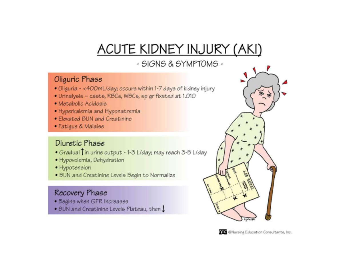
Definition: Sudden, often reversible, onset within hours to days, characterized by an abrupt decline in kidney function where the kidneys cannot maintain fluid, electrolyte, and acid-base balance. This rapid deterioration causes systemic problems, accumulation of nitrogenous waste products (azotemia), and can lead to death if untreated.
Severity Assessment in AKI
Serum creatinine and urine output are critical for determining the severity.
the most current definition of AKI is an increase in serum creatinine by 0.3 mg/dl or more within 48 hrs; or an increase in serum creatinine to 1.5 times or more from baseline, which is known or presumed to have occurred in the previous 7 days; or urine vol of less than 0.5 ml/kg/hr for 6 hrs.
the creatinine level is commonly used in the recognition of AKI.
this value is not ideal because the creatinine level takes time to increase which can create delays in treatment (the BUN is usually elevated 1st)
Glomerular Filtration Rate (eGFR): The best indicator of overall kidney function; however, GFR is not measured during acute and critical illness. Instead, calculated GFR estimations and trends in serum creatinine and urine output are used to infer kidney function.
Causes of Acute Kidney Injury
Decreased Perfusion to the kidneys.
Damage to Kidney Tissue.
Obstruction of Urine Outflow.
Classifications of AKI
Prerenal (before the kidney) Causes: Problems occurring before the kidney, e.g., decreased blood flow to the kidney (must maintain MAP >65 for kidney perfusion).
Examples:
Hypovolemia (hemorrhage, dehydration, excessive diuresis, gastrointestinal losses),
decreased cardiac output (heart failure, myocardial infarction, cardiogenic shock),
systemic vasodilation (sepsis, anaphylaxis),
renal artery stenosis,
severe liver disease (hepatorenal syndrome).
severe dehydration
ASA, NSAIDs, naproxen
blood clot
Intrarenal (Intrinsic) Causes: Issues occurring within the kidney itself, e.g., glomerulonephritis or nephrotoxic agents.
Examples:
multiple myeloma
scleroderma
systemic or local infection
clot in kidney blood vessel
bleeding in kidney, thrombotic thrombocytopenic purpura.
Glomerulonephritis,
Vasculitis/ischemia
Postrenal Causes: Problems occurring after urine has been formed, e.g., obstruction from bladder, cervical, colon, prostate cancer, or kidney stones.
Examples:
Benign prostatic hyperplasia (BPH) and prostate cancer in men,
bladder or cervical cancer,
kidney stones (nephrolithiasis or ureterolithiasis),
blood clots,
nerve damage
strictures in the ureters or urethra,
neurogenic bladder.
AKI Risk Factors:
Commonly occurs in:
Hospitalized older adults with ages 80 to 89
Hypertension (HTN)
Diabetes Mellitus (DM)
Peripheral Vascular Disease (PVD)
Liver disease
Chronic Kidney Disease (CKD, acute on chronic renal failure).
Additional independent risk factors:
Prolonged mechanical ventilation
Sepsis
Cardiac surgery
Hypotension
Shock
The presence of comorbid conditions increases the risk of End-Stage Kidney Disease (ESKD).
Potential Causes of AKI
Changes in urine characteristics or any existing obstructive problems.
Patient history of recent surgery, trauma, drug use, and coexisting conditions.
mycins
chemo drugs: methotrexate
metformin
when a nephrotoxic agent such as contrast medium is used withhold metformin for 24 hrs before and after procedure
give IV fluids before and after exposure to the contrast medium
Acute illnesses that could precipitate AKI (immunity-mediated AKI).
Post-hypotension or shock.
Assessment for AKI
History
ask about any changes in urine appearance, frequency, or volume
drug Hx especially use of antibiotics and NSAIDs
use of contrast medium
coexisting conditions of advanced age, CKD, diabetes, long-term HTN, sepsis, PVD, chronic liver disease, AIDS, prior kidney surgery
acute illness (influenza, colds, gastroenteritis, sore throats)
resent use of IV vasopressors (epinephrine, or norepinephrine)
physical assessment/ Sign/symptoms
brief period of oliguria, defined as less than 0.5 ml/kg/hr of urine output for 2 or more hrs.
buildup of nitrogenous wastes (azotemia) and decreased UO (oliguria)
progression in severity, the pt may have symptoms:
of fluid overload include pulmonary crackles,
dependent and generalized edema (anasarca),
decreased oxygenation (low peripheral oxygenation or Spo2),
confusion, increased RR, and dyspnea.
symptoms of reduced blood vol such as MAP below 65 mmHg,
tachycardia, thready peripheral pulses, or
decreasing cognition may indicate risk for AKI from poor perfusion.
Spo2 below 88%, may indicate potential hypoxemic or ischemic damage to kidney tissue.
KDIGO Classification of AKI:
Stage 1: Creatinine increased by times baseline, OR an increase of or more within 48 hours.
Urine output less than for 6-12 hours.
Stage 2: Creatinine increased by times baseline.
Urine output less than for more than 12 hours.
Stage 3: Creatinine increased by times baseline, OR serum creatinine >4.0\text{ mg/dL}, OR initiation of renal replacement therapy (RRT), OR anuria for more than 24 hours, OR urine output less than for more than 24 hours.
Expected Findings in AKI:
Cardiovascular:
Fluid overload (hypertension, peripheral and pulmonary edema),
dysrhythmias (due to hyperkalemia).
Respiratory:
Crackles,
decreased oxygenation,
shortness of breath,
tachypnea,
Kussmaul respirations (for metabolic acidosis).
Renal:
Varied urine output (scant to normal or excessive),
hematuria,
proteinuria.
Neurological:
Lethargy,
disorientation,
confusion,
muscle twitching,
asterixis,
seizures, coma.
Integumentary:
Dry skin and mucous membranes,
pruritus (uremic frost),
pallor.
Post-Hospital Care: Patients may experience varying recovery levels (return to baseline, partial recovery leading to mild/moderate CKD, or dialysis dependency/ESKD).
Chronic Kidney Disease (CKD)

Definition: Progressive, irreversible condition characterized by a gradual loss of kidney function over months or years, persisting for over 3 months. It's often silent in its early stages.
Transitions to End-Stage Kidney Disease (ESKD) (also known as Stage 5 CKD) when kidney functions become insufficient to sustain life without renal replacement therapy (dialysis or transplant).
This state is marked by severe
azotemia (accumulation of nitrogenous waste products like BUN and creatinine),
uremia (systemic symptoms resulting from the accumulation of these toxins),
metallic taste in the mouth
anorexia
N/V
muscle cramps
uremic frost on skin
fatigue and lethargy
hiccups
edema
dyspnea
paresthesia
uremic syndrome (a constellation of symptoms affecting multiple body systems).
Presence of albumin in urine (albuminuria), particularly persistent albuminuria, is an early and sensitive indicator of kidney damage.
Stages of CKD (based on eGFR):
Stage 1 (At risk): GFR >. Kidney damage with normal or increased GFR. Focus on screening for risk factors and early interventions.
abnormal urine findings, structural abnormalities, or genetic traits that point to kidney disease.
pt is at an increased risk for kidney damage from infection, immunity response with inflammation, pregnancy, dehydration, and hypotension
careful management of conditions: diabetes, HTN, HF can slow the onset and progression of CKD.
Stage 2 (Mild CKD): GFR and albuminuria may be present. Mild decrease in GFR. Focus on risk reduction, monitoring, and blood pressure control.
increased output of dilute urine may occur at this stage and lids to severe dehydration.
teach pts with mild CKD that carefully managing fluid vol, BP, electrolytes, and other kidney- damaging diseases by following prescribed drug and nutrition therapies can slow progression to ESKD.
Stage 3 (Moderate CKD): GFR , and albuminuria is usually present. Moderate decrease in GFR (3a , 3b ). Strategies to slow disease progression are paramount.
nephron damage is greater, and azotemia reflecting poor waste elimination is present.
underly conditions that cause nephron damage is essential, especially DM and BP control.
restriction of fluids, protein and electrolytes is needed
Stage 4 (Severe CKD): GFR . Severe decrease in GFR. Management of complications and preparation for RRT is crucial.
severely reduced kidney function: a noticeable jaundice can occur, particularly around the eyes
manage complications
discuss pt preferences and values
educate about options and prepare for renal replacement therapy
Stage 5 (ESKD): GFR < or initiation of dialysis. Necessitates dialysis or a kidney transplant.
Metabolic Changes in CKD:
Increase in serum creatinine and BUN: These nitrogenous waste products accumulate as the kidneys' filtration capacity diminishes.
a decrease in kidney function leads to a buildup of serum creatinine. urea is made from protein metabolism and is excreted by the kidneys
Potassium rises (Hyperkalemia): Due to impaired excretion, causing life-threatening cardiac dysrhythmias.
any increase in k+ load during the later stages of CKD can lead to hyperkalemia
high K+ levels then develop quickly, reaching 7 to 8 or greater
high K+ levels further reduce kidney ammonium production and excretion.
Sodium levels fluctuate: Initially may drop (hyponatremia), then rise (hypernatremia) with fluid retention.
due to fewer healthy nephrons to reabsorb sodium. thus, sodium is lost in the urine
polyuria of mild-moderate CKD also causes sodium loss
later stages of CKD, kidney excretion of sodium is reduced as urine production decreases.
then sodium retention and high serum sodium levels (hypernatremia) occur with only modest increases in dietary sodium intake
severe disruption of F&E balance. sodium retention causes HTN and edema.
Phosphorus levels increase (Hyperphosphatemia): Due to impaired excretion, leading to decreased serum calcium.
Calcium potentially decreases (Hypocalcemia): Due to hyperphosphatemia and reduced Vitamin D activation, leading to secondary hyperparathyroidism and bone demineralization.
Decrease in Vitamin D activation by the kidneys, contributing to hypocalcemia and bone disease.
Metabolic Acidosis:
Characterized by a decrease in blood pH (acidemia) and bicarbonate () levels. Kidneys fail to excrete ions and cannot adequately reabsorb/generate bicarbonate.
serum bicarbonate measures the extent of metabolic acidosis
Compensation occurs through respiratory mechanisms (Kussmaul respirations) to expel excess .
Cardiac and Hematologic Changes in CKD:
Cardiovascular: Fluid overload leading to chronic hypertension, heart failure, dysrhythmias (from hyperkalemia), and uremic pericarditis.
Hematologic:
Anemia (reduced erythropoiesis, iron deficiency, shortened RBC lifespan, blood loss),
impaired platelet function (bleeding diathesis)
immune dysfunction (increased susceptibility to infections).
Gastrointestinal:
Stomatitis,
anorexia,
nausea, vomiting,
metallic taste,
hiccups,
peptic ulcer disease,
uremic colitis,
halitosis (foul breath)
Neurologic and Respiratory Changes in CKD:
Neurological:
Lethargy,
impaired cognitive function, slurred speech,
asterixis,
peripheral neuropathy (restless legs, paresthesia),
muscle weakness,
seizures, coma.
Respiratory:
Uremic halitosis (ammonia-like odor),
shortness of breath,
tachypnea,
Kussmaul respirations,
crackles (pulmonary edema),
pleuritis.
2. Differentiate between normal/abnormal urinary diagnostic test findings of the patient experiencing acute and chronic renal failure.
Diagnostic Assessment for AKI
Key Indicators for AKI:
Creatinine levels and urine output are primary indicators used to determine the severity and stage of AKI.
Rising creatinine and decreasing urine output signal worsening kidney function.
GFR is NOT reliably measured during acute and critical illness.
Diagnostic Criteria for AKI:
Increase in serum creatinine by or more within 48 hours;
increase of 1.5 times baseline serum creatinine occurring within the last 7 days;
urine volume less than for at least 6 hours.
Urine output: Less than for more than 2 hours is a significant alarm bell.
Laboratory assessments: Creatinine and BUN levels (serial monitoring for rapid increases); electrolyte values (Na, K, Ca, P, bicarbonate).
Urine tests:
Urinalysis (detects proteinuria, hematuria, pyuria, cast formation).
specific gravity assessments (high specific gravity >1.020 suggests prerenal dehydration
low specific gravity <1.010 might indicate tubular damage).
urine may be dilute with a specific gravity near 1.000 or concentrated with a specific gravity greater than 1.030.
Imaging assessments:
Renal ultrasound (detects hydronephrosis, kidney size),
CT scan (without contrast for stones/masses),
X-rays (KUB for large stones).
MAG3 Scan: Nuclear medicine scan to evaluate renal perfusion, tubular function, and GFR.
Kidney Biopsy: May be indicated for unexplained intrarenal AKI (e.g., suspected glomerulonephritis); pre-testing for hypertension management.
Diagnostic Assessment for CKD
diagnostic procedure
GFR: Estimated GFR (eGFR) calculation using creatinine, age, gender, and sometimes race, is the primary and best indicator for assessing the severity and staging of CKD.
cystoscopy
retrograde pyelography
kidney biopsy
Laboratory tests:
BUN, creatinine (for eGFR),
electrolytes (Na, K, Ca, P, bicarbonate),
hemoglobin/hematocrit (H/H) for anemia,
pts with AKI usually don’t have the anemia associated with CKD unless there is blood loss from another condition (surgery and trauma)
albuminuria (UACR or 24-hour urine collection to quantify protein loss – indicates kidney damage),
Parathyroid Hormone (PTH),
Vitamin D levels.
3. Recognize the independent and collaborative nursing interventions focusing on nutrition, fluid balance, infection control, safety and patient/family education.
General Nursing Interventions
Health Promotion (AKI):
Monitoring fluid status: Assess for signs of volume depletion or fluid overload; monitor BUN/creatinine levels.
Noting urine characteristics and any urinary obstructive issues.
Monitoring medication use, especially NSAIDs and nephrotoxic drugs.
Ensuring patients take all prescribed antibiotics.
Disease Prevention (AKI):
Avoiding dehydration.
Quitting smoking, limiting alcohol intake.
Diet and exercise to manage weight and diabetes.
Nursing Care in AKI:
Avoid hypotension, maintain fluid balance (administer IV fluids cautiously to ensure adequate perfusion without fluid overload; use vasopressors if MAP )
maintain a MAP of 80 to 85 mmHg has been shown to lower rate of AKI in pts with preexisting HTN.
Reduce exposure to nephrotoxic agents (review medication lists, avoid NSAIDs, ensure appropriate dosing for renally cleared medications, hydrate patients adequately before/after contrast dye).
Frequently monitor laboratory values and watch intake/output (I/O), daily weights.
Nutrition therapy adjustments (collaborate with dietitians).
Initiating kidney replacement therapy as needed (prepare for dialysis for severe fluid overload, intractable hyperkalemia, or metabolic acidosis).
Common Treatment Options: Diuretics (e.g., furosemide) for fluid overload. this doesn’t preserve kidney function or stop AKI
fluid challenges (IV bolus 500 to 1000 ml infused over 1 hr) in pt without fluid overload to assess prerenal AKI responsiveness.
Health Promotion and Maintenance (CKD):
Control underlying diseases (rigorous management of hypertension and diabetes).
Modify diet, maintain weight, engage in regular exercise, and cease smoking.
observation about new-onset or increased peripheral edema, increased daily weight, and reduced UO
Recommend drinking at least 2 liters of water daily unless restricted and completing all prescribed antibiotics for infections.
assess and monitor vascular access or PD insertion site
restrict dietary sodium, potassium, phosphorous, and magnesium
control protein intake based on the pt’s stage of CKD and type of dialysis prescribed
provide a diet that a high in carb and moderate in fat
restrict intake of fluids based on urinary output
Assessment (CKD):
Noticing: Assess age, gender, weight gain/loss (fluid retention or anorexia from uremia), height, medical and drug history, dietary habits, energy levels, and urine elimination patterns.
chronic HTN, diabetes, inflammatory diseases of SLE or arthritis, cancer, and TB
meat having a metallic taste, sweet foods are not as appealing
fatigue is a common and often profound problem especially in pt receiving dialysis. weakness, drowsiness, SOB suggest impending pulmonary edema!!!
urine frequency, appearance of the urine, and any difficulty starting or controlling urination.
recurrent severe infections
renal artery stenosis
nephrotoxic meds (gentamycin, NSAIDs, ASA)
DM
AKI
PKD, pyelonephritis
Physical Assessment:
nausea, fatigue, lethargy, involuntary movements of legs, depression, intractable hiccups.
neurologic: lethargy, decreased attention span, slurred speech, tremors or jerky movements, ataxia, seizures, coma
cardiovascular: fluid overload (JVD), hyperlipidemia, HTN, dysrhythmias, HF, OH, peaked T-waves hyperkalemia
respiratory: uremic halitosis with deep sighing, yawning, SOB, tachypnea, hyperpnea, Kussmaul respiration, crackles, pleural friction rub, frothy pink sputum
hematologic: anemia (pallor, weakness, dizziness, ecchymoses, petechiae, melena
GI: ulcers in mouth and throat, foul breath, blood in stools vomiting
musculoskeletal: osteodystrophy (thin fragile bones)
renal: urine contains protein, blood, particles; change in the amount, color, concentration
skin: decreased skin turgor, yellow cast to skin, dry, pruritus, urea crystal on skin (uremic frost).
Psychosocial Assessment: Evaluate emotional responses, anxiety, depression, coping styles, social support systems.
Analysis and Planning (CKD):
Interpreting: Identify fluid overload risks, decreased cardiac efficiency, weight loss, potential for infection, risk for injury, fatigue, anxiety, and depression.
Planning and Implementation: Develop a comprehensive care plan targeting fluid volume management, electrolyte balance, nutrition, medication management, and symptom control.
Nutrition Interventions
Nutrition Therapy in AKI:
Protein intake: Without dialysis: or .
With dialysis: .
pts who have AKI often have high rate of catabolism (protein breakdown). which causes the breakdown of muscle protein and increases azotemia
the rate of protein breakdown correlates with the severity of uremia and azotemia
Sodium: Typically restricted to () to manage fluid retention and hypertension.
Potassium, Magnesium, and Phosphorus: These electrolytes commonly accumulate in AKI and almost always require restriction in the diet.
renal diet
if high potassium levels are present, dietary potassium is restricted to 60 to 70 mEq/kg.
Fluid intake: Generally restricted to urine volume + per day.
Use of kidney-specific nutritional formulas (e.g., Nepro, Suplena, and Nova source Renal) may be necessary.
most specialty formulas for pts with kidney problems are lower in sodium, potassium, and phosphorus and higher in cal.
Fluid Balance Interventions
Fluid Status Monitoring: Closely assess for signs of volume depletion (thirst, dry mucous membranes, postural hypotension) or fluid overload (e.g., peripheral edema, crackles in lungs, dyspnea).
Fluid intake restriction: Crucial for AKI and CKD, typically urine volume + , or as prescribed.
Fluid Overload Risks in CKD: Identify and manage due to impaired sodium and water excretion, leading to pulmonary and peripheral edema.
Infection Control and Safety Interventions
Patient Safety in Peritoneal Dialysis:
Employ strict aseptic technique during catheter site care, bag exchanges, and dressing maintenance (hand hygiene, masks, clean environment).
Assess for signs of infection (peritonitis: abdominal pain, cloudy dialysate outflow, fever, nausea, vomiting).
Ensure correct dressing maintenance: Keep exit site clean, dry, and covered with a sterile dressing.
Complications of Dialysis (Hemodialysis) - Infections:
Serious risk, particularly bloodstream infections (BSIs), related to vascular access (catheter, fistula, or graft) contamination.
Rigorous aseptic technique is essential.
Complications of Long-Term Dialysis - Infections:
Remain a critical lifelong risk (vascular access-related in hemodialysis, peritonitis in peritoneal dialysis).
Proactive monitoring for blood-borne infections like hepatitis B/C and HIV.
Safety with Vascular Access (Hemodialysis):
Avoid blood pressure measurements, venipunctures, or IV insertions in the arm with the access to prevent damage or clotting.
Protect the access from injury or compression.
Patient/Family Education (Collaborative Intervention)
Developing and Implementing Education: Empower patients and families with knowledge about disease management, dietary restrictions, medication adherence, recognizing complications, self-monitoring for fluid overload or worsening function, and when to seek medical help (e.g., for AKI, CKD, and transplant care).
Preoperative Education for Transplant Patients: About postoperative expectations, lifelong immunosuppressive therapy, signs of rejection/infection, and adherence to follow-up care.
Education on Immunosuppressive Therapy: Regarding strict adherence to complex medication regimens (multiple drugs, specific timing, monitoring of drug levels) is critically important for graft survival and preventing complications.
4. Recognize desired patient outcomes and complications for a patient with a renal transplant.
Kidney Transplant
Ideal candidates: Must be free of significant systemic conditions that may increase surgical risks. A comprehensive medical and psychosocial evaluation is performed.
indications of ESKD
anuria
proteinuria
marked azotemia (elevated BUN) and creatinine
severe electrolyte imbalance (hyperkalemia, hypernatremia)
fluid vol excess conditions (HF, pulmonary edema)
uremia lungs
expected findings of ESKD
anorexia, N/V
fatigue, dyspnea
numbness and tingling of extremities
dry, itchy skin
metallic taste in the mouth
seizures, tremors, HF
edema of hands and feet
JVD
Halitosis (ammonia odor to breath)
diminished or dark-colored urine
HTN, cardiac dysrhythmias,
pallor, anemia
Contraindications:
Advanced, untreatable cardiac disease.
active systemic infection or malignancy.
age older than 70 yrs (older adults are at risk for developing advanced heart disease and malignancies, which increases the risk for complication with kidney transplantation surgery.
active alcohol or chemical dependency.
significant pulmonary or gastrointestinal issues.
untreated psychiatric illness.
morbid obesity (BMI > ) in some centers.
Donor Criteria:
Living related donors (highest graft survival rates due to optimal HLA matching and shorter cold ischemia time);
non-heartbeat donors (DCD) and cadaveric donors (DBD).
Rigorous screening for infectious diseases,
malignancies (no current or active cancer)
adequate kidney function,
blood type compatibility
human leukocyte antigen (HLA) tissue typing.
no
Preoperative Care:
Immunologic compatibility tests (cross-matching, tissue typing)
to increase the chance of graft survival, blood from the live kidney donor is often transferred into the pt receiving transplant.
dialysis immediately before surgery (within 24 hr of surgery) to optimize fluid/electrolyte balance.
extensive patient education about postoperative expectations,
smoking cessation, BP and BG control
lifelong immunosuppressive therapy,
administer preop meds (prophylactic antibiotics, immunosuppressants therapy)
corticosteroids decrease the immune system response of inflammation and rejection of the donor kidney
Cyclosporine, azathioprine, tacrolimus, sirolimus, everolimus, mycophenolate mofetil, belatacept; immunosuppressant meds prevent rejection of the donor kidney
signs of rejection/infection, and adherence.
Desired Outcomes (Postoperative Care):
assess I&O at least hourly
UO should be greater than 30 ml/hr. notify HCP of oliguria UO less than 30ml/hr
monitor for abrupt decrease in UO, indicating rejection, tissue injury, thrombosis of the renal artery, or obstruction in the renal system
assess urine appearance and odor hrly (initially pink and bloody), gradually returning to clear in a few days to several wks
daily weight assists in monitoring fluid status
monitor for F&E imbalances (hypervolemia, hypovolemia, hypokalemia, hyponatremia)
monitor for manifestations of infection (dyspnea, fever, incisional drainage, redness)
monitor for early manifestation of organ rejection (fever, HTN, pain at the transplant site)
provide urinary cath (indwelling urinary cath)
maintain continuous bladder irrigation as prescribed to prevent obstruction from blood clots
intervene for oliguria as prescribed. diuretics or dialysis
Mannitol, preferred because it draws water into the nephrons of the kidney and promotes diuresis
thiazides and loop diuretics less effective
monitor for excessive diuresis, which can result in hypovolemia and hypotension and cause reduced blood flow to graft. notify HCP
administer immunosuppressive meds to prevent rejection (corticosteroids, cyclosporine, monoclonal antibodies (basiliximab or daclizumab)
monitor for complications (infection, hypovolemia, retention)
immediately notify surgeon if any manifestation of organ rejection appears
administer stool softener
client education
monitor and report manifestation of infection
adhere to the pharmacological regimen (corticosteroids, antilymphocyte preparation, cyclosporine, monoclonal antibodies)
TCDB and ambulate early to prevent atelectasis and pneumonia
low-fat to decrease cholesterol
high fiber to avoid constipation
increased protein healing and rebuild and maintain muscle mass
adequate intake K, calcium, and phosphorus
restrict Na intake to prevent fluid retention and HTN especially when taking prednisone
Post-Transplant Complications:
Rejection: The leading cause of graft loss. Types include
hyperacute (minutes-hours, rare): occurs 48 hrs after surgery
findings: fever, HTN, pain at the transplant site
treatment: immediate removal of the donor kidney
acute: occurs 1 wk to 2 yrs after surgery)
oliguria, anuria, low-grade fever, HTN, tenderness over the site, lethargy, azotemia, fluid retention
often reversible with increased immunosuppression)
chronic (months-years, slow deterioration).
gradual return of azotemia, fluid retention, electrolyte imbalance and fatigue
treatment: conservative (monitor kidney status; continue immunosuppressive therapy) until dialysis is required.
rejection is dx through a kidney scan and kidney biopsies.
Thrombosis: Clotting of the renal artery or vein in the early postoperative days (1st 2-3 days), a critical complication leading to acute graft loss, requiring immediate surgical intervention.
prepare pt for emergency surgery requiring an emergency transplant nephrectomy (removal of transplanted kidney)
renal artery stenosis: due to scarring of surgical anastomosis
monitor for and report HTN, bruit over artery anastomosis site, and decreased kidney function such as oliguria and elevated BUN and creatinine
prepare pt for a kidney scan to verify the status of renal blood flow
angioplasty and/or surgical intervention might be needed
Other complications: Infection (due to immunosuppression), cardiovascular disease (accelerated atherosclerosis), malignancy (increased risk with long-term immunosuppression), and recurrence of original kidney disease.
Immunosuppressive Therapy:
corticosteroids- increased risk of infection, elevated BG, bone loss, weight gain
following drugs also increased risk of infection and death
T-Cell inhibitors
azathioprine
mycophenolic acid
cyclosporine
tacrolimus
mTor inhibitors
Monoclonal antibodies
5. Recognize the name, dosages, side effects/adverse reactions of drugs used to treat acute and chronic renal failure.
Medications in CKD Management
Diuretics (e.g., furosemide or bumetanide): Used to manage fluid overload and hypertension.
avoid administering to pt who has ESKD
monitor K+
output > intake by 500 - 1000 ml. greater than intake over 12 to 24 hrs or other goal for intake and output
Side effects include electrolyte imbalances (hypokalemia, hyponatremia), dehydration, hypotension.
Phosphate binders (e.g., sevelamer, calcium acetate, calcium carbonate, lanthanum carbonate): Taken with meals to bind dietary phosphate in the GI tract, preventing its absorption and reducing hyperphosphatemia.
may bind with other drugs (cardiac, antibiotics)
Side effects include constipation (give stool softener), nausea, diarrhea. Calcium-based binders can cause hypercalcemia.
Multivitamins (renal-specific): To replace water-soluble vitamins lost during dialysis and address specific deficiencies, often B vitamins and folic acid. Generally well-tolerated.
Iron salts (e.g., ferrous sulfate): To treat iron deficiency anemia, often given orally or intravenously.
Side effects include GI upset, constipation, dark stools (oral); anaphylaxis, hypotension (IV).
Vitamin D supplements/analogs (e.g., calcitriol, paricalcitol, doxercalciferol): To treat secondary hyperparathyroidism and hypocalcemia by promoting calcium absorption and suppressing PTH secretion.
Side effects include hypercalcemia, hyperphosphatemia.
Erythropoietin-stimulating agents (ESAs) (e.g., epoetin alfa, darbepoetin alfa): Administered subcutaneously or intravenously to stimulate red blood cell production in the bone marrow and treat anemia.
Adverse reactions include hypertension, thrombosis (increased risk of stroke, MI), pure red cell aplasia (rare).
Antihypertensives (e.g., ACE inhibitors, ARBs, beta-blockers, calcium channel blockers): To control blood pressure and reduce proteinuria.
Side effects are class-specific (e.g., ACE inhibitors: cough, hyperkalemia, angioedema; ARBs: hyperkalemia; beta-blockers: bradycardia, fatigue; calcium channel blockers: edema, dizziness).
digoxin: a cardiac glycoside that increases contractility of the myocardium and promotes cardiac output
administer digoxin after dialysis
monitor carefully for manifestations of digoxin toxicity (N/V, anorexia, visual changes). monitor potassium level
Medication for AKI
administer IV fluids therapy as a fluid challenge to promote kidney perfusion or as fluid replacement if the client is in the diuretic phase
administer diuretics (furosemide, mannitol, ethacrynic acid) to promote increased filtration of blood by kidney
AKI caused by medication nephrotoxicity, administer CCB to prevent the movement of calcium into the kidney cells and to maintain cell integrity and increase the GFR
sodium polystyrene sulfonate replaces sodium with potassium in the intestinal tract to promote excretion
sorbitol induces a bowel movement to promote excretion of excess potassium
in an emergency, IV medications (dextrose, insulin and calcium) can be required to reduce potassium
administer sodium bicarbonate if the client has severe metabolic acidosis
for hyperphosphatemia administer phosphate-binding agents
Timing of Medications Related to Dialysis
Be mindful that some medications may be removed during dialysis (dialyzable drugs), influencing administration timing. Medications that are dialyzable should ideally be given after dialysis to ensure their full effect and appropriate therapeutic levels.
This is especially critical for antihypertensives, which can cause severe hypotension during or immediately after dialysis if given before treatment, due to rapid fluid removal compounding their vasodilatory effects.
6. Develop and implement patient and family education of expected outcomes in the plan of care for renal failure.
Patient/Family Education (General):
Empower patients and families with knowledge about disease management, including explaining the purpose and implications of AKI/CKD diagnoses.
Provide clear instructions on dietary restrictions (protein, sodium, potassium, phosphorus) and the rationale behind them.
Emphasize the importance of strict medication adherence for all prescribed drugs, including how to take them, potential side effects, and warning signs.
Educate on recognizing signs of complications (e.g., fluid overload, infection, worsening uremic symptoms) and when to seek medical help.
Teach self-monitoring skills, such as daily weight monitoring, fluid intake tracking, and assessing urine output changes.
Education for AKI Patients:
Reinforce the importance of avoiding dehydration and completing all prescribed antibiotics.
Educate on identifying and avoiding nephrotoxic drugs (e.g., NSAIDs, certain antibiotics, contrast dyes).
Discuss the variable recovery levels post-AKI and the need for frequent follow-up visits and adherence to diet/fluid restrictions to prevent recurrence or progression to CKD.
Education for CKD Patients:
Stress the critical importance of controlling underlying diseases like hypertension and diabetes through medication adherence, diet, and lifestyle modifications (weight management, regular exercise, smoking cessation).
Educate on the stages of CKD, expected progression, and the role of GFR in monitoring their kidney health.
Provide information on the options for renal replacement therapy (dialysis, transplant) as the disease progresses, including preparing for access placement.
Education for Transplant Patients:
Deliver extensive preoperative education regarding immediate postoperative expectations (e.g., urinary output, pain management), lifelong immunosuppressive therapy (names, dosages, timing, side effects, importance of adherence), and signs of rejection or infection.
Emphasize the need for strict adherence to complex medication regimens for graft survival.
7. Discuss special need for the geriatric patient with renal failure.
8. Understand the difference between hemodialysis and peritoneal dialysis.
Kidney/Renal Replacement Therapy (Dialysis)
Utilized when there is a significant loss of kidney function, resulting in the inadequate elimination of waste products and severe fluid/electrolyte imbalances that are resistant to conservative medical management.
Indications for dialysis: Symptomatic uremia (e.g., pericarditis, encephalopathy, neuropathy); persistent or rapidly rising hyperkalemia (>6.5 mEq/L); severe metabolic acidosis (pH <7.1); severe fluid overload inhibiting tissue perfusion (e.g., pulmonary edema); treatable intoxications (e.g., salicylates, lithium).
Hemodialysis (HD)
Principle: Blood is pumped through an external artificial kidney (dialyzer) to filter waste products and excess fluid from the blood. This process involves diffusion, osmosis, and ultrafiltration.
restores F&E balance and acid-base balance.
HD involves passing the pt’s blood through an artificial semipermeable membrane to perform the kidney’s filtering and excretion functions.
blood clotting can occur during dialysis. anticoagulation, usually with heparin, is delivered into the blood circuit via a pump.
Patient selection
starting HD depends on symptoms from disruptions of F&E balance and waste and toxin accumulation, not GFR alone.
normally the decision to start dialysis is made by a nephrologist who has been monitoring a pts decreasing GFR and increasing symptoms. some indications for emergent dialysis include
pulmonary edema
severe uncontrollable HTN
symptomatic Hyperkalemia with ECG changes
other severe electrolyte or acid-base disturbances.
most commonly, HD for CKD is started when Uremia symptoms (intractable N/V, confusion, seizures, or severe bleeding from platelets dysfunction) occur
length of survival with HD therapy depends on pt’s age, cause of CKD, and presence of other diseases, such as cardiovascular conditions or diabetes. selection criteria include:
irreversible kidney failure when other therapies are unacceptable or ineffective
no disorders that would seriously complicate HD
pt values and preferences
expected ability to continue or resume roles at home, work, or school
Access: Requires vascular access capable of high blood flow rates.
Arteriovenous (AV) fistula: Surgical connection of an artery and vein, preferred due to lower infection and thrombosis rates, matures over weeks/months, sustains high flow rates.
forearm and upper arm
rotate needle insertion sites with each hemodialysis treatment.
Arteriovenous (AV) graft: Synthetic tube connecting an artery and vein, used when natural vessels are unsuitable, usable sooner than a fistula (2-3 weeks post-surgery), but has higher risk of infection and thrombosis.
forearm, upper arm, inner thigh
Central vein catheter: Temporary double-lumen catheter (e.g., subclavian, internal jugular, femoral) for short-term access, high infection risk.
don’t use the Cath for blood sampling, IV fluids, or drug administration.
Location: Typically performed in outpatient clinics (most common), hospitals, or at home with specialized equipment and training.
Frequency:
Intermittent Hemodialysis (IHD) is typically performed over 3-6 hours, 3-4 times a week.
Continuous Renal Replacement Therapy (CRRT), including continuous hemofiltration, is performed continuously over 24 hours for unstable patients in ICU settings.
Complications:
Dialysis disequilibrium syndrome: results from too rapid a decrease of BUN and circulating fluid vol. it can result in cerebral edema and increased ICP. it characterized by mental status changes and can include seizures or coma.
early signs: N/V, changes in LOC, seizures, and agitation
advanced age is a risk factor for dialysis disequilibrium and hypotension due to rapid changes in fluid and electrolyte status
slow dialysis exchange rate, especially in older pt and 1st-time hemodialysis
hypotension, cardiac dysthymias
hypotension carefully replaces fluid volume by infusing IV fluids or colloid. slow the dialysis exchange rate
lower the HOB
for severe hypotension that doesn’t respond to fluid replacement discontinue dialysis
when this occurs, reduce the temp of dialysis to 35 C (95F)
respond to modest declines in BP by adjusting the rate of dialyzer blood flow and placing the pt in a legs-up (Trendelenburg) position.
respond to sustained or symptomatic hypotension by giving a fluid bolus of 100 to 250 ml of NS, albumin, or mannitol (prescribed)
if hypotension persists, new-onset myocardial injury or pericardial disease may be a contributing factor; respond by applying O2, reducing the blood flow, notifying PHCP urgently.
discontinue HD when hypotension continues despite 2 bolus infusions
itching
muscle cramps and back pain
bloodstream infections (BSIs) related to vascular access,
thrombosis of access.
infection
anemia
administer erythropoietin to stimulate the production of RBC
monitor Hgb and RBC level
monitor for hypotension and tachycardia
transfuse blood products
access site complication
increased risk for subdural and intracranial hemorrhage from anticoagulation and changes in blood pressure during dialysis.
contraindication
hemodynamic instability or severe cardiac disease
severe vascular disease that prevents vascular access
serious bleeding disorders
Nursing Care for Vascular Access:
Check distal pulses and capillary refill, auscultate for a "bruit" and palpate for a "thrill" for patency
first assess for adequate circulation in the fistula or graft and in the lower portion of the arm. check distal pulses and cap refills in the arm with the fistula or graft
avoid blood pressure measurements/venipunctures/IV insertions in the access arm, protect access from injury.
palpate for thrills and auscultate for bruits over the vascular access site every 4 hrs while the pt is awake
elevate the affected extremity after surgery
encourage ROM exercises
check for bleeding at needle insertion site
instruct pt not to carry heavy objects or anything that compresses the extremity in which the vascular access is placed
Don’t use an AV fistula or graft for general delivery of IV fluids or drugs.
instruct the pt not to sleep with their body weight on top of the extremity in which the vascular access is placed.
complications with Grafts
thrombosis or stenosis: most common
infection: during cannulation caused by staphylococcus aureus
aneurysms: from repeated sticks. watch for large aneurysms may cause loss of fistula’s function and require surgical repair.
ischemia: below the fistula (steal syndrome).
HF: blood is shunted
preprocedure
assess VS, lab values (BUN, creatinine, electrolytes, Hct) and weight
discuss with the provider meds to withhold until after dialysis. withhold any dialyzable meds and meds that lower BP
intraprocedure
have protamine sulfate ready to reverse heparin
postprocedure
monitor VS and lab values (BUN, creatinine, electrolytes, Hct). decrease in BP and changes in lab values are common following dialysis
compare pt’s preprocedure weight with the postprocedure weigh as a way to estimate the amount of fluid the procedure removed. 1 L fluid equals 1 kg (2.2 lb)
assess for the following
hypotension, clotting of vascular access, headaches, muscle cramps, bleeding
indications of bleeding or infection at the access site
finding of disequilibrium syndrome (N/V, headaches)
hypovolemia (hypotension, dizziness, tachycardia)
avoid invasive procedures for 4 to 6 hr after dialysis due to risk of bleeding as a result of anticoagulation
reinforce AV fistula or AV graft precautions
eat well-balanced meals to include foods high in folate (beans, green vegetables) and take supplements
Peritoneal Dialysis (PD)
Principle: Uses the patient's peritoneal membrane in the abdominal cavity as a natural filter. A specialized dialysate solution is infused into the cavity, where waste products and fluid diffuse from the blood across the peritoneal membrane into the dialysate due to concentration gradients and osmotic pressure (aided by dextrose). After a dwell time, the dialysate is drained.
PD is slower than hemodialysis and more time needed to achieve the same effect.
PD is the treatment of choice for the older adults who require dialysis
PD treats pts requiring dialysis who
are unable to tolerate anticoagulation
have difficulty with vascular access
have chronic infections or are unable
have chronic diseases (DM, HF, severe HTN)
Access: Peritoneal catheter surgically placed into the abdominal cavity.
Location: Primarily home-based, allowing greater patient independence and flexibility.
Methods:
Continuous Ambulatory Peritoneal Dialysis (CAPD): Manually performed by the patient/caregiver, is performed by the pt with the infusion of 4 2L exchanges of dialysate into the peritoneal cavity
each time, the dialysate remains for 4 to 8 hrs, and these exchanges occur 7 days a week
Automated Peritoneal Dialysis (APD): Performed overnight by a cycler machine for dialysate inflow (how much we put in), dwell (how long it sits in the there), and outflow (is how much comes out) according to preset times and volume
a warming chamber for dialysate is part of the machine. the functions are programmed for the pts specific needs.
a typical prescription calls for 30 min exchanges (10/10/10 for inflow, dwell, and outflow) for a period of 8 to 10 hrs.
Advantages:
Fewer interruptions to daily life,
greater patient independence,
better preservation of residual kidney function,
less cardiovascular stress,
avoidance of vascular access.
flexible schedule for exchanges
few hemodynamic changes during and following exchanges
fewer dietary and fluid restrictions
Disadvantages:
Higher risk of peritonitis (infection of the peritoneum),
weight gain due to dextrose absorption carrying 1-2L in abdomen during dwell time; potential for back pain or development of hernia
body image concerns,
less efficient solute clearance than hemodialysis.
respiratory distress
protein loss
bowel perforation
Patient Safety:
mask pt and everyone present in room including yourself
sterile gloves-remove old dressing and your gloves
assess for infection (swelling, redness or discharge)
aseptic dressing- precut 4×4, 3 cotton swabs with povidone-iodine, sterile gloves
clean site circular motion, center out x3
apply pre-cut gauze and only tape the outer edges
Complications
peritonitis: PD can allow micro-organisms into the peritoneum and cause peritonitis. cloudy or opaque effluent is the earliest indication of peritonitis
maintain surgical asepsis during the procedure. monitor for infection (fever, purulent drainage, redness, swelling, cloudy or discolored drained dialysate).
infection at the site: can result from leakage of dialysate. access-site infections can cause peritonitis. advanced age is a risk factor for access site complication due to chronic illness and/or fragile veins
maintain aseptic technique at the access site
assess the site for wetness from a leaking cath
monitor for infection (fever, purulent drainage, redness, swelling)
notify provider of any indications of infection
poor dialysate inflow or outflow
causes include:
obstruction or twisting of the tubing
constipation
client positioning
fibrin clot formation
cath displacement
reposition pt if inflow or outflow is inadequate or lower/raise the dialysate bag to improve flow
milk the tubing to break up fibrin clot
check the tubing for kinks or closed clamps
lie supine with head slightly elevated during CCPD and APD treatment
prevention of constipation with diet and stool softener
hyperglycemia (give insulin and monitor BG)
Nursing Care:
baseline vitals, respirations and breath sounds
pt weight-before and after drain, every 24 hrs
electrolytes and glucose before and daily
check dressing
monitor dwell time and initiate outflow time
observe outflow-clear, continuous flow, record amount
Hemodialysis vs. Peritoneal Dialysis Summary
Feature | Hemodialysis (HD) | Peritoneal Dialysis (PD) |
|---|---|---|
Principle | Blood pumped through an external dialyzer ("artificial kidney") to filter waste and fluid. | Dialysate infused into peritoneal cavity; peritoneal membrane acts as natural filter. |
Access | Arteriovenous (AV) fistula/graft or central venous catheter. | Peritoneal catheter surgically placed in the abdomen. |
Location | Outpatient clinic, hospital, or home. | Primarily home-based; can be done in hospital. |
Frequency | 3-4 times/week, 3-6 hours per session. | Daily exchanges (CAPD) or overnight cycles (APD). |
Blood Exposure | Direct contact with blood outside the body. | No direct contact with blood. |
Rapidness of fluid/solute removal | Rapid and efficient. | Slower, continuous removal. |
Impact on Lifestyle | Restrictive schedule, travel limitations, dietary/fluid limitations. | More flexible, greater autonomy, fewer restrictions. |
Complications | Hypotension, muscle cramps, disequilibrium syndrome, vascular access complications (thrombosis, infection, ischemia). | Peritonitis (infection), catheter obstruction, fluid leakage, weight gain (dextrose absorption), hernia. |
Dietary Restrictions | Stricter (fluids, potassium, phosphorus, sodium) due to intermittent nature. | Less rigid, but still important to manage (protein, sodium, potassium, phosphorus, calories due to dextrose). |
Patient Suitability | Hemodynamically stable, requires rapid clearance, limited independence. | Preference for home therapy, good manual dexterity, stable home environment, some residual renal function beneficial. |
Residual Renal Function | Often diminishes faster. | Tends to preserve residual renal function longer. |
Complications of Long-Term Dialysis
Long-term dialysis (both hemodialysis and peritoneal dialysis) is associated with several chronic complications beyond those encountered acutely:
Infections: Remain a critical lifelong risk (vascular access-related in HD, peritonitis in PD). Major cause of morbidity and mortality.
Cardiovascular disease: Accelerated atherosclerosis, heart failure, dysrhythmias; leading cause of death.
Anemia: Chronic issue despite ESA therapy.
Bone and mineral disorders: Renal osteodystrophy (weakening of bones) due to persistent hyperphosphatemia, hypocalcemia, and altered Vitamin D metabolism.
Malnutrition/Cachexia: Due to anorexia, metabolic acidosis, protein losses.