Microscopy
Types of microscope
Light microscope
Laser scanning confocal microscope
Electron microscope
Scanning electron microscope (SEM)
Transmission electron microscope (TEM)
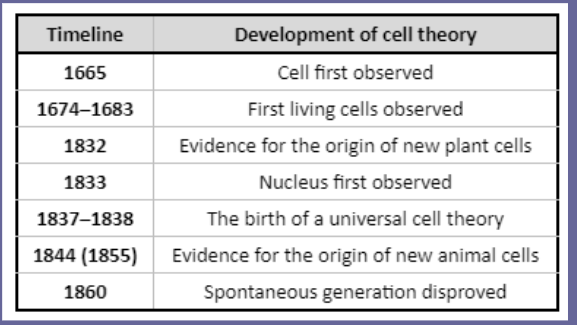
History of microscopy
Light microscopes
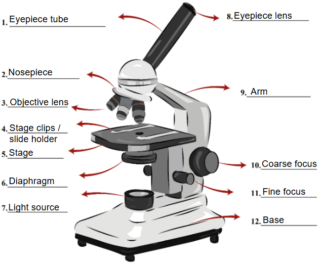
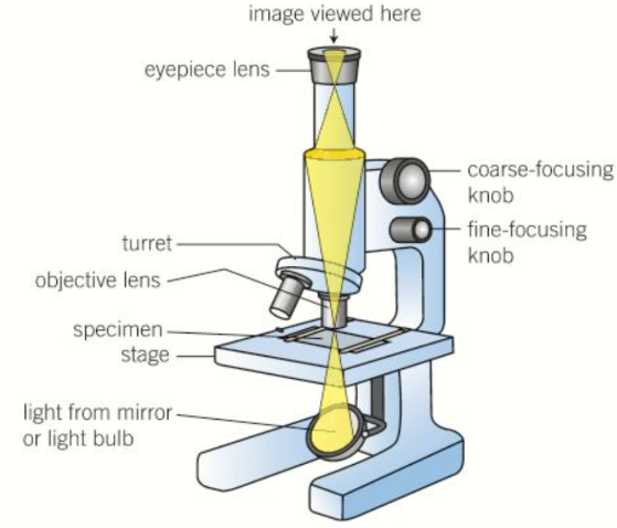
Compound microscope
2 lenses
objective lens magnifies the image
eyepiece lens magnifies further
allows higher magnification vs simple light microscope
less chromatic aberration
some opaque specimen can be viewed from above
Magnification vs resolution
magnification
How many times larger the image is compared to the object
resolution
the ability to distinguish separate objects
higher resolution = more detail
Making permanent slides
Fixing
chemicals like formaldehyde used to preserve specimen in as close to natural state as possible
Sectioning
specimen dehydrated with alcohol then mounted in wax to form hard block
microtome (special sharp knife) used to cut very thin sections
Staining
specimen often treated with multiple stains to highlight different structures
Mounting
specimen secured onto a microscope slide and a coverslip placed on top
Sample preparation methods
dry mount
object to be viewed is placed on a slide and covered with a cover slip
wet mount
specimen to be viewed is placed on a slide and a drop of liquid added
cover slip lowered carefully to avoid air bubbles
smear slide
sample added to one end of the slide and a second slide pulled back until it reaches the sample
slide is then pushed across the surface of the lower side to create a thin smear spreading out sample in a thin layer
squash slide
wet mount slide prepared
lens tissue placed over the coverslip and gently pressed
Methods of staining
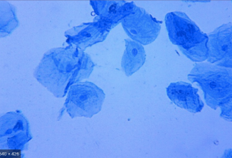
Positive staining
crystal violet or methylene blue
positively charged dyes so attracted to negatively charged cell structures
leads to staining of cell parts so they are more easily distinguished from cytosol (cell cytoplasm)
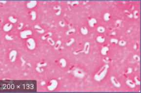
negative staining
congo red or nigrosin
negatively charged so repelled by cell structures
leave cells unstained but colour the background so cells stand out
Differential staining can distinguish between different organisms or different organelles in the same organism
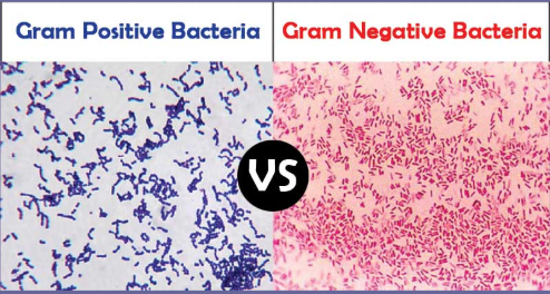
Gram staining
used to separate bacteria into two groups
gram positive
gram negative
method
add crystal violet stain
add iodine (fixes dye)
wash with alcohol
counterstain with safranin
gram positive bacteria retain the crystal violet stain and appear blue
gram negative bacteria lose the crystal violet stain when washed with alcohol and so appear red due to counterstain
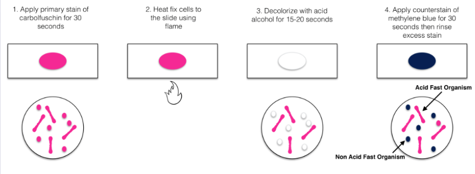
Acid fast
used to identify mycobacterium species from other bacteria
mycobacteria are red
other bacteria species are blue