Skeletal Muscle
Alpha Motor Neurones:
α-motor neurones (α-motoneurones) are neurones of the spinal cord. Their cell bodies are located in the ventral horn, with their axons projecting out to skeletal muscle.
Motor Units:
Skeletal muscles are made up of bundles of muscle fibres.
A motor unit is a single α-motoneurone and all the muscle fibres it innervates.
Each α-motor neurone can synapse with multiple muscle fibres, but the fibres only receive input from a single motoneurone (focal innervation).
A safety feature of nerve terminals is that they always release 8-10x more ACh than necessary in order to ensure that a muscle contraction with a single AP occurs.
Neuromuscular Junction:
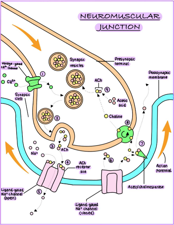
1. An action potential arrives at the presynaptic terminal and causes voltage-gated Ca2+ ion channels in the presynaptic membrane to open.
2. Calcium ions then enter the presynaptic terminal and initiate the release of ACh from synaptic vesicles.
3. Acetylcholine is released into the synaptic cleft by exocytosis.
4. ACh diffuses across the synaptic cleft and binds to ligand-gated Na+ ion channels on the postsynaptic membrane.
5. Ligand-gated sodium ion channels open and Na+ ions enter the postsynaptic cell, depolarising it. If it passes the threshold, an action potential is generated.
6. ACh unbinds from the sodium ion channels, which then close.
7. Acetylcholinesterase (AChE) removes ACh from the synaptic cleft by breaking it down into acetic acid and choline.
8. Choline is symported with sodium ions into the presynaptic terminal, where it is used to make ACh. Acetic acid diffuses away from the synaptic cleft.
9. ACh is reformed within the presynaptic terminal using acetic acid generated from metabolism, and from recycled choline.
Smooth Contraction and Contractile Precision:
Each α-motoneurone innervates nerve fibres that are spread throughout the muscle, and fire asynchronously.
The innervation ratio is the number of muscle fibres innervated by each individual α-motoneurone. It is inversely correlated with contractile precision.
Fingers are dextrous, with 5-15 fibres per α-motoneurone.
Abdominal muscles have coarse movements, with 200-1500 fibres.
The lower the innervation ratio, the finer the control.
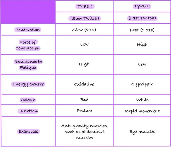
Slow and Fast Twitch Muscle Fibres:
Increasing Force of Contraction:
Generally, slow twitch muscle fibres are suited for endurance activities due to their ability to sustain contractions over longer periods, while fast twitch fibres are designed for high-intensity movements.
Heavy loads require fast-twitch fibres, meaning that glycolytic fatigue occurs quickly.
Increasing the summation of α-motoneurones leads to increased force.
The tetanus (maximum summation) is 3.5kgcm-2 in all muscles.
Ventral Horn Neurones:
α-motoneurones innervate muscle fibres, release ACh and cause contraction of muscle fibres via nAChRs.
Renshaw Cells are inhibitor interneurones which release glycine, and synapse with α-motoneurones to inhibit their activity.
Lateral inhibition prevents overactivity and enables fine control of movement.
Y-motoneurones innervate muscle spindles.
Muscle Spindles:
Intrafusal muscle fibres are attached to extrafusal muscle fibres.
Y-motoneurones innervate the ends of intrafusal muscles, whereas sensory neurones innervate the middle section.
Sensory Neurones of Muscle Spindles:
Dynamic nuclear bag fibres have only annulospiral sensory endings.
Static nuclear bag fibres have annulospiral and flower spray sensory endings.
Nuclear chain fibres have annulospiral and flower spray endings.
Knee Jerk Reflex:
Strike the patellar tendon with a hammer.
This stretches the muscle spindle in the quadricep.
Action potentials project along la afferent sensory fibres to the spinal cord.
Synapse (glutamate, which is excitatory) with alpha motor neurones in the ventral horn that innervates the quadricep.
The α-motoneurones release ACh at the neuromuscular junction causing contraction of the quadricep.
Collateral synapse (glutamate, which is excitatory) with Renshaw cell in the ventral horn.
The Renshaw cell (glycine, which is inhibitory) synapses with α-motoneurones which innervate the hamstring. This causes reciprocal inhibition of the hamstring.
The leg raises.
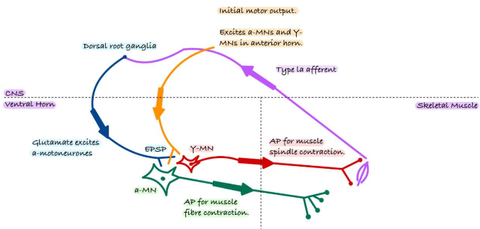
The Gamma (Y) Loop:
The gamma loop is a neural feedback mechanism that helps regulate muscle tone and maintain postural stability by controlling the sensitivity of muscle spindles.
Golgi Tendon Organs:
They are sensory mechanoreceptors located within tendons.
They inhibit muscle activation when tension becomes too high.
They are innervated by lb sensory neurones.
They enact the inverse stretch reflex which causes a muscle to relax if it is stretched.