Unit 3 - Human Physiology
Ch. 12 - Fundamentals of the Nervous Systems and Nervous Tissue
Nervous System
Master Control
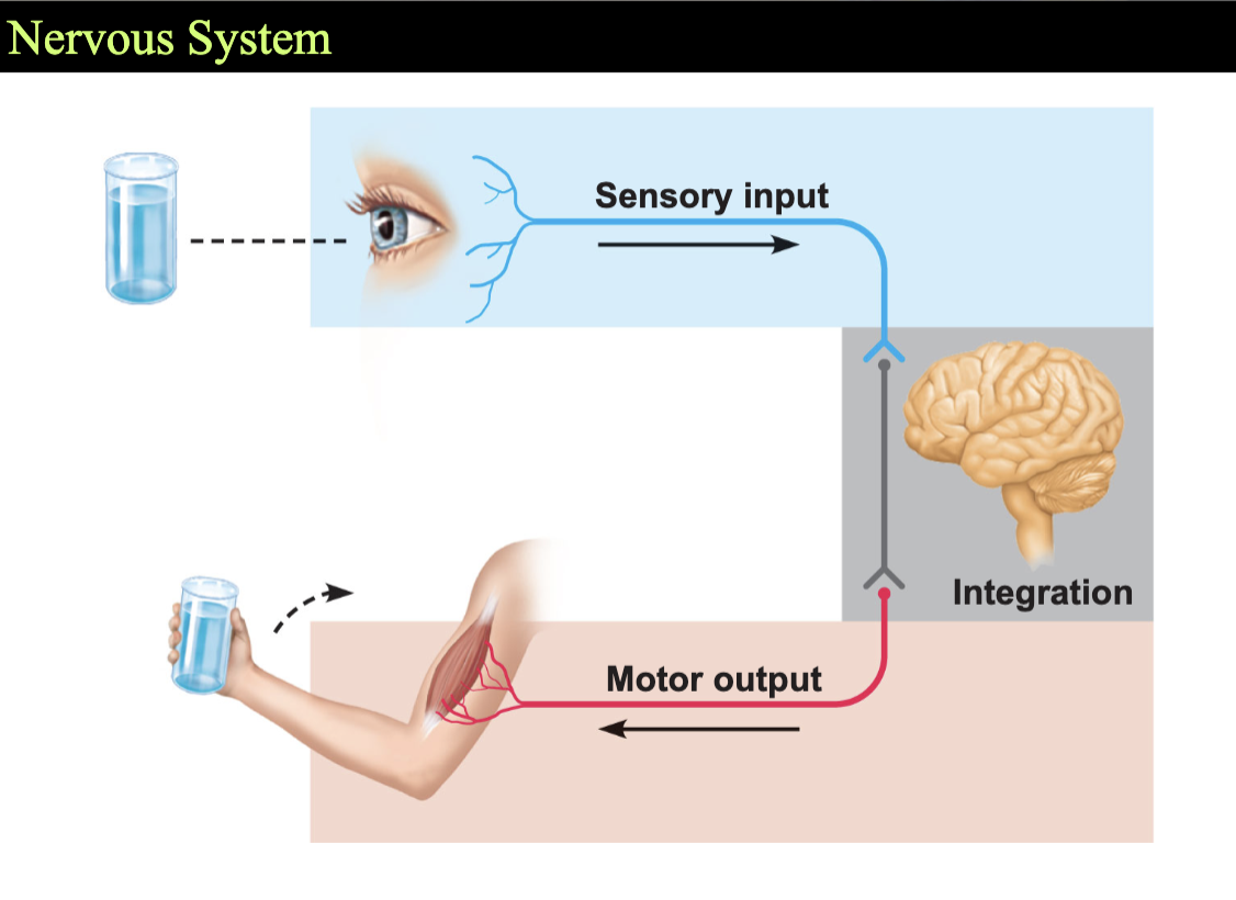
Has 3 overlapping functions
Sensory input - Sensory receptors - monitor inside and outside the body
from PNS to CNS
Integration - Processes and interprets sensory input
Makes decisions - integration
Motor Output - Dictates a response by activating effector organs
response —> muscles and glands
From CNS to PNS
Basic Divisions of the Nervous System
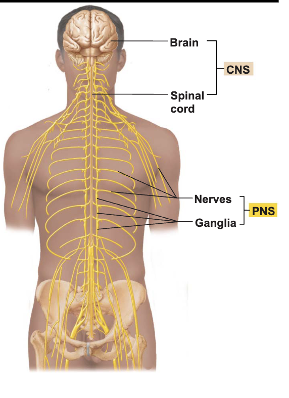
Central Nervous System (CNS)
brain and spinal cord
integrating and command center; processes information
Peripheral Nervous System
outside the CNS
carries sensory input to CNS and motor output away
nerves from brain and spinal cord
cranial nerves and spinal nerves
peripheral nerves link all regions of the body to CNS
Sensory Input and Motor Output
Divided according to region they serve
Somatic body region (outer body structures: skin, joints, muscles and bones)
Visceral body region (inner body structures: heart, lung, and intestines)
Results in four main subdivisions
Somatic sensory
Visceral sensory
Somatic motor
Visceral motor
Types of Sensory and Motor Information
Sensory (afferent) division
Somatic Sensory
General: touch, pain, pressure, vibration, temperature, and proprioception in skin, body wall, and limbs
Special: hearing, equilibrium and vision
Visceral Sensory
General: stretch, pain, temperature, chemical changes, and irritation in viscera; nausea and hunger
Special: taste and smell
Motor (efferent) division
Somatic motor (voluntary motor)
General: motor innervation of all skeletal muscles
Visceral motor (involuntary motor)
General: motor innervation of smooth muscle, cardiac muscle, and glands; equivalent to autonomic nervous system (ANS)
STUDY TIP: Afferent = Arrives (Sensory) ; Efferent = Exits (Motor)
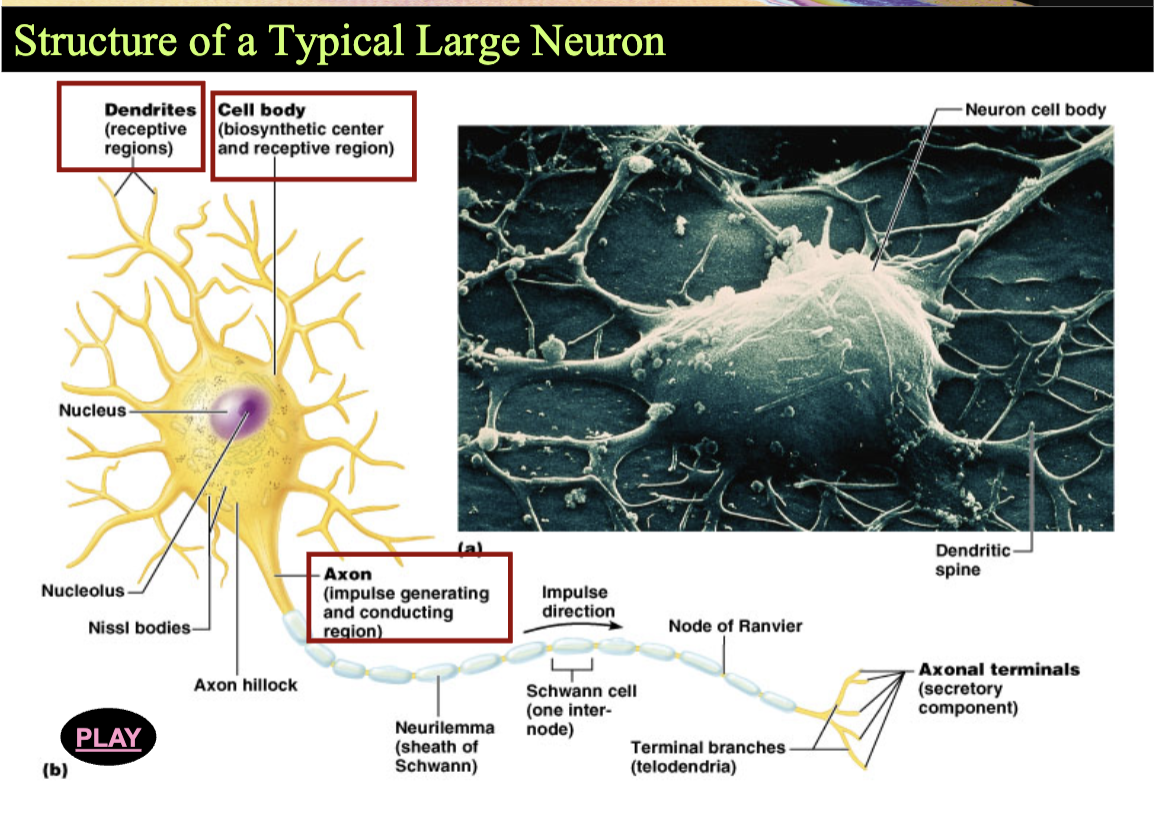
Structure of a Typical Large Neuron:
Anatomy of a Neuron
Dendrites —> Receive Signals
Cell Body (Soma) —> Integrates Information
Axon —> Carries Impulses Away
Myelin —> Insulates Axon; Speeds Conduction
Nodes of Ranvier —> Impulse jumps node to node
Axon Terminals —> Release Neurotransmitters
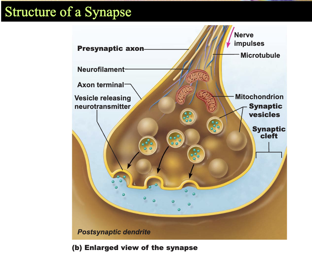
Synapses
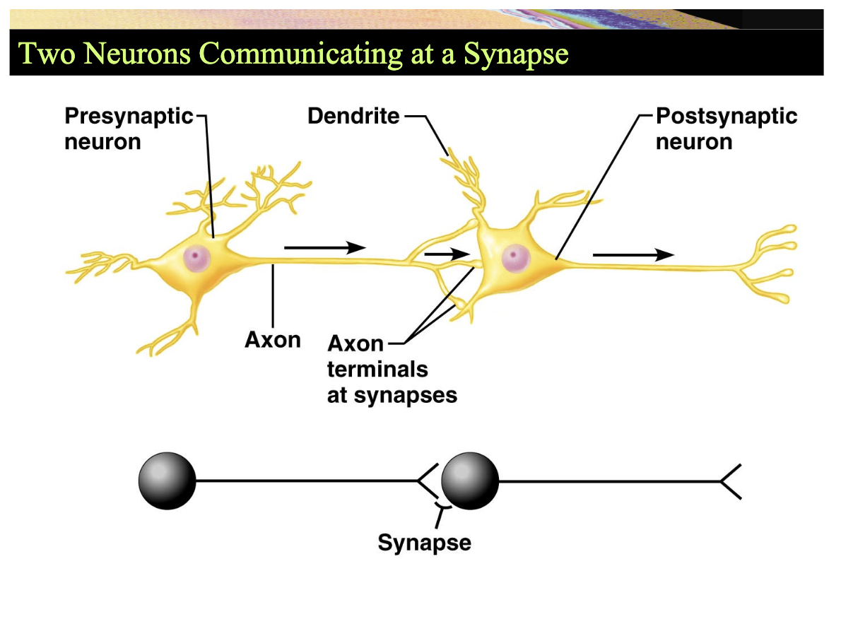
Site at which neurons communicate
Signals pass across synapse in one direction
Presynaptic neuron
Conducts signal toward a synapse
Postsynaptic neuron
Transmits electrical activity away from a synapse
Two Neurons Communicating at a Synapse
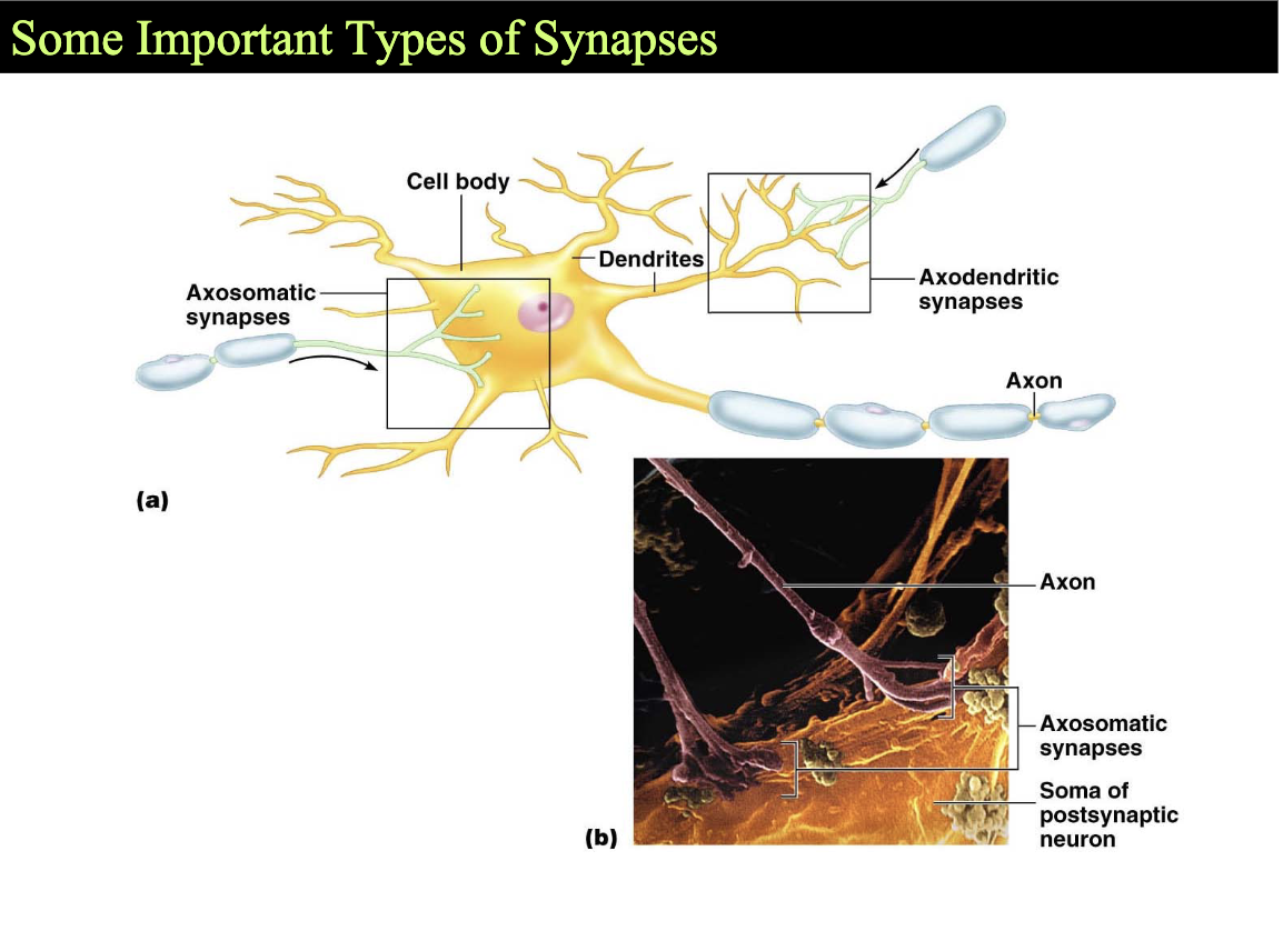
Axosomatic Synapses:
forms between the axon terminal of one neuron and the cell body (soma) of another neuron
has a strong influence on whether the post synaptic neuron fires an action potential because they are close to axon hillock
often inhibitory, help reduce or prevent the neuron from generating an action potential
Axodendritic Synapses:
form between axial terminal of one neuron and the dendrites of another
most common type of synapse in the nervous system and receive most incoming signals
usually excitatory, transmitting information that contributes to whether the neuron will fire an action potential
Structure of a Synapse
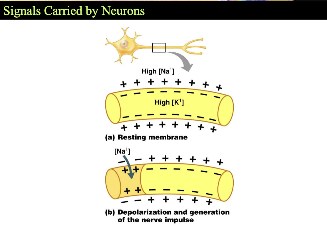
Signals Carried by Neurons*
plasma membranes of neurons conduct electrical signals
resting neuron
membrane is polarized
inner, cytoplasmic side is negatively charged
stimulation of the neuron —> depolarization
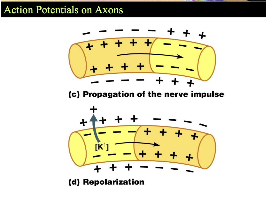
Action Potentials on Axons
strong stimulus applied to the axon triggers
nerve impulse or action potential
membrane becomes negative externally
impulse travels the length of the axon and membrane repolarizes (neutralizes) itself
Synaptic Potentials*
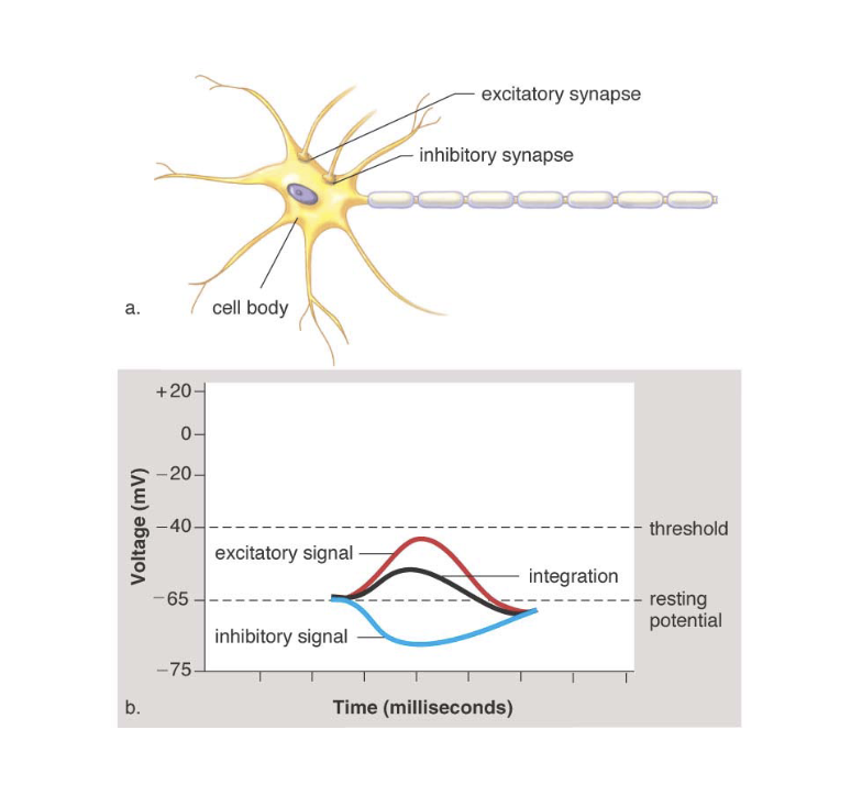
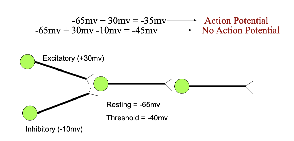
Excitatory synapses
leads to an inflow of positive ions
depolarizes the postsynaptic membrane
drives the postsynaptic neuron toward impulse generation
Inhibitory synapses
inside becomes more negative
action potential less likely
Classification of Neurons
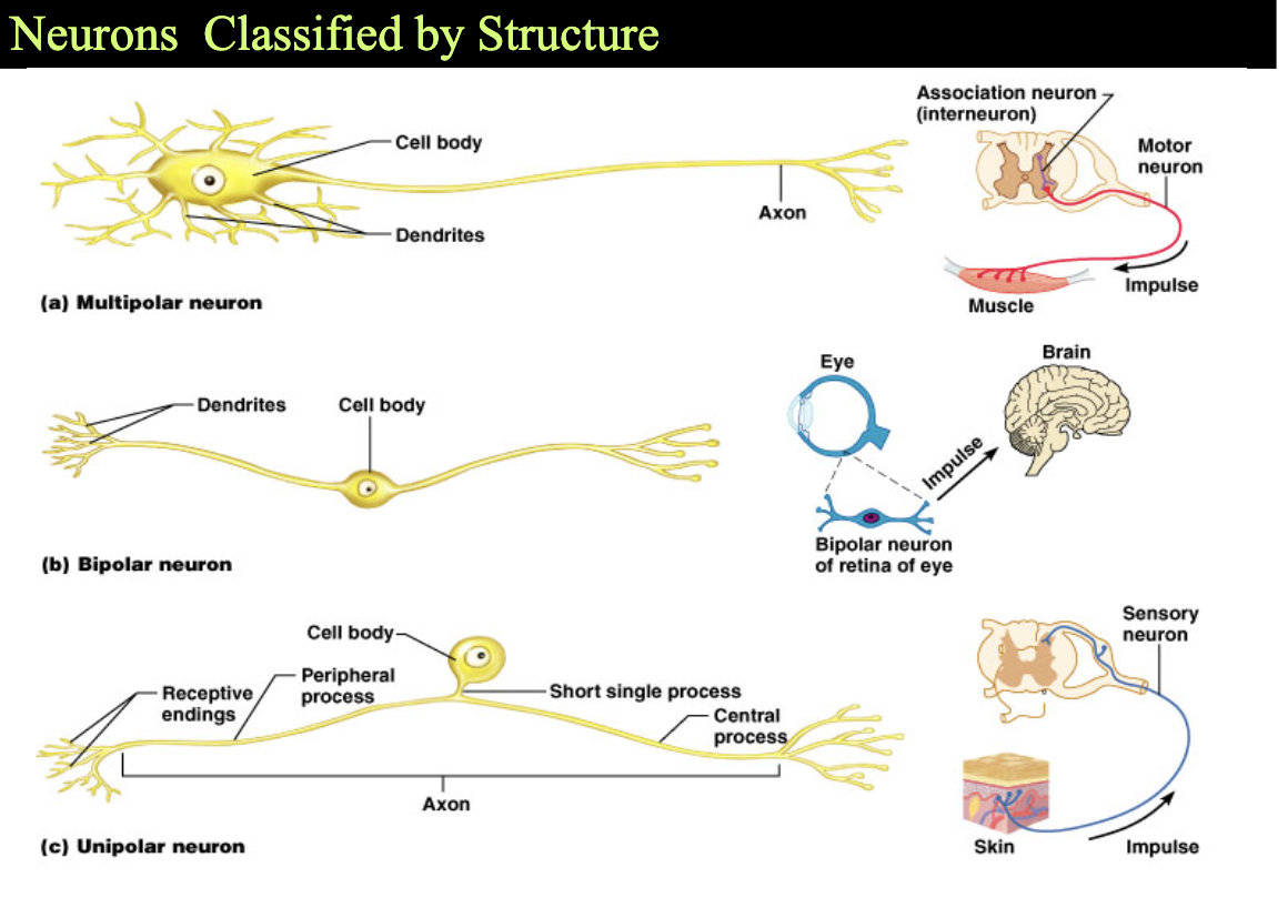
Structural Classification
Multipolar - possess more than two processes
Numerous dendrites and one axon
Bipolar - possess two processes
Rare neurons - found in some special sensory organs
Unipolar (pseudounipolar) - possess one short, single process
Functional Classification of Neurons*
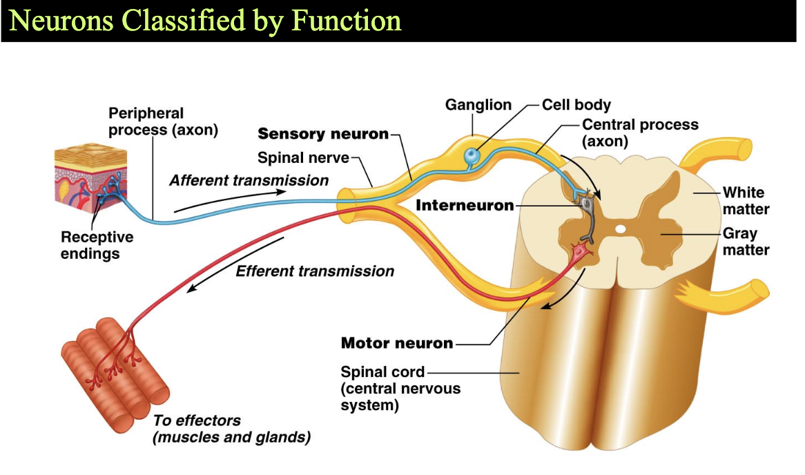
According to the direction the nerve impulse travels
Sensory (afferent) neurons - transmit impulses toward the CNS
virtually all are unipolar neurons
cell bodies in ganglia outside the CNS
Motor (efferent) neurons
carry impulses away from the CNS to effector organs
most are multipolar
cell bodies are within the CNS (ex: nucleus)
form junctions with effector cells
Interneurons (association neurons) - most are multipolar
lie between motor and sensory neurons
confined to the CNS
Supporting Cells
Six types of supporting cells
Four in the CNS
Two in the PNS
Provide supportive functions for neurons
non-excitable
maintenance
insulation
Cover nonsynaptic regions of the neurons
Neuroglia
Function:
support, protect, and feed neurons to keep nervous system going
forming myelin insulation
maintaining chemical balance
defending against damage
outnumber neurons 10 - 1
make up half the mass of the brain
Types of Neuroglial Cells*
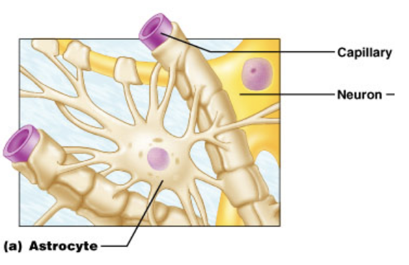
Astrocyte
CNS - Support neurons; Blood Brain Barrier
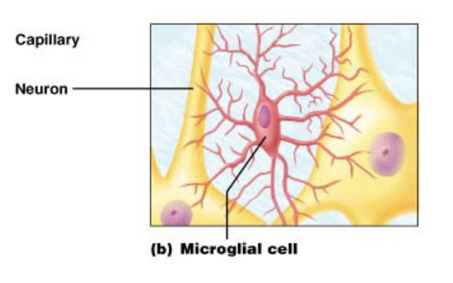
Microglia
CNS - Immune defense
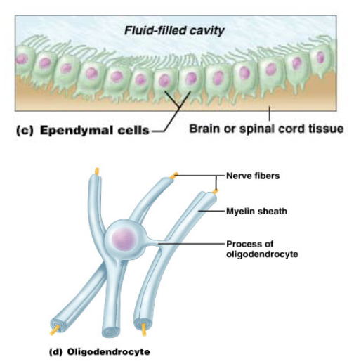
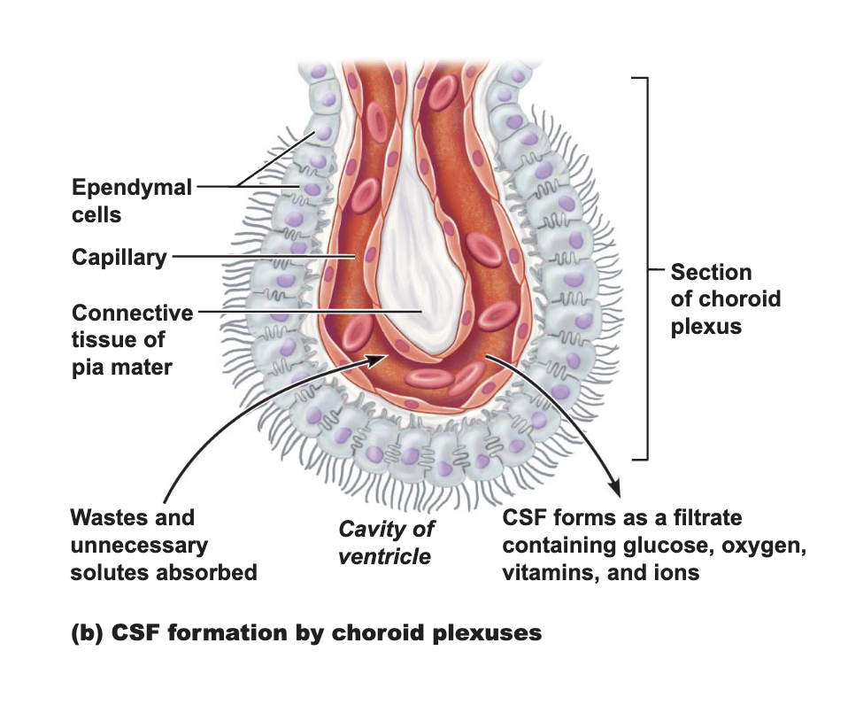
Ependymal Cells*
CNS - Produce/Circulate CSF
Oligodendrocytes*
CNS - Myelin for multiple axons
Supporting Cells in the PNS
Satellite cells
surround neuron cell bodies within ganglia
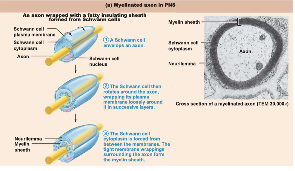
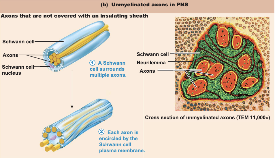
Schwann cells (neurolemmocytes)
surround axons in the PNS
form myelin sheath around axons of the PNS
Myelin Sheath
segmented structures composed of the lipoprotein myelin
surround thicker axons
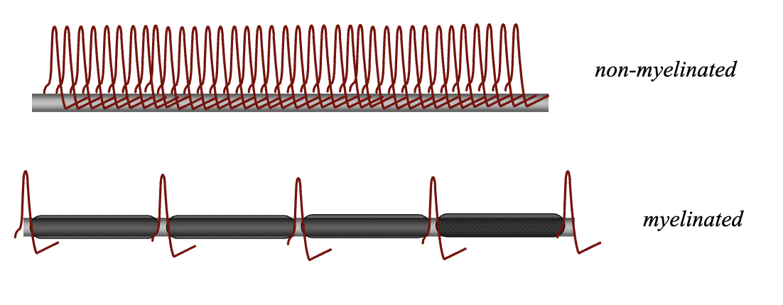
form an insulating layer
prevent leakage of electrical current
increase the speed of impulse conduction
Myelin Sheaths in the CNS
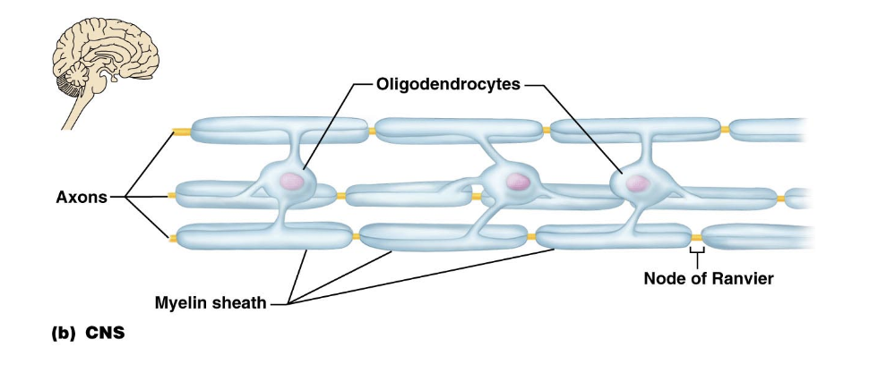
Oligodendrocytes form the myelin sheaths in the CNS
have multiple processes (multipolar)
coil around several different axons
Disorders of the Nervous System
Multiple sclerosis
An autoimmune disease
Immune system attacks the myelin around axons in the CNS
Nerves
Nerves - cordlike organs in the PNS
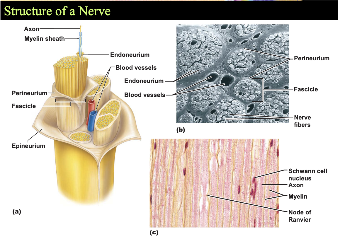
Consists of numerous axons wrapped in connective tissue
Axon is surrounded by Schwann cells
KNOW - Structure of a Nerve*
Axon —> Myelin Sheath —> Endoneurium —> bundle of axons in Perineurium —> Fascicle with Blood Vessels in —> Epineurium
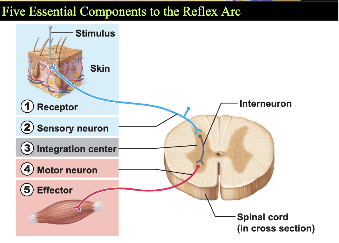
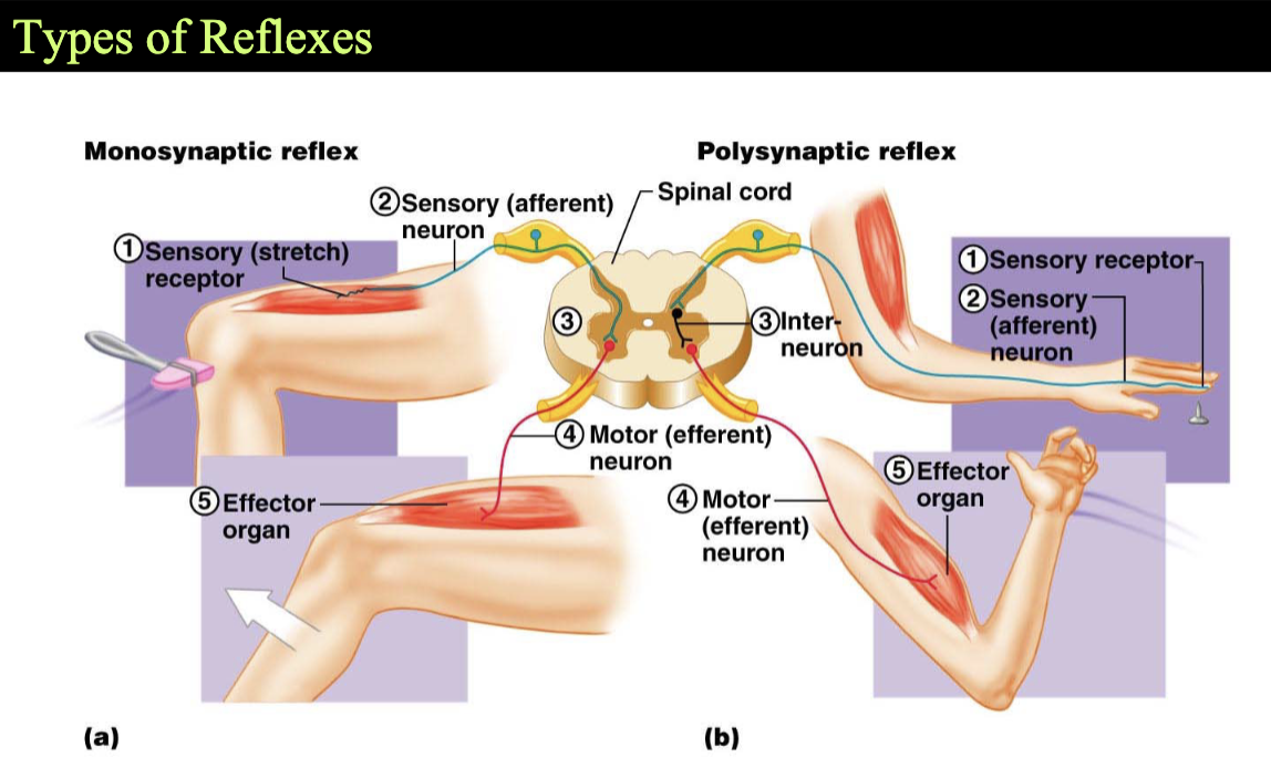
Reflex Arcs
explain and are responsible for reflex behaviors
rapid, autonomic motor responses
can be visceral or somatic
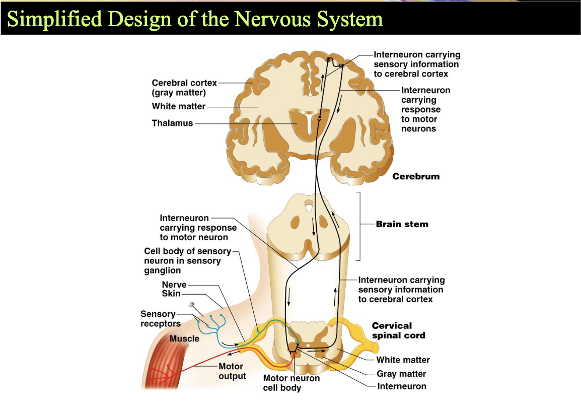
Simplified Design of the Nervous System
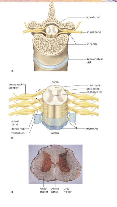
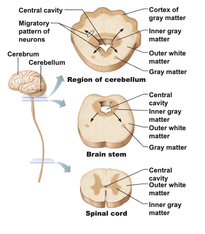
Gray matter in the spinal cord
H-shaped region - surrounds central cavity
Cell bodies are clustered in the gray matter
Nucleus vs. ganglia
White matter in the spinal cord
External to gray matter
No neuronal cell bodies, but millions of axons
Myelin sheath - white color
Consists of axons running between different parts of the CNS
Tracts - bundles of axons traveling to similar destinations
vs. nerves
Neuronal Regeneration
Neural injuries may cause permanent dysfunction
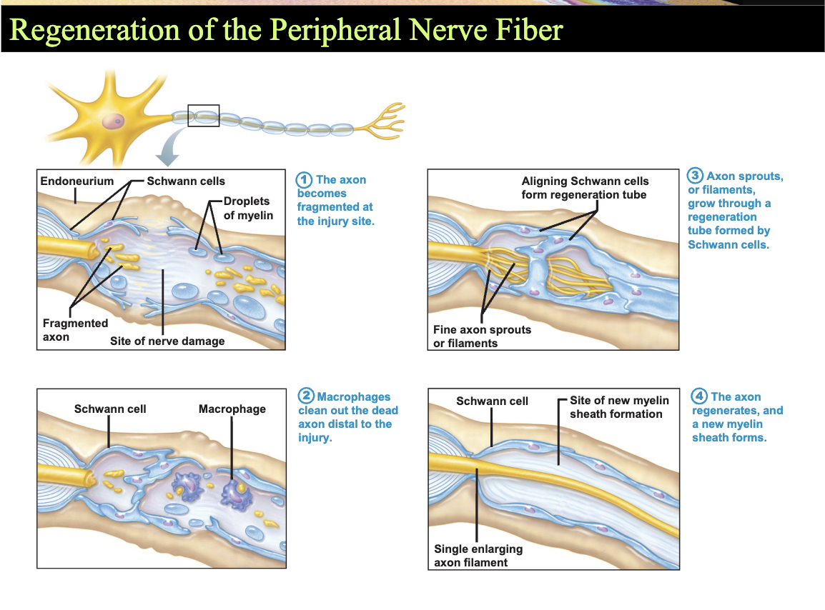
If axons alone are destroyed, cells bodies often survive and the axons may regenerate
PNS - macrophages invade and destroy axon distal to the injury
Axon filaments grow peripherally from injured site
Partial recovery is sometimes possible
CNS - neuroglia never form bands to guide regrowing axons may hinger axon growth with growth - inhibiting chemicals
No effective regeneration after injury to the spinal cord and brain
Chapter 13 - The Central Nervous System
The Central Nervous System
Consists of the brain and spinal cord
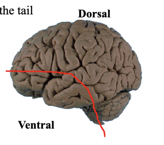
Directional terms unique to the CNS
Rostral - toward the nose
Caudal - toward the tail
Brain Development
Brain Development from Week 5 to Birth
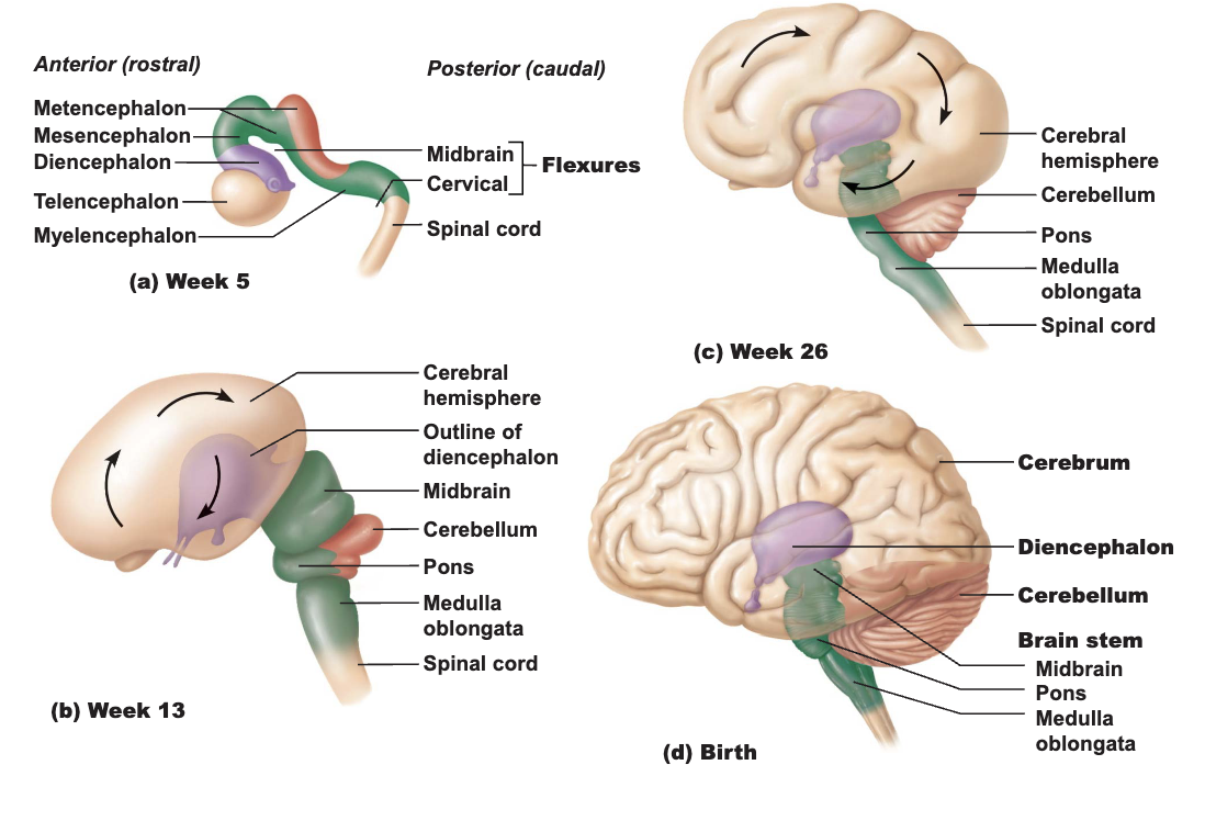
Basic Parts and Organization of the Brain
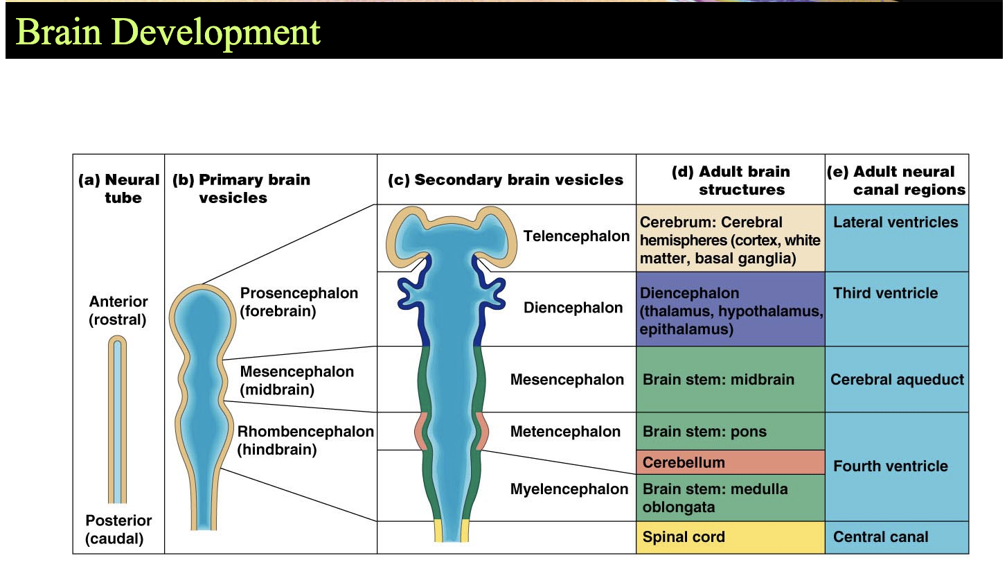
Divided into four regions
1) Cerebrum
2) Diencephalon
3) Brain stem: midbrain, pons, and medulla
4) Cerebellum
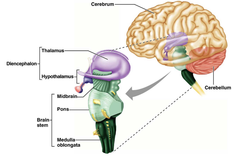
Basic Parts and Organization of the Brain
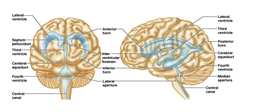
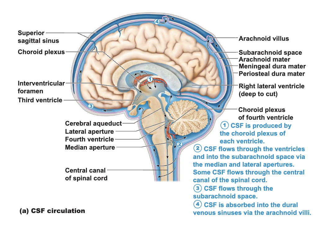
Ventricles of the Brain
Expansions of the brain’s central cavity
Filled with cerebrospinal fluid
Lined with ependymal cells
Continuous with each other
Continuous with the central canal of the spinal cord
Lateral ventricles - located in cerebral hemispheres
Horshoe-shaped from bending of the cerebral hemispheres
Third ventricle - lies in diencephalon
Connected with lateral ventricles by interventricular foramen
Cerebral aqueduct - connects 3rd and 4th ventricles
Fourth ventricle - lies in hindbrain
connects tho the central canal of the spinal cord
Protection of the Brain
The brain is protected from injury by
The skull
Meninges
Cerebrospinal fluid
Blood-brain barrier
Meninges
cover and protect the CNS
enclose and protect the vessels that supply the CNS
contain CSF
The Dura Mater - strongest of the meninges
largest sinus - the superior sagittal sinus
extends inward to subdivide the cranial cavity
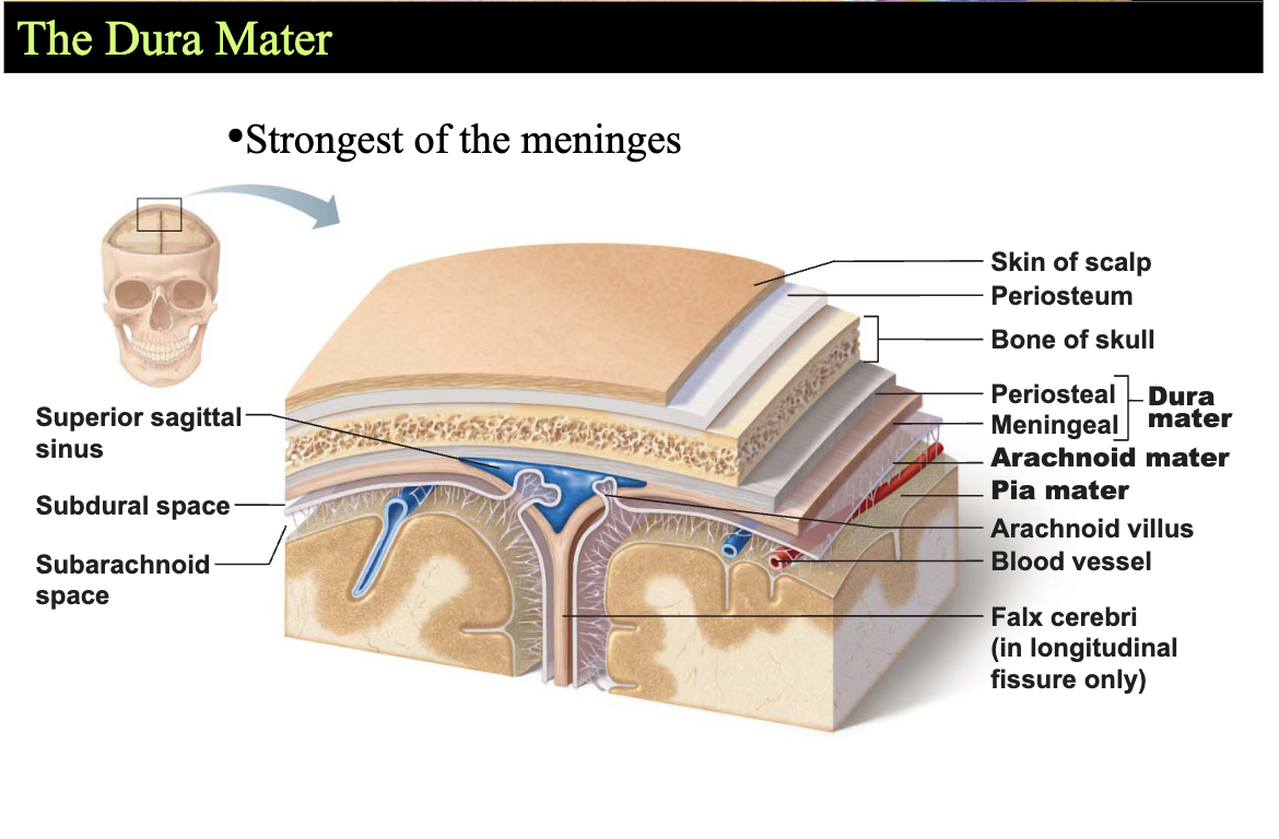
The Arachnoid Mater
located beneath the dura mater
Subdural space
potential space between dura and arachnoid mater
Subarachnoid space
filled with CSF (cerebralspinal fluid)
contains the blood vessels that supply the brain
Arachnoid villi
allow CSF to pass into the dural blood sinuses
The Pia Mater
Delicate connective tissue
Clings tightly to the surface of the brain
follows all convolutions of the cortex
Protection of the Brain - Cerebrospinal Fluid (CSF)
Provides a liquid cushion for the brain and spinal cord
The brain “floats” in CSF
Formed in choroid plexuses in the brain ventricles
Protection of the Brain - Blood-Brain Barrier
Prevents most blood-borne toxins from entering the brain
Impermeable capillaries
Not an absolute barrier
Nutrients such as oxygen pass through
Allows alcohol, nicotine, and anesthetics through
The Cerebral Hemispheres
Account for 83% of brain mass
Fissures - deep grooves - separate major regions of the brain
Transverse fissure - separates cerebrum and cerebellum
Longitudinal fissure - separates cerebral hemispheres
Sulci - grooves on the surface of the cerebral hemispheres - grooves between gyri
Gyri - twisted ridges between sulci - raised folds
Prominent gyri and sulsi are similar in all people
Deeper sulci divide cerebrum into lobes
Lobes are named for the skull bones overlying them
Central sulcus separates frontal and parietal lobed
Bordered by two gyri
Precentral gyrus
Postcentral gyrus
Parieto - occipital sulcus
Separates the occipital from the parietal lobe
Lateral sulcus
Separates temporal lobe from parietal and frontal lobes
Insula - deep within the lateral sulcus
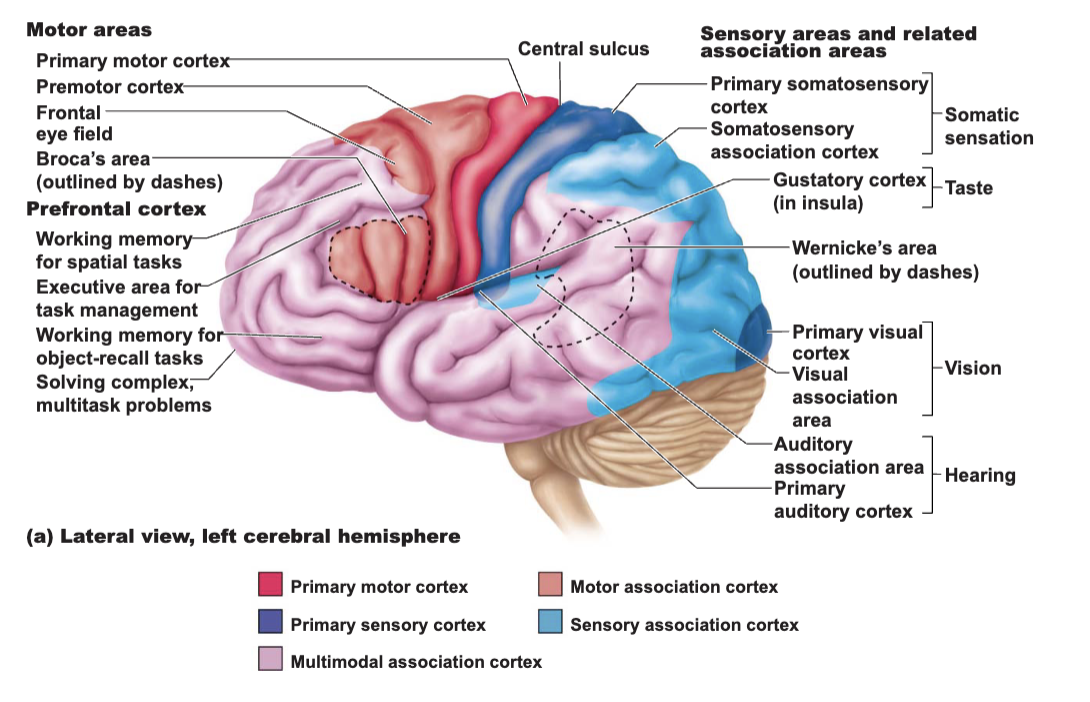
Functional and Structural Areas of the Cerebral Cortex (conscious part of brain)
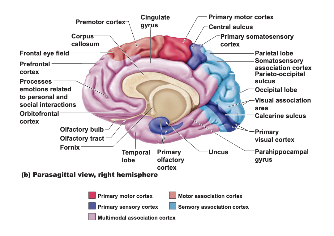
The Cerebral Cortex
Three kinds of functional areas
1) Motor areas
2) Sensory areas
3) Association areas
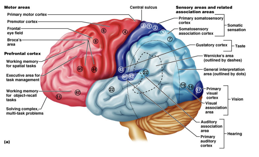
Motor Areas - Primary Motor Cortex
Control motor functions
Primary motor cortex (somatic motor area)
Located in precentral gyrus (Brodmann area 4)
Pyramidal cells - large neurons of primary motor cortex
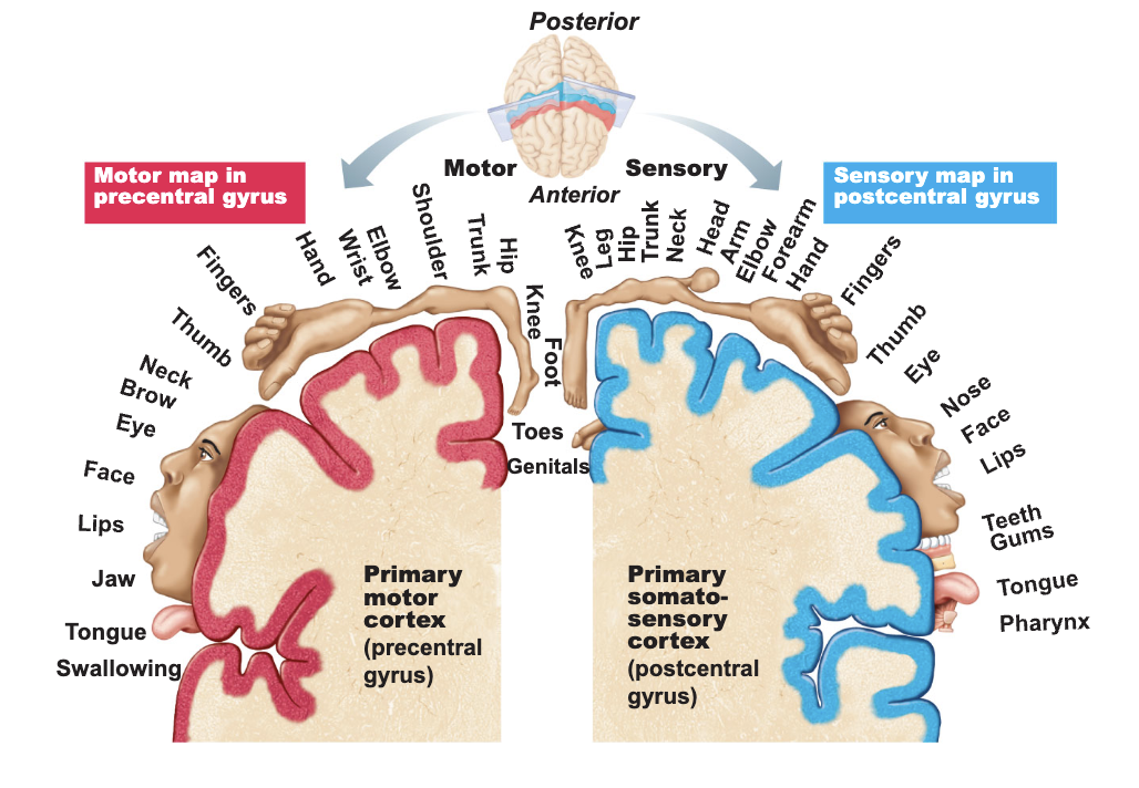
Motor Areas - Premotor Cortex
located anterior to the precentral gyrus
controls more complex movements
receives processed sensory information
visual, auditory, and general somatic sensory
controls voluntary actions dependent on sensory feedback
involved in the planning of movements
Sensory Areas
cortical areas involved in conscious awareness of sensation
located in parietal, temporal, and occipital lobes
distinct area for each of the major senses
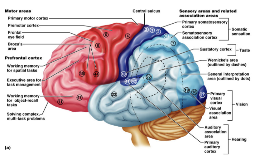
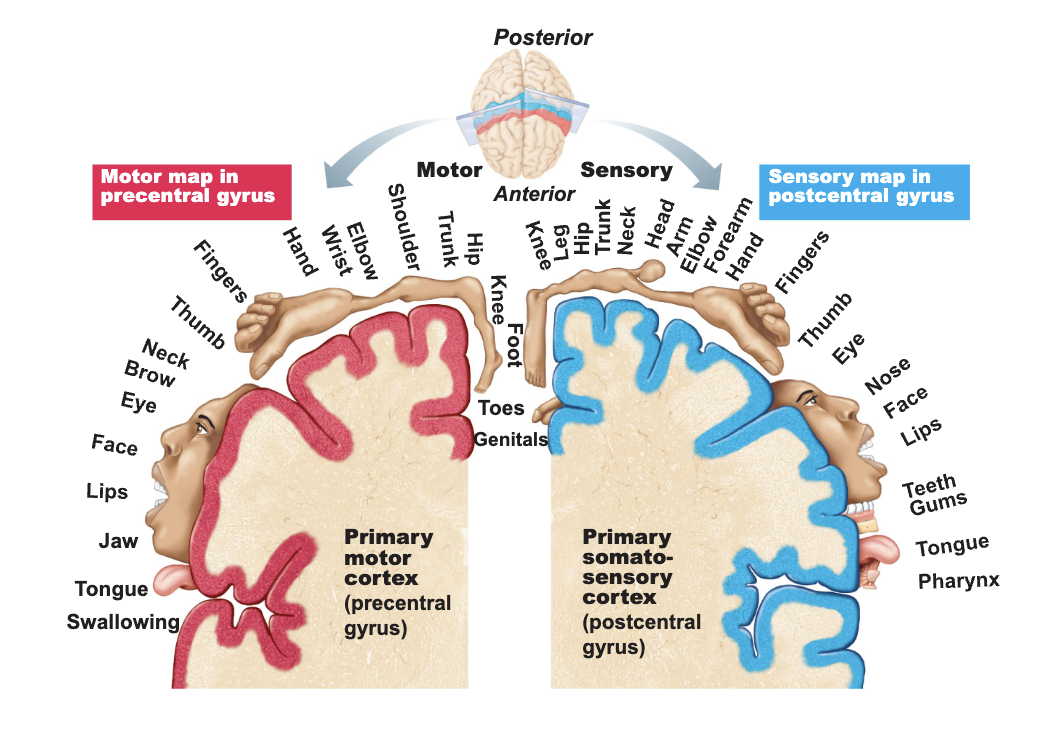
Sensory Areas - Somatosensory Association Area
Lies posterior to the primary somatosensory cortex
Corresponds to Brodmann areas 5 and 7
Integrates different sensory inputs
Touch, pressure, and others
Draws upon stored memories of past sensory experiences
Interprets new with respect to old
Language and Speech
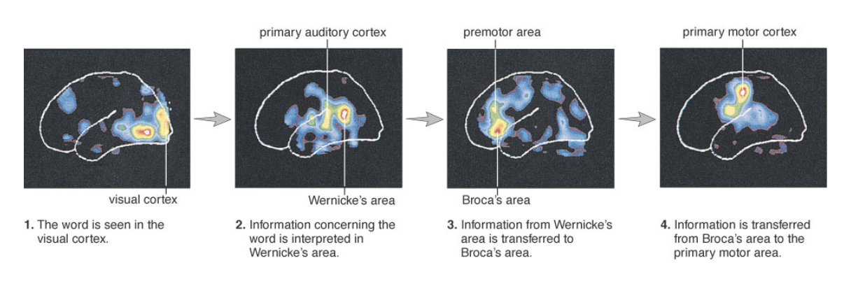
Lateralization of Cortical Functioning
The two hemispheres control opposite sides of the body
Hemispheres are specialized for different cognitive functions
More pronounced in men
Females generally have larger connections between 2 sides
Damage to left side produces aphasia
Damage to same area on right side lead to speech with little emotional inflection
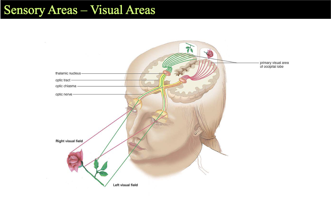
Sensory Areas - Visual Areas
Cerebral White Matter
Types of tracts
Commissures - composed of commissural fibers
Allows communication between cerebral hemispheres
Corpus callosum - the largest commissure
Association fibers
Connect different parts of the same hemisphere
Projection fibers - run vertically
descend from the cerebral cortex
ascend to the cortex from lower regions
Projection Tracts
Internal capsule - projection fibers form a compact bundle
passes between the thalamus and basal nuclei
Corona radiata - superior to the internal capsule
fibers run to and from the cerebral cortex
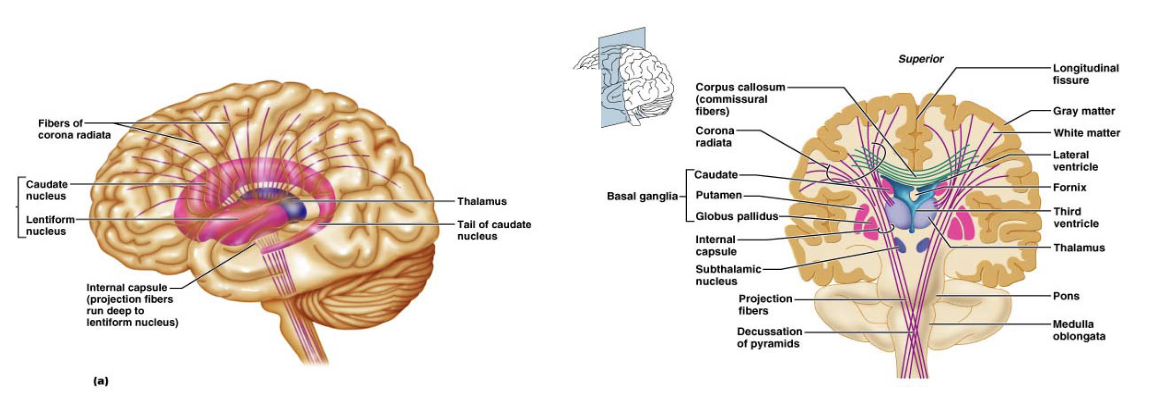
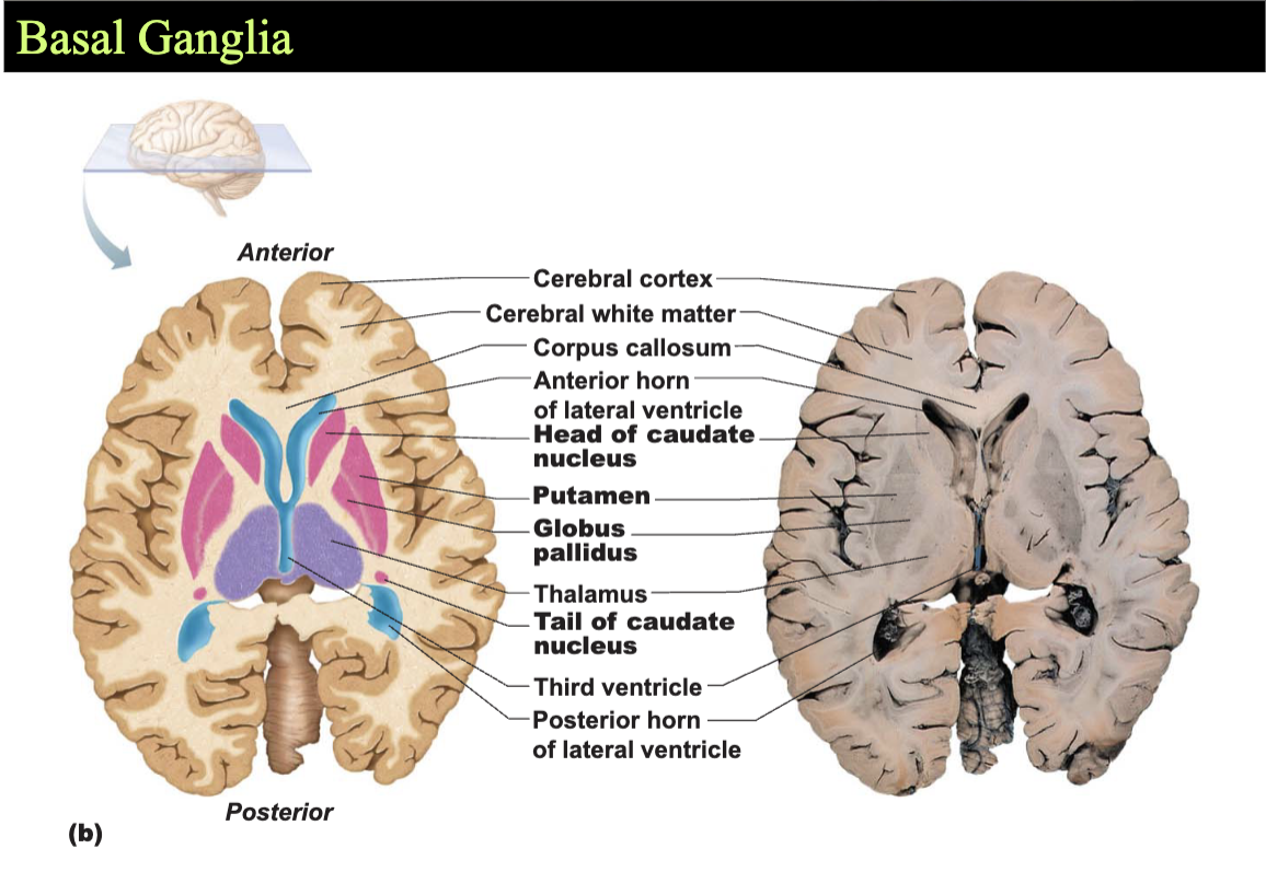
Basal Ganglia
Cooperate with the cerebral cortex in controlling movements
Receive input from many cortical areas
Evidence shows that they:
start, stop, and regulate intensity of voluntary movements
in some way estimate the passage of time
Function: motor control and cognitive and emotional processing
Five primary structures:
Striatum (putamen and caudate nucleus)
Globus Pallidus
Subthalamic Nucleus
Substantia Nigra
The Diencephalon
Composed of three paired structures:
Thalamus, hypothalamus, and epithalamus
Border the third ventricle
Primarily composed of gray matter
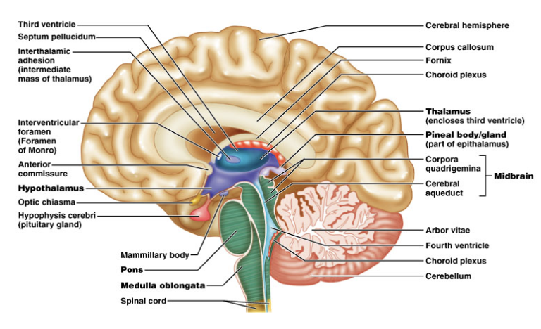
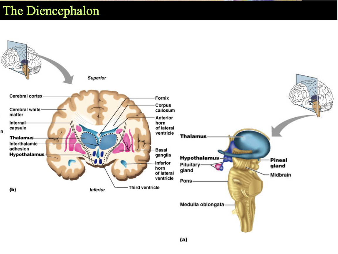
The Diencephalon - The Thalamus
makes up 80% of the diencephalon
contains approx. a doxen major nuclei
send axons to regions of the cerebral cortex
nuclei act as relay stations for incoming sensory messages
“gateway” to the cerebral cortex
nuclei organize and amplify or tone down signals
The Diencephalon - The Hypothalamus
lies between the optic chiasm and the mammilary bodies
pituitary gland projects inferiorly
contains approx a dozen nuclei
main visceral control center of the body
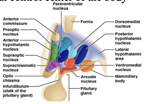
Functions include the following:
Control of the autonomic nervous system
ex: HR, BP
Control of emotional responses
Limbic system
Regulation of body temperature
Regulation of hunger and thirst sensations
Control of behavior
ex: motivation for feeding
Regulation of sleep-wake cycles
optic nerve inpute
Control of endocrine system
Pituitary gland
Formation of memory
The Diencephalon - The Epithalamus
Forms part of the “roof” of the third ventricle
Consists of a tiny group of nuclei
Includes hte pineal gland (pineal gland)
Secrets the hormone melatonin
Under the influence of the hypothalamus
The Brain Stem
Structures include:
Midbrain
Pons
Medulla Oblongata
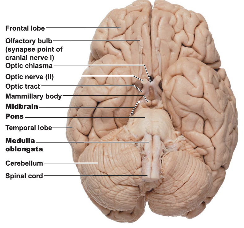
The Brain Stem - The Midbrain
Lies between the diencephalon and the pons
Central cavity - the cerebral aqueduct
Cerebral peduncles located on the ventral surface of the brain
contain pyramidal (corticospinal) tracts
Superior cerebellar peduncles
connect midbrain to the cerebellum
Periaqueductal gray matter surrounds the cerebral aqueduct
Involved in two related functions
fright-and-flight reaction
mediates response to visceral pain
Corpora quadrigemina - the largest nuclei
Superior colliculi - nuclei that act in visual reflexes
Inferior colliculi - nuclei that act in reflexive response to sound
Imbedded in the white matter of the midbrain
Two pigmented nuclei
Substantia nigra - neuronal cell bodies contain melanin
Functionally linked to the basal nuclei
Red nucleus - lies deep to the substantia nigra
Largest nucleus of the reticular formation
The Brain Stem - The Pons
Located between the midbrain and medulla oblongata
Contains the nuclei of cranial nerves V, VI, and VII
Pontine nuclei
Middle cerebellar peduncle
The Brain Stem - The Medulla Oblongata
Most caudal level of the brain stem
Continuous with the spinal cord
Choroid plexus lies in the roof of the fourth ventricle
Pyramids of the medulla - lie on its ventral surface
Decussation of the pyramids - crossing over of motor tracts
Cranial nerves VIII - XII attach to the medulla
Inferior cerebella peduncle
The core of the medulla contains:
Much of the reticular formation
Nuclei influence autonomic functions
Visceral centers of the reticular formation include:
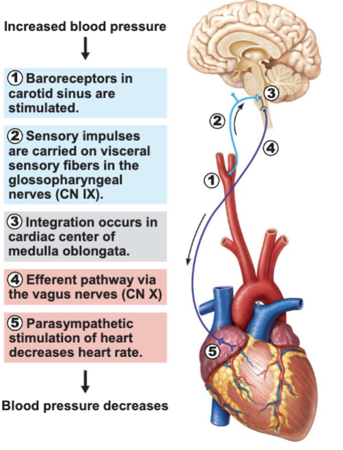
Cardiac center
Vasomotor center
The medullary respiratory center
Centers for hiccupping, sneezing, swallowing, and coughing
The Cerebellum
Located dorsal to the pons and medulla
Smoothes and coordinates body movements
Helps maintain equilibrium
Consists of two cerebella hemispheres
Surface folded into ridges called folia (gyri)
separated by fissures
Hemispheres each subdivided into:
anterior lobe
posterior lobe
flocculonodular lobe
Composed of 3 regions:
Cortex - gray matter
Internal white matter
Deep cerebellar nuclei - deeply situated gray matter
Cerebellum must receive information
On equilibrium
On current movements of limbs, neck, and trunk
From the cerebral cortex
The Cerebellum - Cerebellar Peduncles
Thick tracts connecting the cerebellum to the brain stem
Superior cerebellar peduncles
Middle cerebellar peduncles
Inferior cerebellar peduncles
Fibers to and from the cerebellum are ipsilateral
Run to and from the same side of the body
Functional Brain Systems
Networks of neurons functioning together
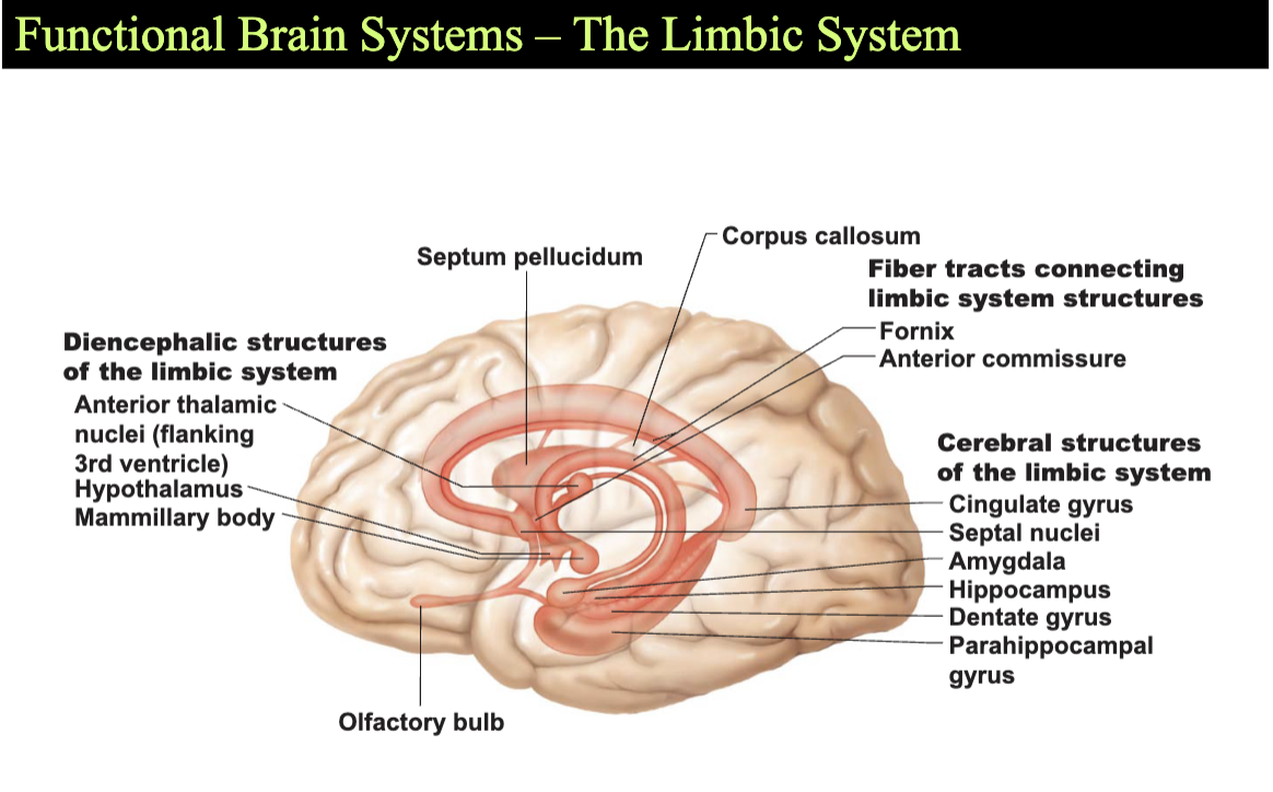
The limbic system - spread widely in the forebrain
The reticular formation - spans the brain stem
The Limbic System
Location
Medial aspect of cerebral hemispheres
Also within the diencephalon
Composed of:
Septal nuclei, cingulate gyrus, and hippocampal formation
Part of the amygdala
The fornix and other tracts link the limbic system together
The “emotional brain”
Cingulate Gyrus
allows us to shift between thoughts
Interprets pain as unpleasant
Encodes, consolidates, and retrieve memories
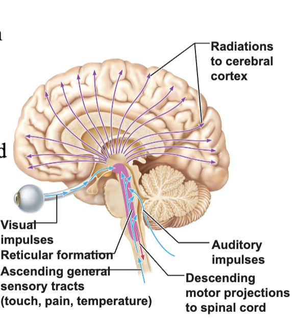
The Reticular Formation
Runs through the central core of the medulla, pons, and midbrain
Widespread connection
Ideal for arousal of the brain as a whole
Reticular Activating System (RAS)
Maintains consciousness and alertness
Functions in sleep and arousal from sleep
The Spinal Cord
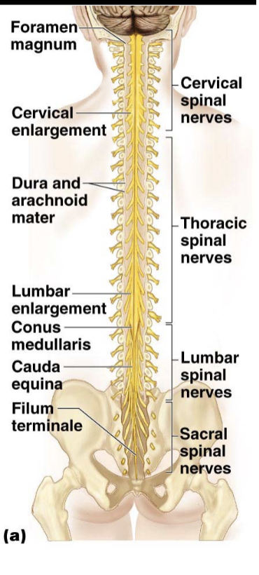
Runs through the vertebral canal
Extends from the foramen magnum to the level of the vertebra L1, or L2
Protected by bone, meninges, and CSF
Conus Medullaris - the inferior end of the spinal cord
Filum Terminale - long filament of connective tissue
Attaches to the coccyx inferiorly
Cervical and lumbar enlargements
Where nerves for upper and lower limbs arise
Cauda equina - collection of nerve roots
Add Diagram
Denticulate ligaments - anchor spinal cord to vertebrae
Two deep grooves run the length of the cord
Posterior median sulcus
Anterior median fissure
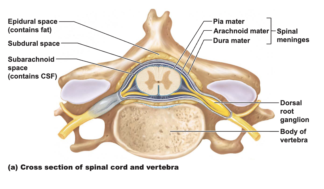
Gray Matter of the Spinal Cord and Spinal Roots
shaped like the letter H
gray commissure - contains the central canal
anterior horns - contain cell bodies of motor neurons
posterior horns - consist of interneurons
gray matter - divided according to somatic and visceral regions
White Matter of the Spinal Cord
Composed of myelinated and unmyelinated axons
3 types of fibers
Ascending
Descending
Commissural
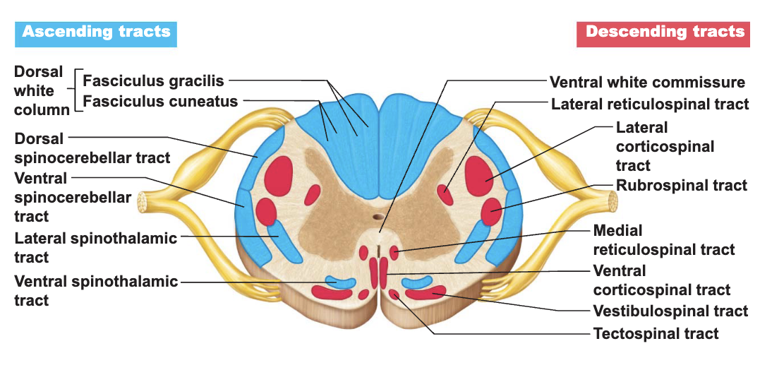
Sensory and Motor Pathways
Most motor pathways:
Decussate at some point along their course
Consist of a chain of two or three neurons
Exhibit somatotopy
Tracts arranged according to the body region they supply
All pathways are paired
One of each on each side of the body
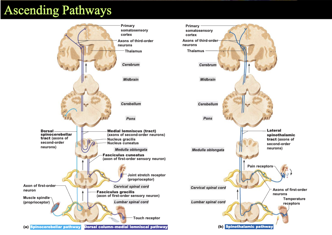
Ascending (Sensory) Pathways
Conduct general somatic sensory impulses
Chains of neurons
4 main ascending pathways
Dorsal column pathway
discriminative tough
Spinothalamic pathway
pain and temperature
Posterior and Anterior spinocerebellar pathway
subconscious proprioception
Descending (Motor) Pathways
Deliver motor instructions from the brain to the spinal cord
Divided into two groups
Pyramidal, or corticospinal tracts
to skeletal muscle
Other motor pathways
Tectospinal tracts
Vestibulospinal tract
Rubrospinal tract
Reticulospinal tract
Chapter 14 - The Peripheral Nervous System
Basic Structure Components of the PNS
Sensory receptors
inside or outside the body
Motor endings
innervate effetors (muscle fibers and glands)
Nerves and ganglia
Nerves
bundles of peripheral axons
Ganglia
clusters of peripheral neuronal cell bodies
Cranial Nerves
Attach to brain
Pass through skull foramina
I-XII
I to II attach to forebrain
Others attach to brain stem
Primarily serve head and neck structure
Exception
X
The 12 Pairs of Cranial Nerves!!
Filaments of Olfactory nerve - Sensory
Optic Nerve - Sensory
Oculomotor Nerve - Motor
Trochlear Nerve - Motor
Trigeminal Nerve - Both
Abducens Nerve - Motor
Facial Nerve - Both
Vestibulocochlear Nerve - Sensory
Glassopharyngeal Nerve - Both
Vagus Nerve - Both
Accessory Nerve - Motor
Hypoglossal Nerve - Motor
Mnemonic: Oh Oh Oh To Touch And Feel Very Good Velvet AH!
Mnemonic: Some Say Marry Money But My Brother Says Big Brains Matter More
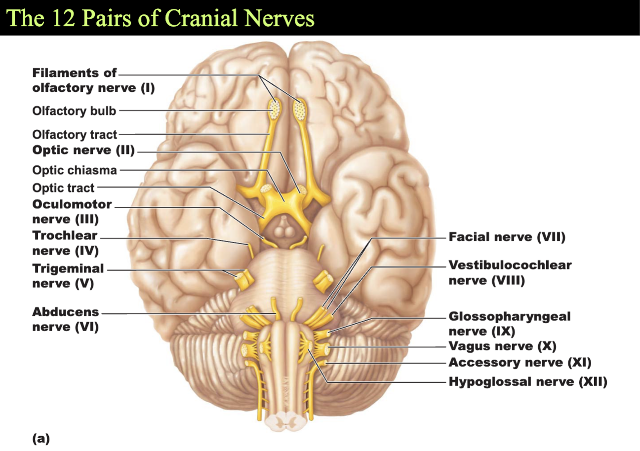
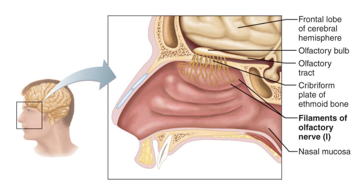
Ofalctory Nerves
Sensory nerves of smell
Sensory
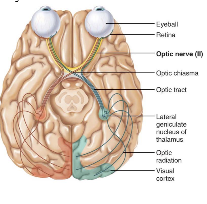
Optic Nerve
Sensory nerve of vision
sensory
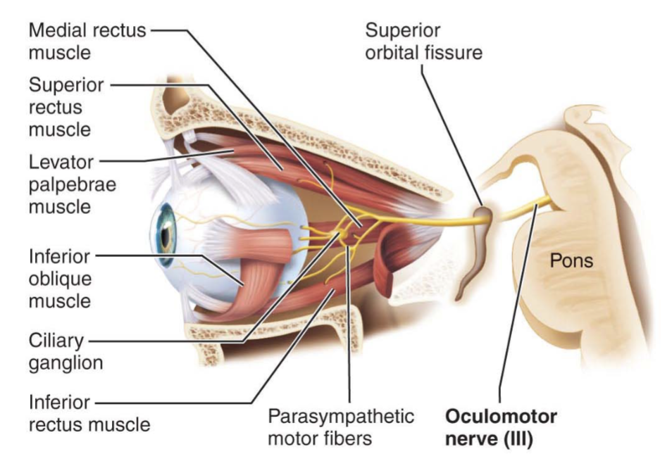
Oculomotor Nerve
Innervates four of the extrinsic eye muscles
motor
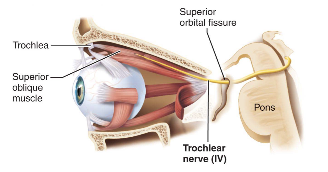
Trochlear Nerve
Innervates an extrinsic eye muscle
motor
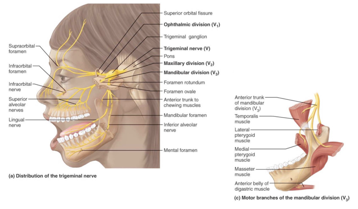
Trigeminal Nerve
Provides sensory innervation to the face
Motor innervation to chewing muscles
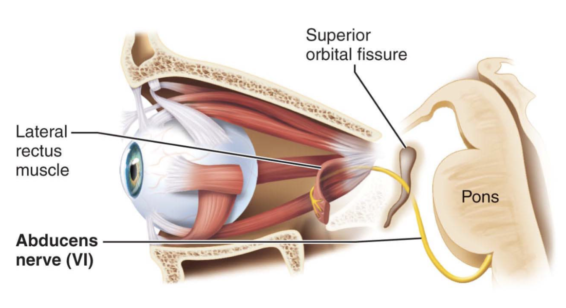
Abducens Nerve
Abducts the eyeball
motor
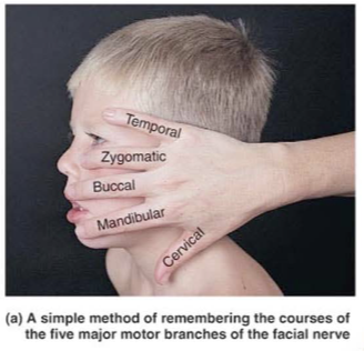
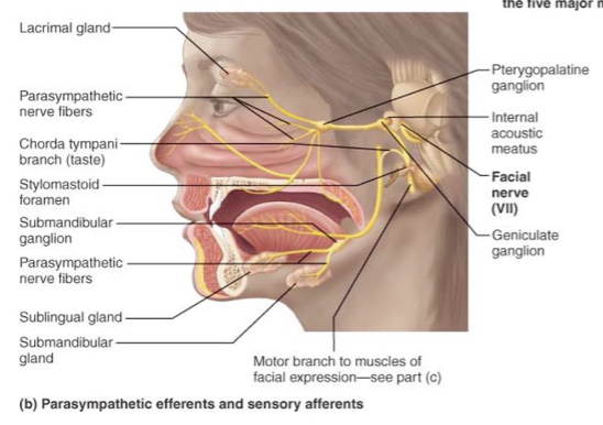
Facial Nerve
Innervates muscles of facial expression
Some taste and some sensation of skin on ear
mixed
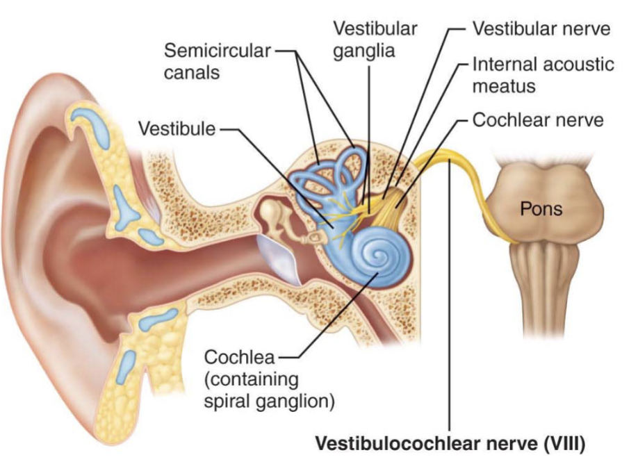
Vestibulocochlear Nerve
Sensory nerve of hearing and balance
sensoory
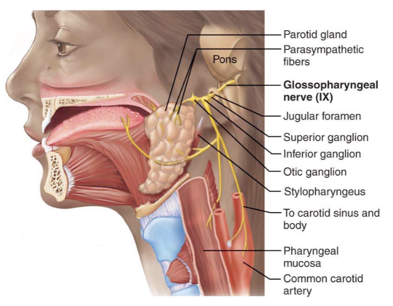
Glossopharyngeal Nerve
Innervates structures of the tongue and pharynx (motor and somatosensory)
Some taste
mixed
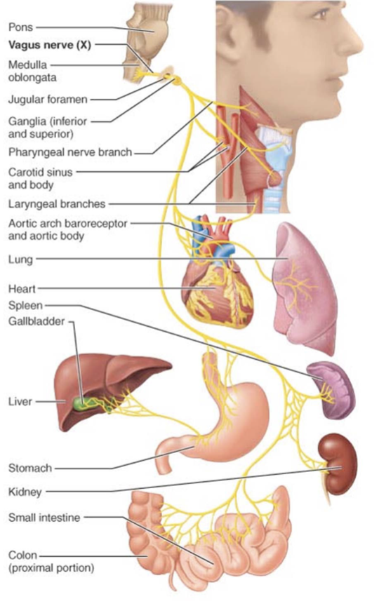
Vagus Nerve
A mixed sensory and motor nerve
“Wanders” into thorax and abdomen
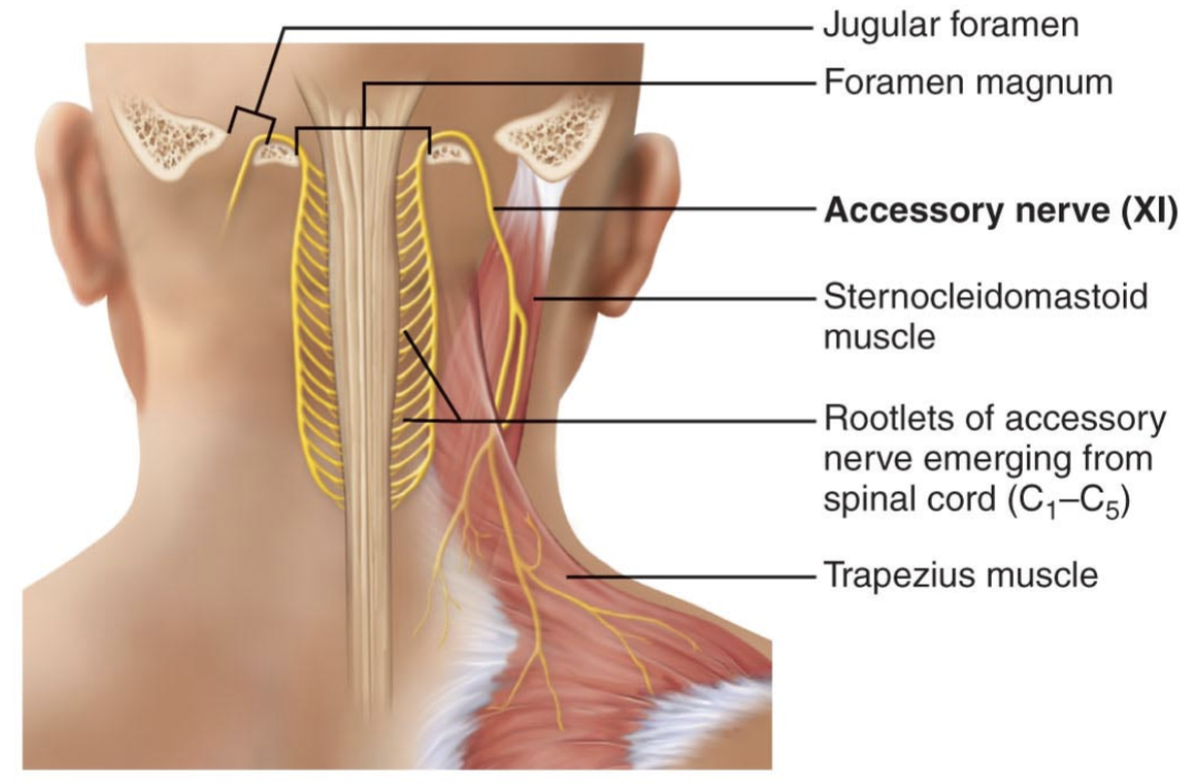
Accessory Nerve
An accessory part of the vagus nerve
motor
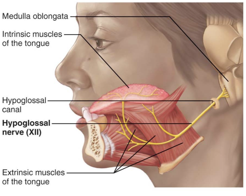
Hypoglossal Nerve
Runs inferior to the tongue
Innervates the tongue muscles
motor
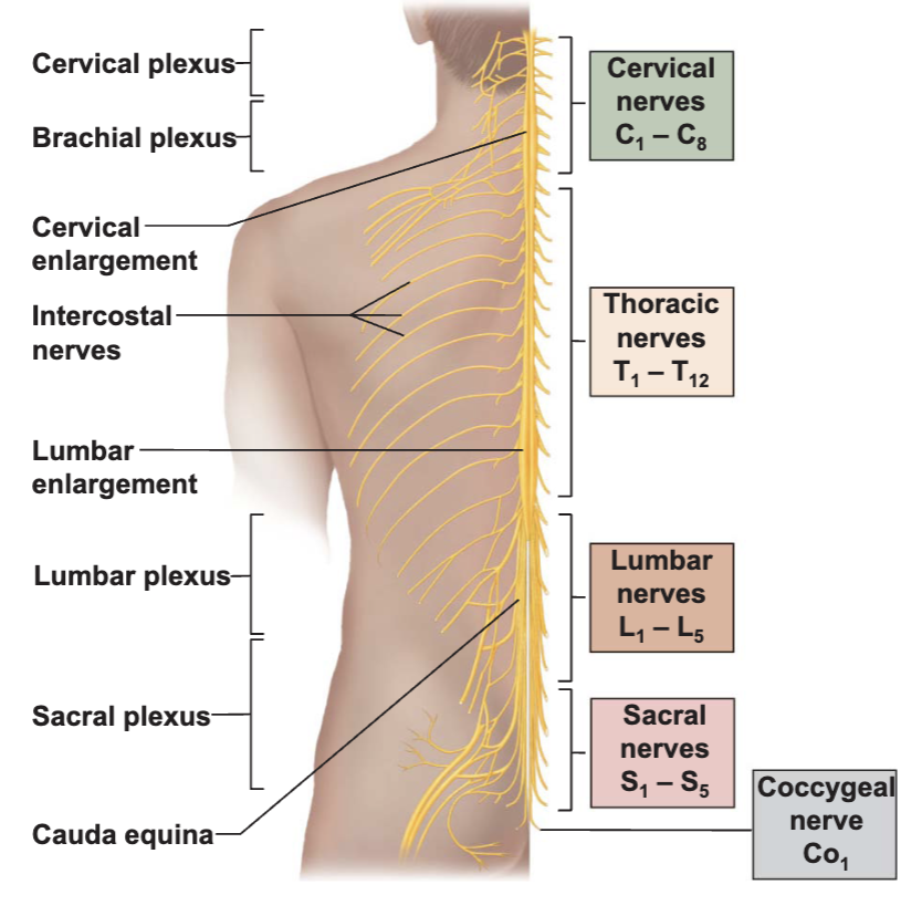
Spinal Nerves
31 pairs
Named for point of tissue from the spinal cord
8 pairs of cervical nerves (C1-C8)
12 pairs of thoracic nerves (T1-T12)
5 pairs of lumbar nerves (L1-L5)
5 pairs of sacral nerves (S1-S5)
1 pair of coccygeal nerves (Co1)
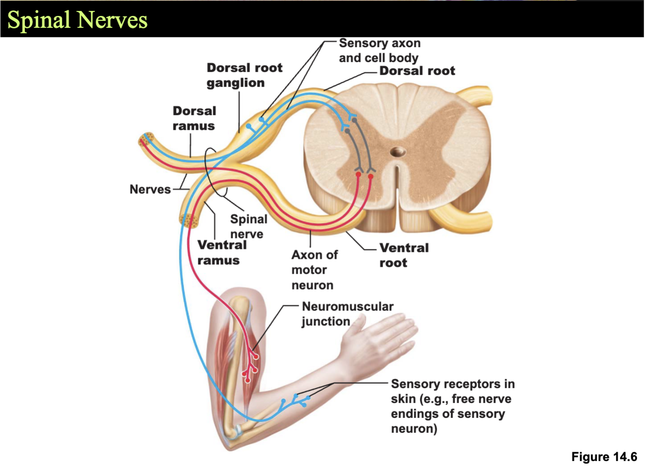
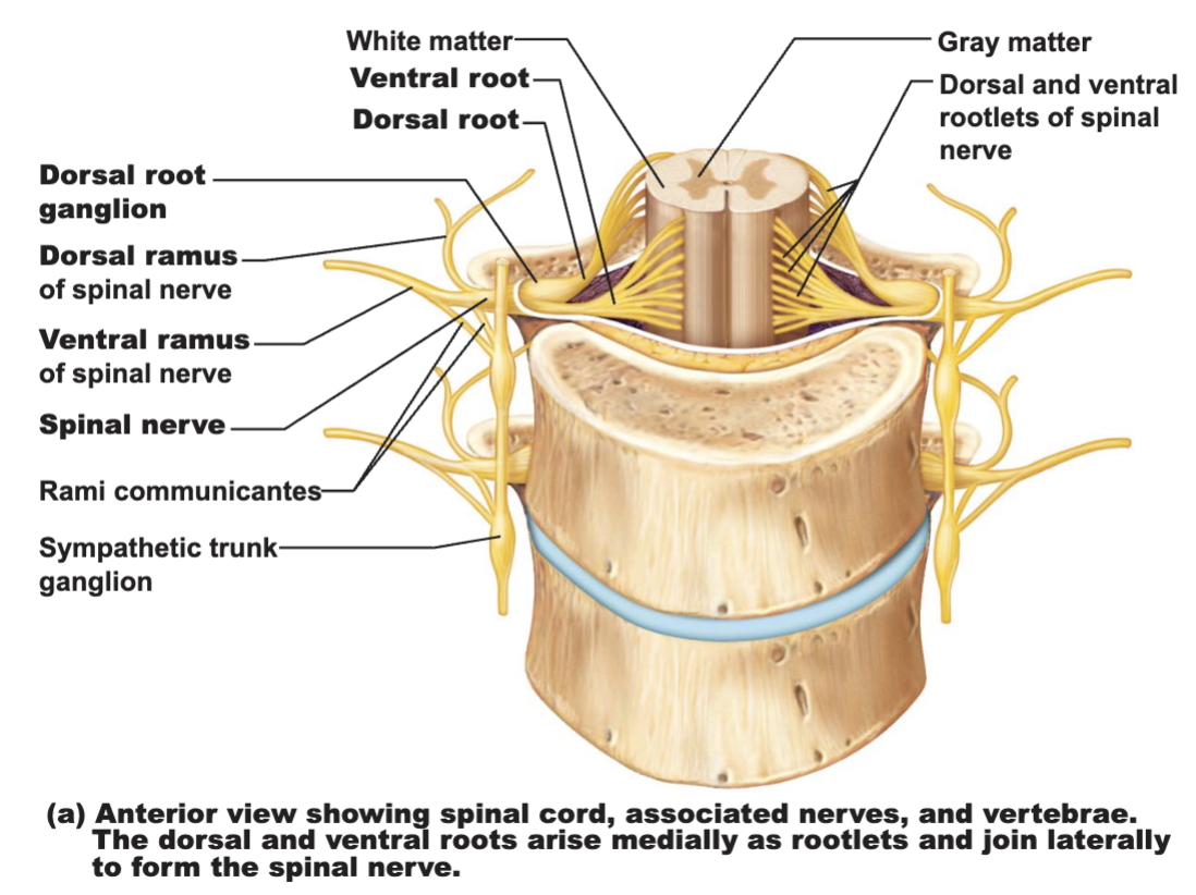
Connect ot the spinal cord by the dorsal root and ventral root
Dorsal root
sensory fibers
cell bodies
dorsal root ganglion
Ventral root
motor fibers
Branchs
dorsal ramus
ventral ramus
Sensory and motor fibers in each
Innervation of the Back
Dorsal rami
Follow a neat, segmented pattern
Innervate a horizontal strip of muscle and skin
In line with emergence point from the vertebral column
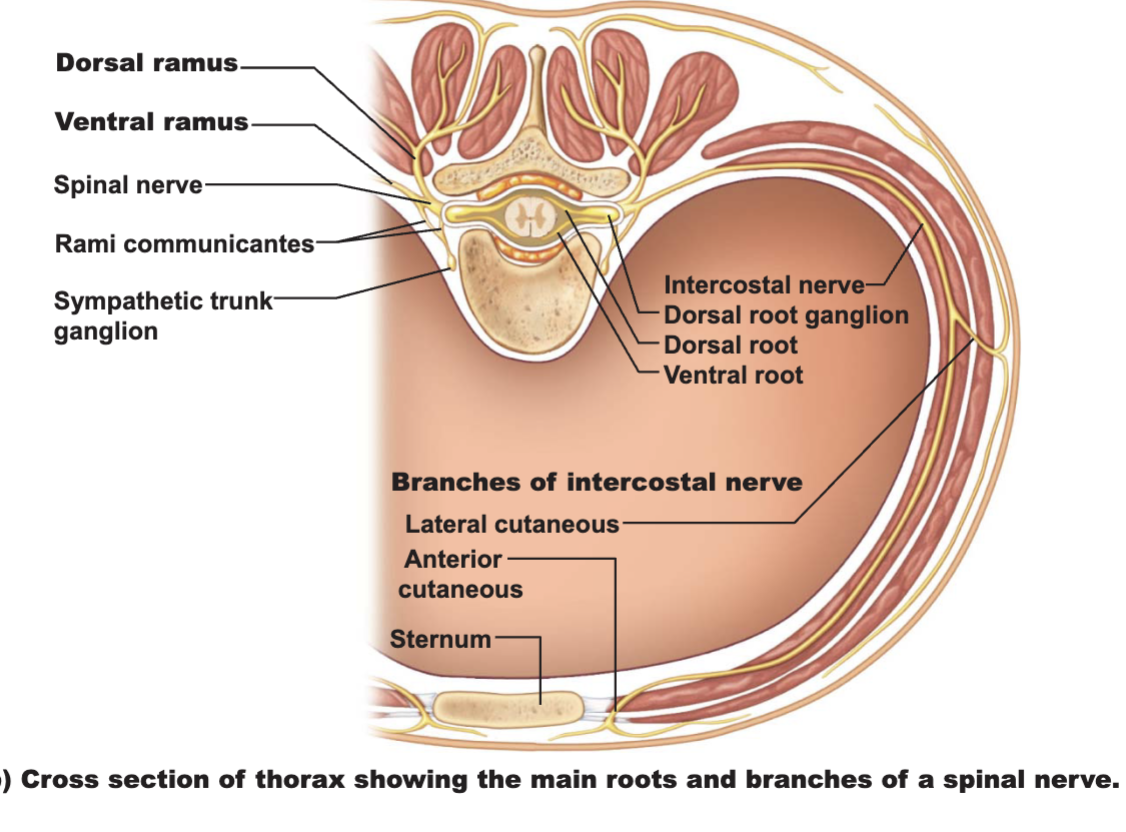
Thoracic region
Ventral rami arranged in simple, segmented pattern
Intercostal nerves - supply intercostal muscles, skin, and abdominal wall
Each gives off lateral and anterior cutaneous branches
Introduction to Nerve Plexuses
Nerve plexus - a network of nerves
Ventral rami (except T1-T12)
branch and join with one another
form nerve plexuses
cervical, brachial, lumbar, and sacral
primarily serve limbs
fibers from ventral rami crisscross
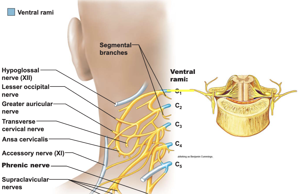
The Cervical Plexus
Deep in neck
under sternocleidomastoid
Formed by ventral rami of first four cervical nerves
Most are cutaneous nerves
Some innervate muscles of anterior neck
Phrenic nerve
most important of cervican plexus
The Brachial Plexus and Innervation of the Upper Limb
Brachial plexus lies in the neck and axilla
Formed by ventral rami of
C5-C8
Most of T1
Sometimes part C4 or T2
Cords give rise to main nerves of the upper limb
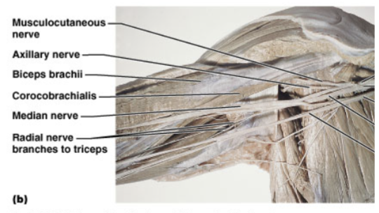
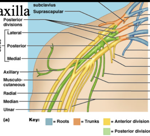
Nerves from the Lateral and Medial Cords
Musculocutaneous
main branch of the lateral cord
innervates the biceps brachii and brachialis
Median
originates from both lateral and medial cords
innervates anterior forerm muscles and lateral palm
Ulnar
branches from the medial cord
innervates intrinsic hand muscles and skin of the medial hand
Nerves from the Posterior Cord
Radial
continuation of the posterior cord
largest branch of the brachial plexus
innervates muscles of the posterior upper limb
Axillary
innervates deltoid and teres minor
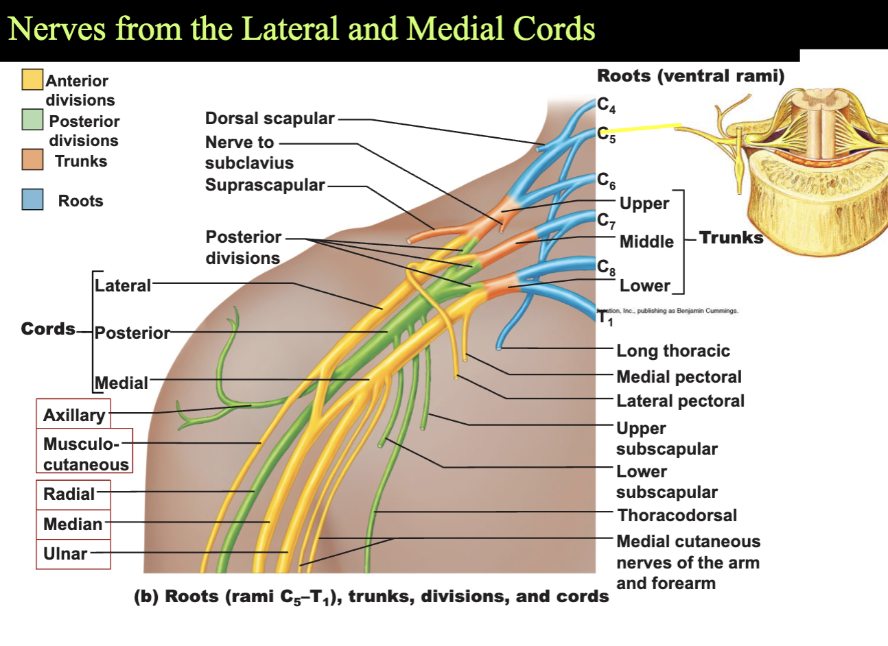
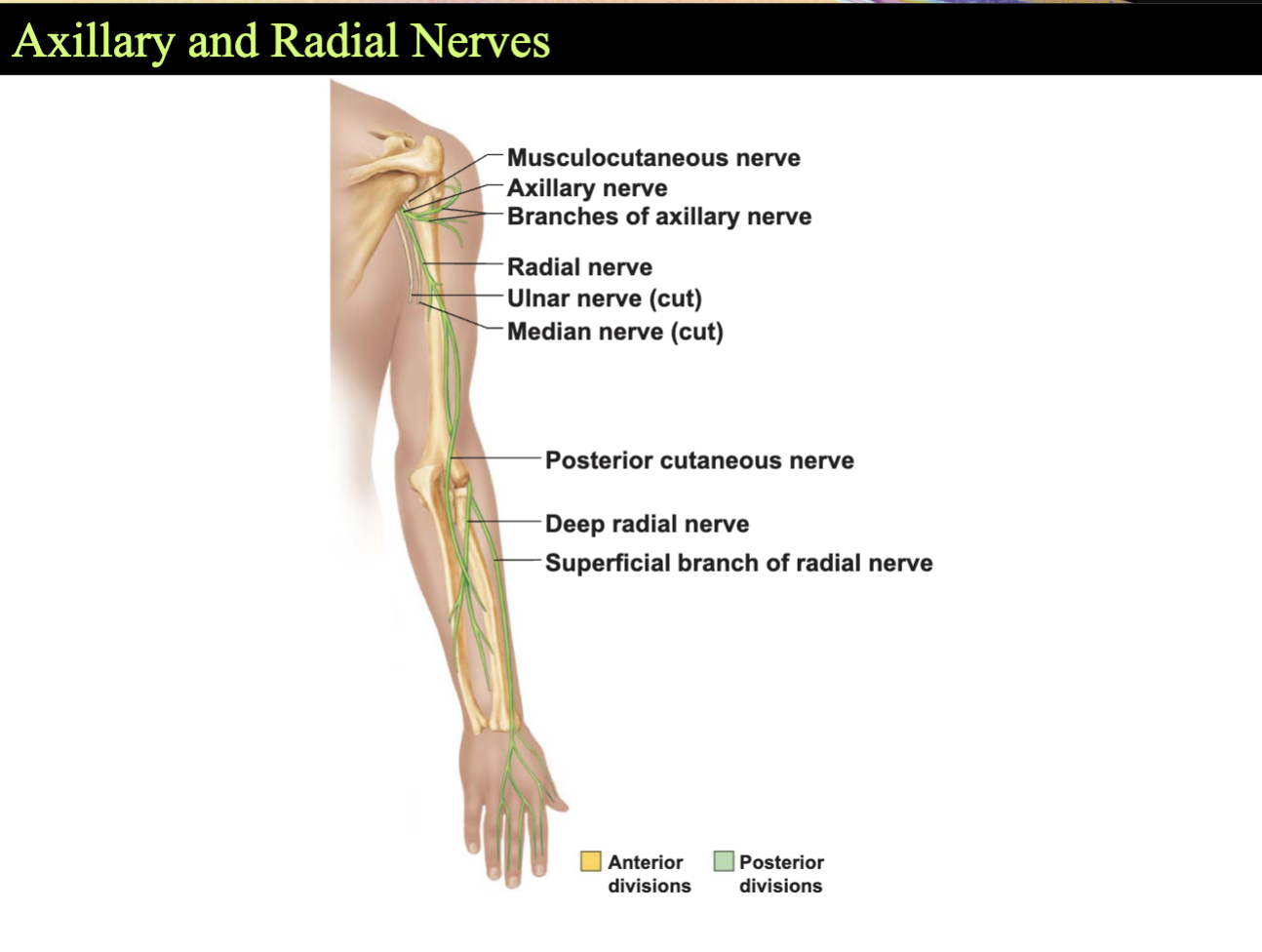
Brachial Plexus
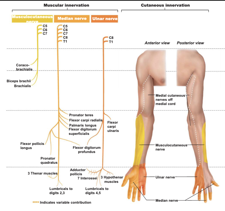
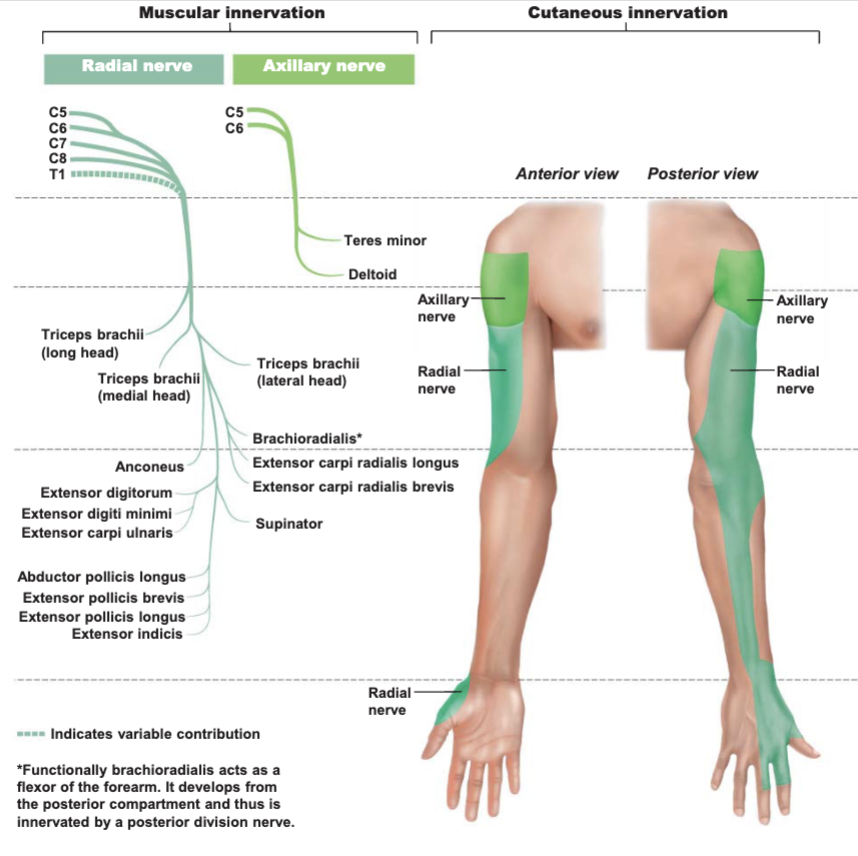
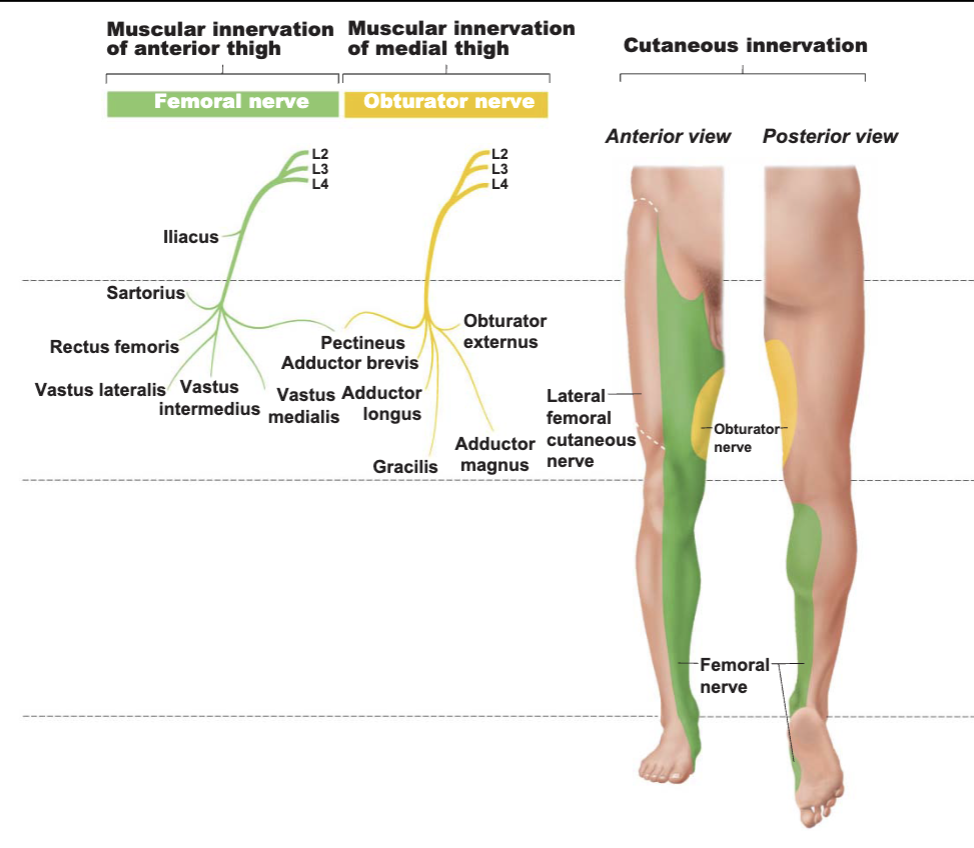
The Lumbar Plexus and Innervation of the Lower Limb
Lumbar plexus
L1-L4
Main branches innervate the anterior and medial thigh
Femoral nerve
anterior thigh muscles
Obturator nerve
adductor muscles
The Sacral Plexus
L4-S4
Caudal to lumbar plexus
Often considered with the lumbar plexus
Lumbosacral plexus
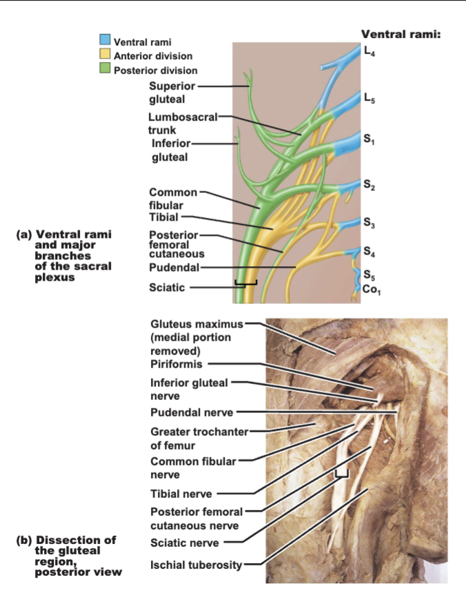
Innervation of the Lower Limb
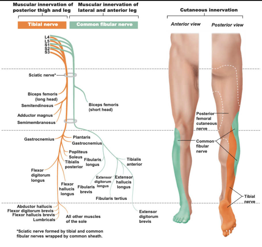
Sciatic nerve
largest nerve of the sacral plexus
actually two nerves in one sheath
Tibial nerve
most of the posterior lower limb
Common fibular (peroneal) nerve
muscles of the anterolateral leg
Superior and inferior gluteal nerves
innervate the gluteal muscles
Pudendal nerve
innervates muscles of the perineum
The LumboSacral Plexus
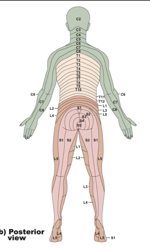
Innervation of the Skin: Dermatomes
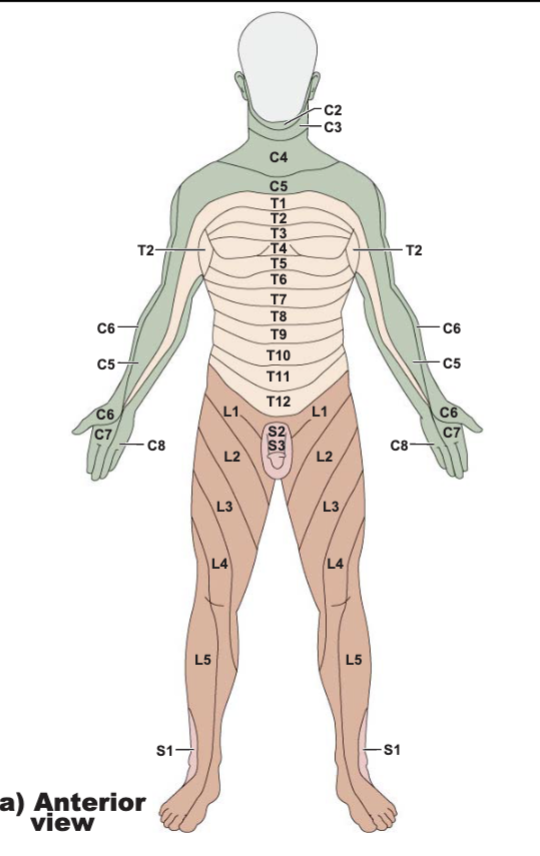
Dermatome
innervated by cutaneous branches of a single spinal nerve
Upper limb
brachial plexus
Lower limb
Lumbar nerves - anterior surface
Sacral nerves - posterior surface
Map of Dermatomes - Anterior View
Map of Dermatomes - Posterior View
Spastic vs. Flaccid Paralysis
Flaccid
when communication between spinal cord and muscle is the site of injury
Spastic
when communication between the brain and spinal cord is the site of injury
Chapter 16 - The Special Senses
The Special Senses
Taste, smell, sight, hearing, and balance
Special sensory receptors
Localized
head region
Receptors are not free endings of sensory neurons
Special receptor cells
The Chemical Senses: Taste and Smell
Taste
gustation
Smell
olfaction
Receptors
chemoreceptors
respond to chemicals
Taste - Gustation
Taste receptors
Occur in taste buds
Most are found on the surface of the tongue
Located within tongue papillae
Collection of 50-100 epithelial cells
Taste Buds
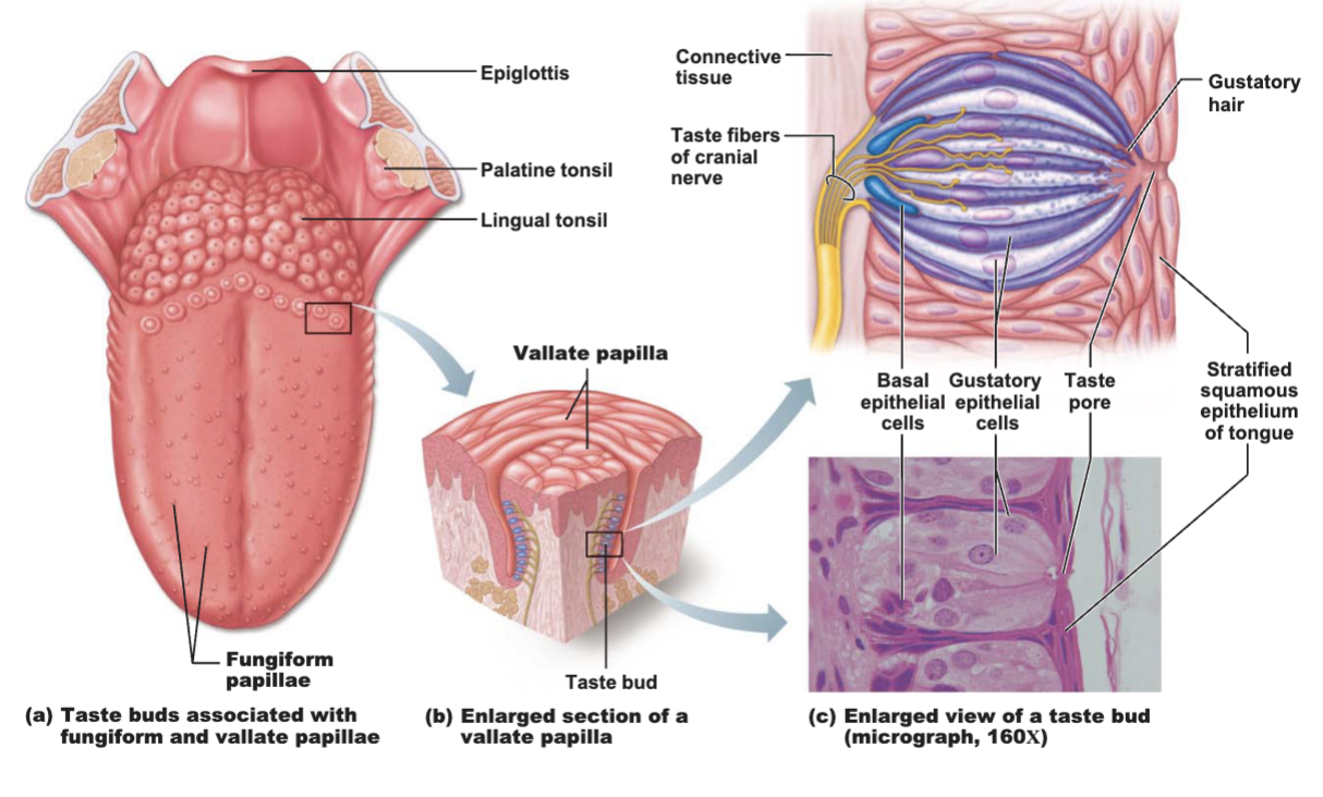
Taste Sensation and Gustatory Pathway
Four basic qualities of taste
Sweet
Salty
Sour
Bitter
A fifth taste - umami - “deliciousness”
The “taste map” is a myth
All taste modalities can be elicited from all areas containing taste buds
No structural difference among taste buds
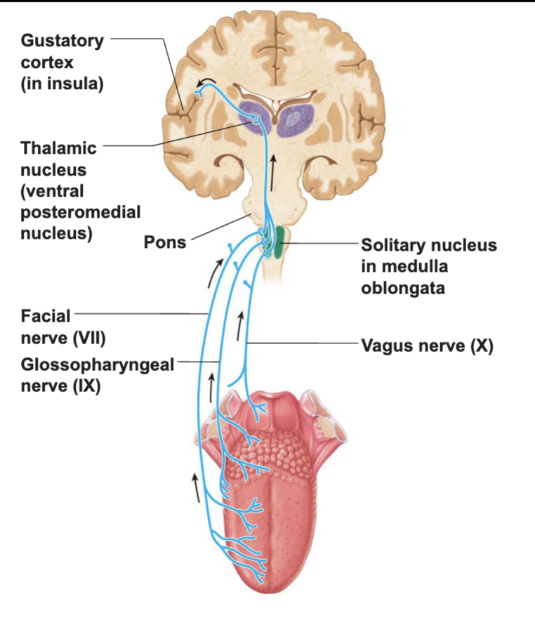
Gustatory Pathway
Taste information reaches the cerebral cortex
Primarily through
facial (VII)
anterior
glossopharyngeal (IX)
posterior
Some through:
vagus nerve (X)
epiglottis and lower pharynx
Sensory neurons synapse in the medulla
solitary nucleus to thalamus to parietal cortex
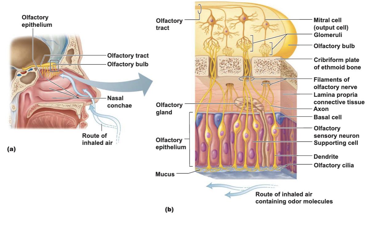
Olfactory Receptors
The Eye and Vision
Visual organ - the eye
70% of all sensory receptors are in the eyes
40% of the cerebral cortex is involved in provessing visual information
Anterior one-sixth of the eye’s surface is visible
Anatomy of the Eyeball
Components of the eye
Protect and support the photoreceptors
Gather, focus, and process light into precise images
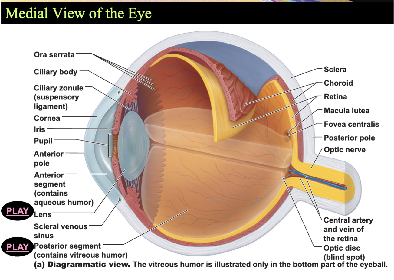
Anterior pole - most anterior part of the eye
Posterior pole - most posterior part of the eye
External walls - composed of three tunics
Fibrous, vascular, sensory
Internal cavity - contain fluids (humors)
The Fibrous Tunic
Most external layer of the eyeball
Composed of two regions of connective tissue
Sclera - posterior five-sixths of the tunic
White, opaque region
Provides shapes and an anchor for eye muscles
Cornea - anterior one-sixth of the fibrous tunic
Medial View of the Eye
The Vascular Tunic
The middle coat of the eyeball
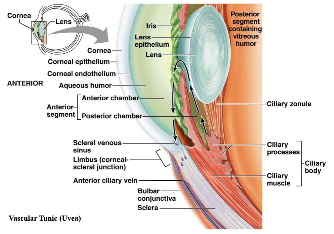
Composed of choroid, ciliary body, and iris
Choroid - vascular, darkly pigmented membrane
posterior 5/6
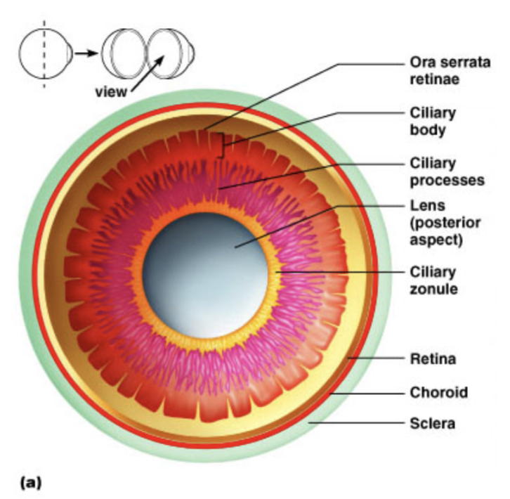
Posterior View of the Anterior Half of the Eye
The Vascular Tunic
Ciliary body - thickened ring of tissue - encirles the lends
Composed of ciliary muscle
The Iris
Visible colored part of the eye
Attached to the ciliary body
Composed of smooth muscle
Pupil - the round, central opening
Sphincter pupillae muscle
Dilator pupillae muscle
Act to vary the size of the pupil
The Sensory Tunic (Retina)
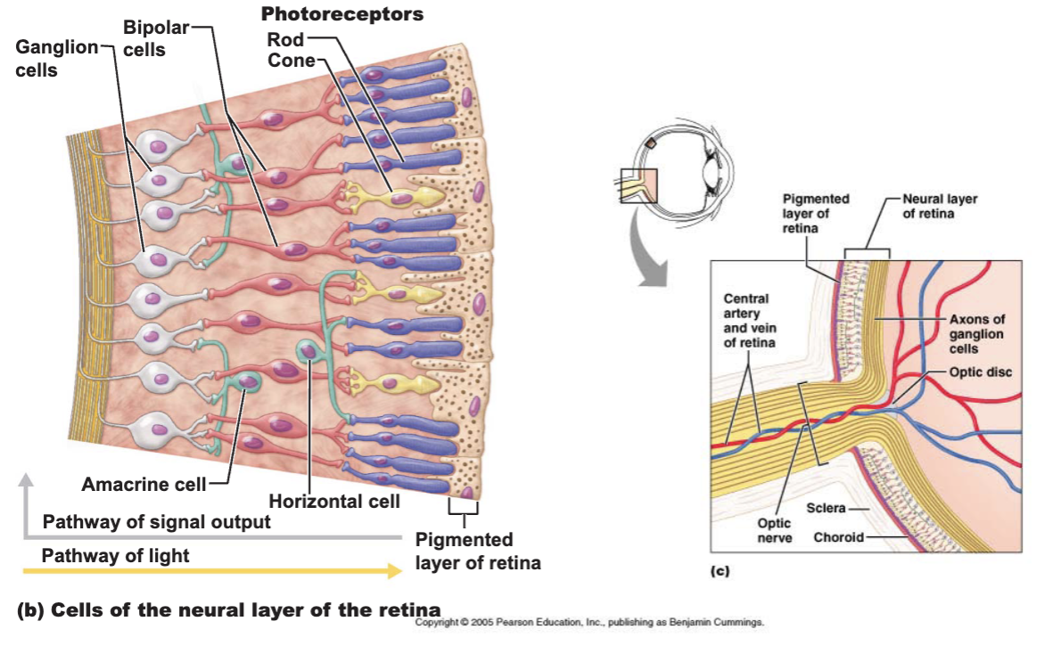
Retina - the deepest tunic
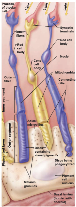
Photoreceptors
Two main types
Rod cells - more sensitive to light
Allow vision in dim light
Cone cells - operate best in bright light
Enable high - acuity, color vision
Internal Chambers and Fluids
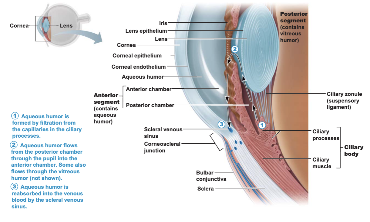
Posterior segment (cavity)
Filled with vitreous humor
Clear, jelly-like substance
Transmits light
Supports the posterior surface of the lens and hold neuronal layer against pigmented layer
Helps maintain intraocular pressure
Anterior segment
Filled with aqueous humor
Renewed continuously
Formed as a blood filtrate
Supplies nutrients to the lends and cornea
Avascular
Glaucoma
The Eye as an Optical Device
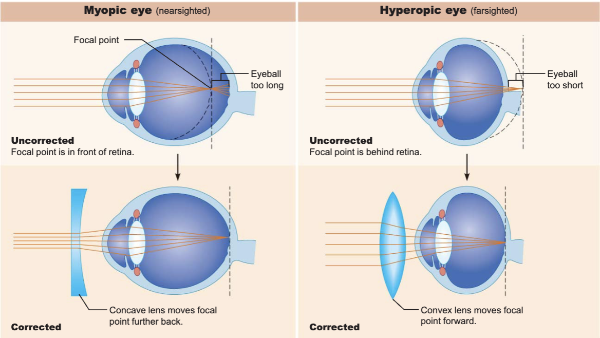
Lens
A thick, transparent, biconvex disc
Cataract
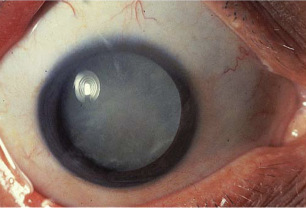
Inadequate delivery of nutrients to part of lens
Possible causes
congenital
sunlight
smoking
medications
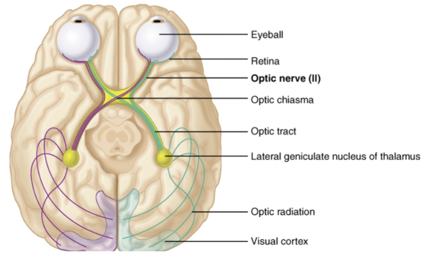
Visual Pathway
Most visual information travels to the cerebral cortex
Responsible for conscious “seeing”
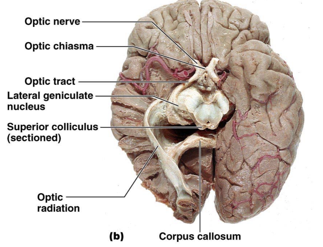
Visual Pathways to the Cerebral Cortex
Pathway begins at the retina
Light activates photoreceptors
Leads to activation of optic nerve (II)
Optic tracts send axons to:
Thalamus
Synapse
Fibers of the optic radiation reach the primary visual cortex
The Ear: Hearing and Equilibrium
The ear
receptor organ
hearing and equilibrium
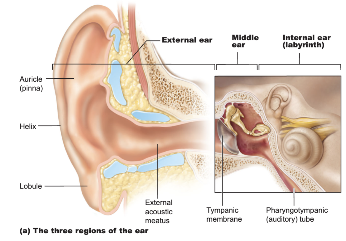
3 main regions
Outer ear
hearing
Middle ear
hearing
Inner ear
both hearing and equilibrium
The Outer (External) Ear
Composed of:
The auricle (pinna)
helps direct sounds
External acoustic meatus
Tympanic membrane
forms the boundary between the external and middle ear
The Middle Ear
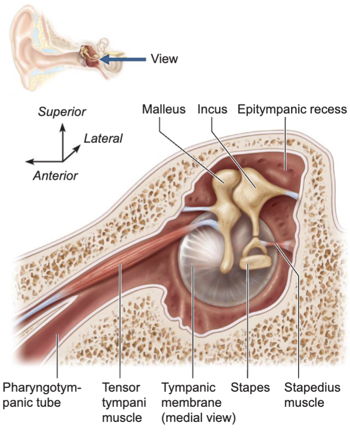
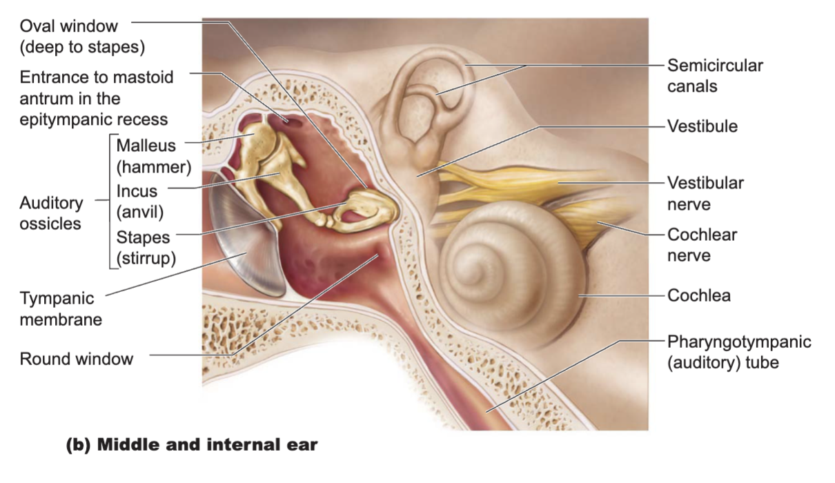
The tympanic cavity
A small, air-filled space
Located within the petrous portion of the temporal bone
Pharyngotympanic tube (auditory or eustachian tube)
Links the middle ear and pharynx
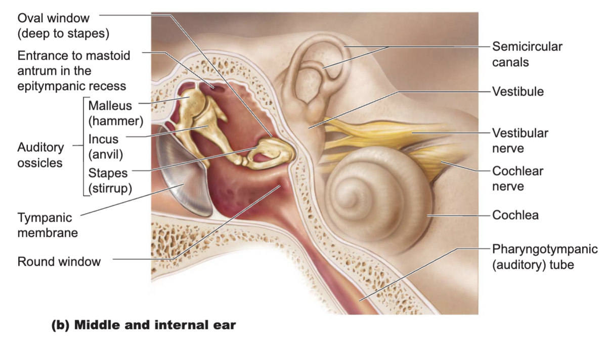
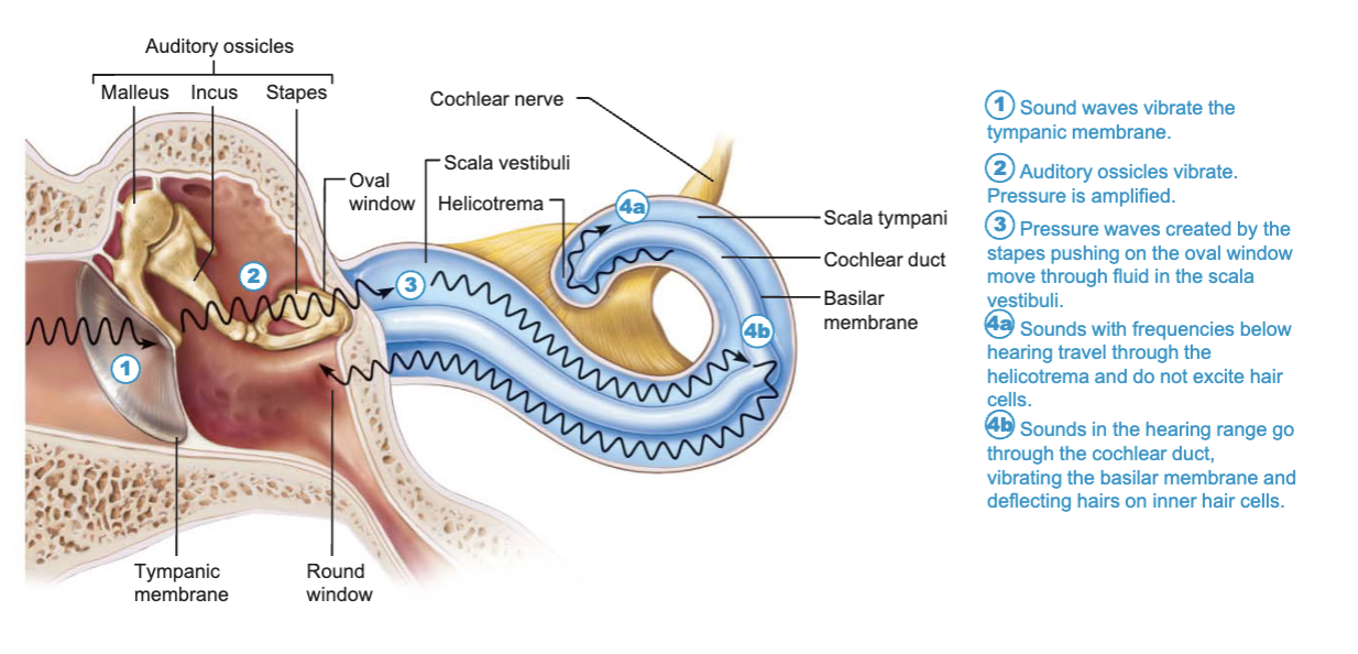
Ear ossicles - smallest bones in the body
Malleus
attaches to the eardrum
Incus
between the malleus and stapes
Stapes
vibrates against the oval window
The Inner (Internal) Ear
Inner ear - also called the labyrinth
Lies within the petrous portion of the temporal bones
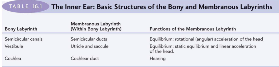
Boney labyrinth - a cavity consisting of 3 parts
semicircular canals
vestiblue
cochlea
Membranous labyrinth
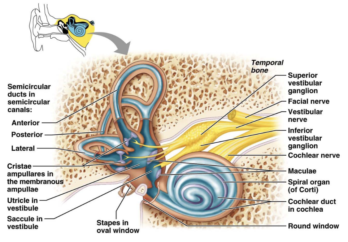
Series of membrane-walled sacs and ducts
Fit within the bony labyrinth
Consists of 3 main parts
semicircular ducts
utricle and saccule
cochlear duct
Filled with a clear fluid - endolymph
confined to the membranous labyrinth
Bony labyrinths is filled with perilymph
continuous with cerebrospinal fluid
The Vestibule
The central part of the bony labyrinth
Lies medial to the middle ear
Utricle and saccule - suspended in perilymph
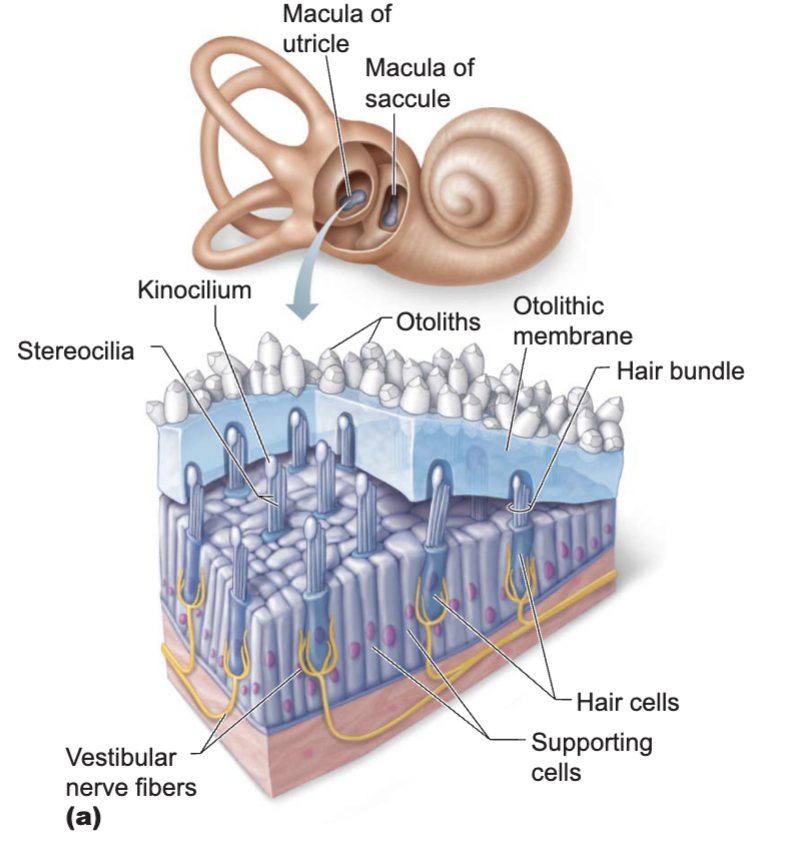
Two egg-shaped parts of the membranous labyrinth
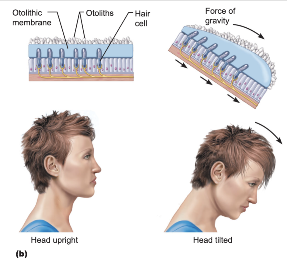
House the macula
a spot of sensory epithelium
Macula - contains receptor cells
Monitor the position of the head when the head is still
Receptor cells - called hair cells
synapse with the vestibular nerve
The Semicircular Canals
Lie posterior and lateral to the vestibule
Anterior and posterior semicircular canals
Lie in the vertical plane at right angles
Lateral semicircular canal
Lies in the horizontal plane
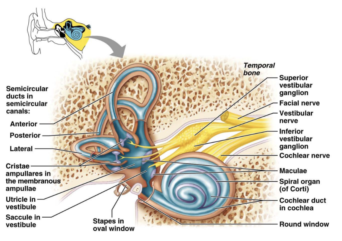
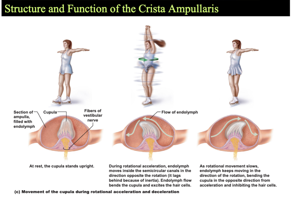
Semicircular duct - snakes through each semicircular canal
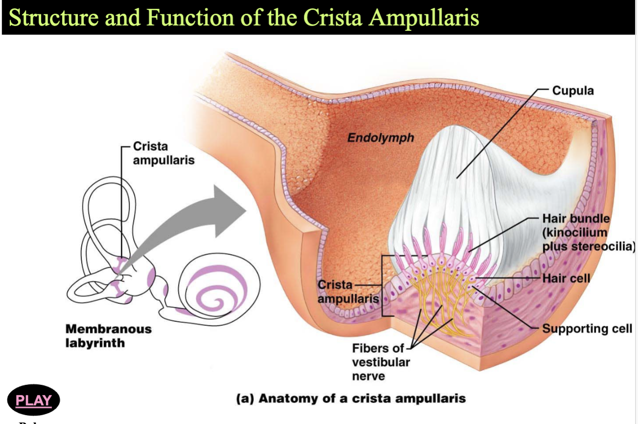
Membranous ampulla - located within bony ampulla
Houses a structure called a crista apullaris
Cristae contain receptors cells of rotational acceleration
Epitheliumm contains supporting cells and receptor hair cells
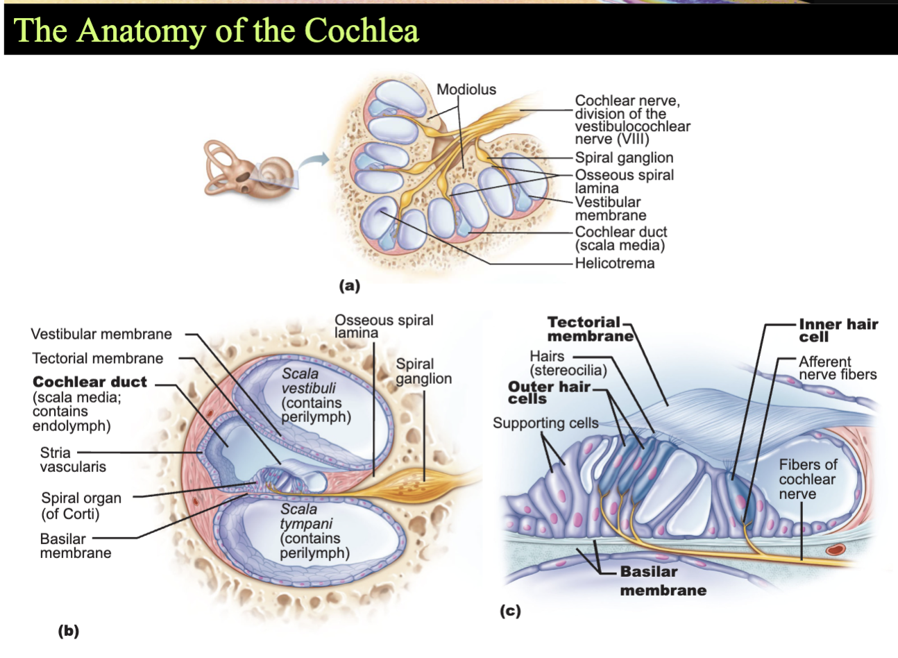
The Cochlea
The cochlear duct (scala media) - contains receptors for hearing
Organ of Corti - the receptor epithelium for hearing
The Role of the Cochlea in Hearing
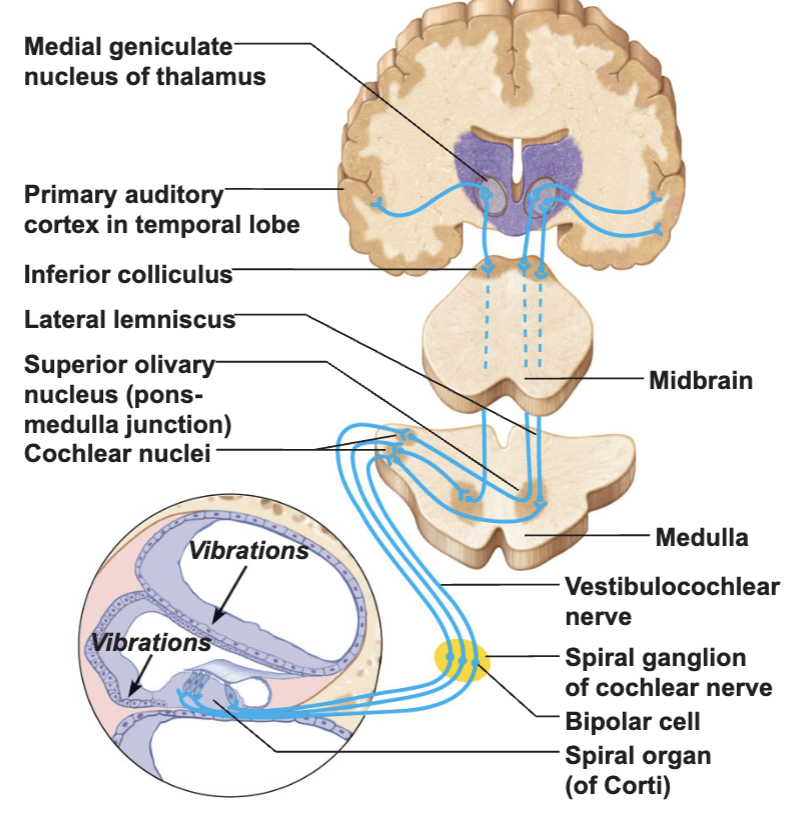
Equilibrium and Auditory Pathways
The equilibrium pathway
Transmits information on the position and movements of the head
Most information goes to lower brain centers (reflex centers)
The ascending auditory pathway
Transmits information from cochlear receptors to the cerebral cortex
Auditory Pathway from the Organ of Corti
Chapter 15 - The Autonomic Nervous System and Visceral Sensory Neurons
The Autonomic Nervous System
Innervates
smooth muscle
cardiac muscle
glands
Regulates visceral functions
heart rate
blood pressure
digestion
urination
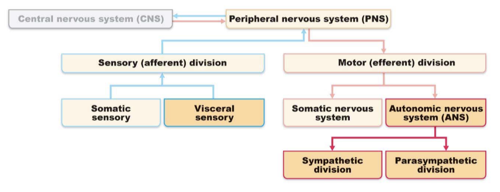
Comparison of Autonomic and Somatic Motor Systems
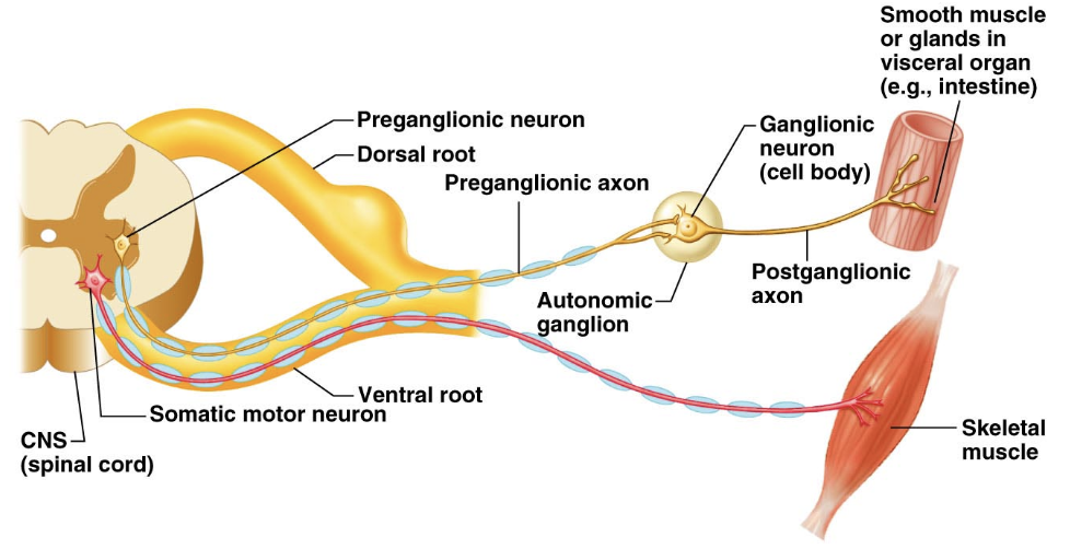
Somatic motor system
One motor neuron
from the CNS
to skeletal muscle
Axons are well myelinated, conduct impulses rapidly
Autonomic nervous system
Chain of two motor neurons
Preganglionic neuron
Ganglionic/Postganglionic neuron
Conduction is slower due to thinkly or unmyelinated axons
Autonomic and Somatic Motor Systems
Divisions of the Autonomic Nervous System
Sympathetic and Parasympathetic Divisions
Chains of two motor neurons
Innervate mostly the same structures
Cause opposite effects
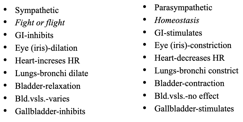
Sympathetic - “fight, flight, or fright”
Activated during exercise, excitement, and emergencies
Parasympathetic - “rest and digest:
Concerned with conserving energy
Anatomical Differences in Sympathetic and Parasympathetic Dvisions
Sympathetic - also called thoracolumbar division
Parasympthetic - also called the craniosacral division
Length of postganglionic fibers
Sympathetic - long postganglionic fibers
Parasympathetic - short postganglionic fibers
Branching of axons
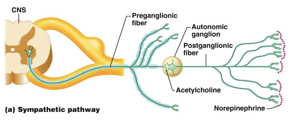
Sympathetic axons - highly branched
influences many organs
Parasympathetic axons - few branches
localized effect
Neurotransmitter released by postganglionic axons
Sympathetic
Most norepinephrine (adrenergic)
Parasympathetic
Acetylcholine
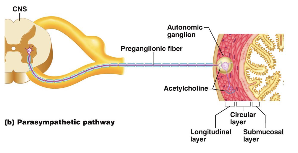
The Parasympathetic Division
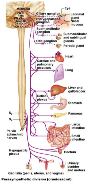
Path of the Vagus Nerve
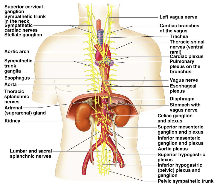
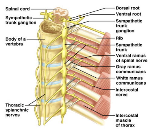
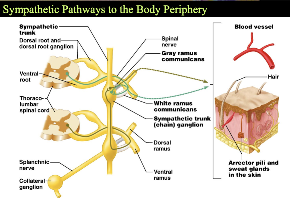
Sympathetic Trunk Ganglia
Both sides of vertebral column
Linked into sympathetic trunks
Joined to ventral rami by white and gray rami communicates
Fusion of ganglia —> fewer ganglia than spinal nerves
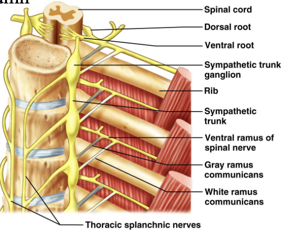
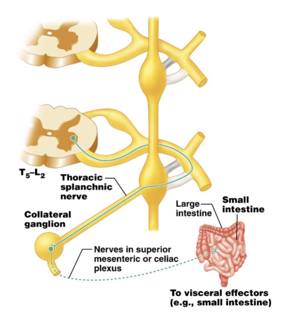
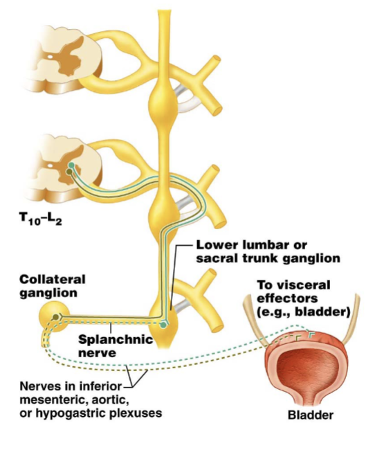
Prevertebral Ganglia
Unpaired, not segmentally arranged
Occur only in abdomen and pelvis
Lie anterior to the vertebral column
Main ganglia
Celiac, superior mesenteric, inferior mesenteric, inferior hypogastric ganglia
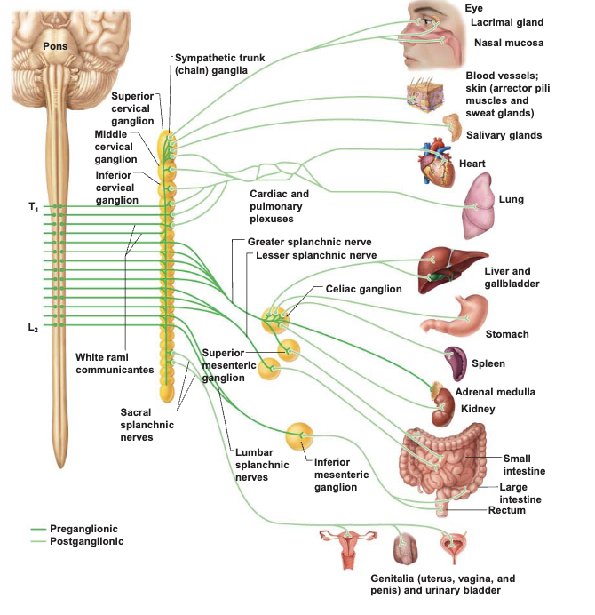
Sympathetic Divison of the ANS
Sympathetic vs. Parasympathetic Effects
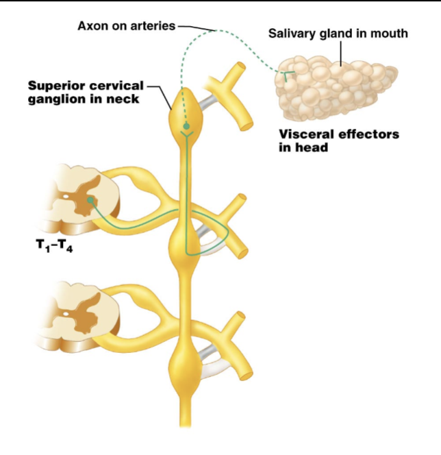
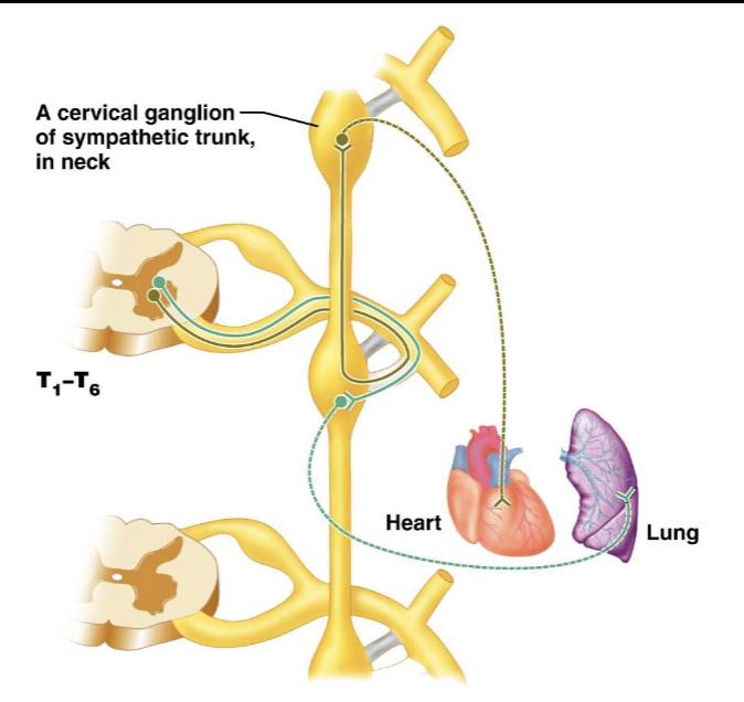
Sympathethic Pathways to the Head
Sympathetic Pathways to Thoracic Organs
Sympathetic Pathways to the Abdominal Organs
Sympathetic Pathways to the Pelvic Organs
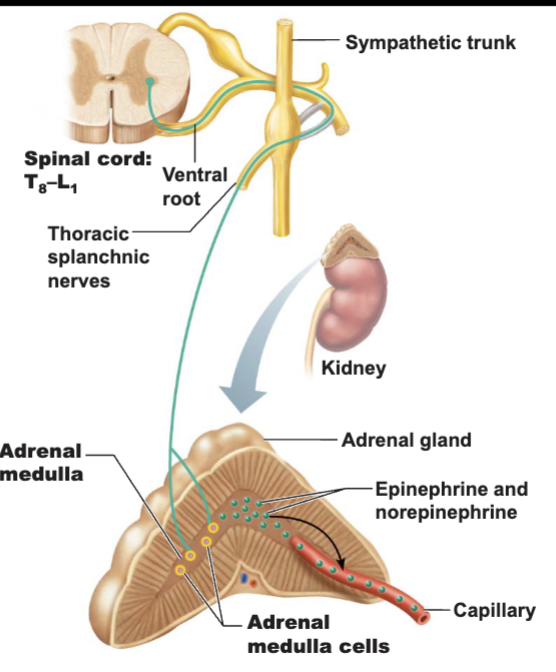
The Role of the Adrenal Medulla in the Sympathetic Division
Major organ of the sympathetic nervous system
Constitutes largest sympathetic ganglia
Secretes great quantities of:
norepinephrine
adrenaline
Stimulated to secrete by preganglionic sympathetic fibers
The Adrenal Medulla
Visceral Sensory Neurons
General visceral sensory neurons monitor:
stretch, temperature, chemical changes, and irritation
Cell bodies are located in the dorsal root ganglia
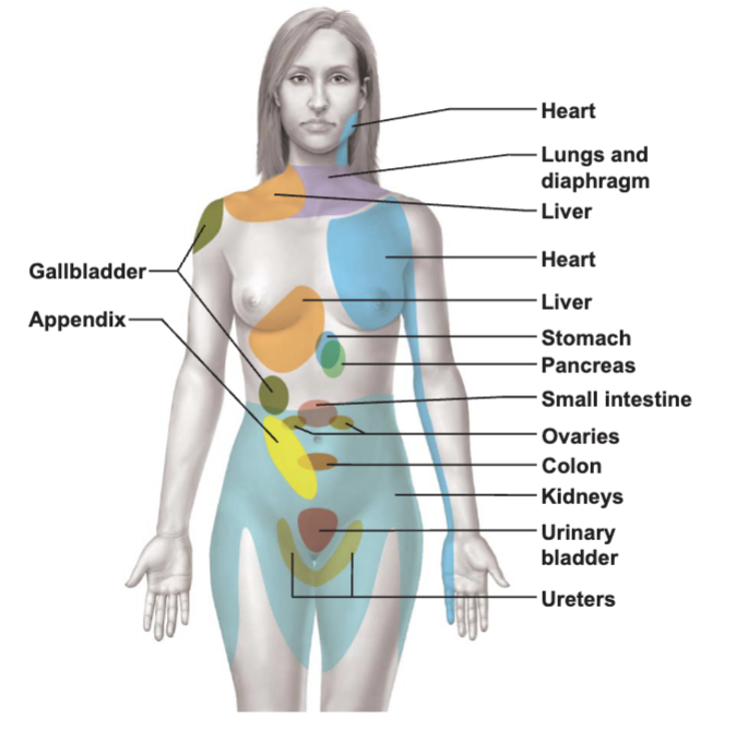
Visceral pain - perceibed to be somatic in origin
Referred pain
A Map of Referred Pain
Visceral Reflexes
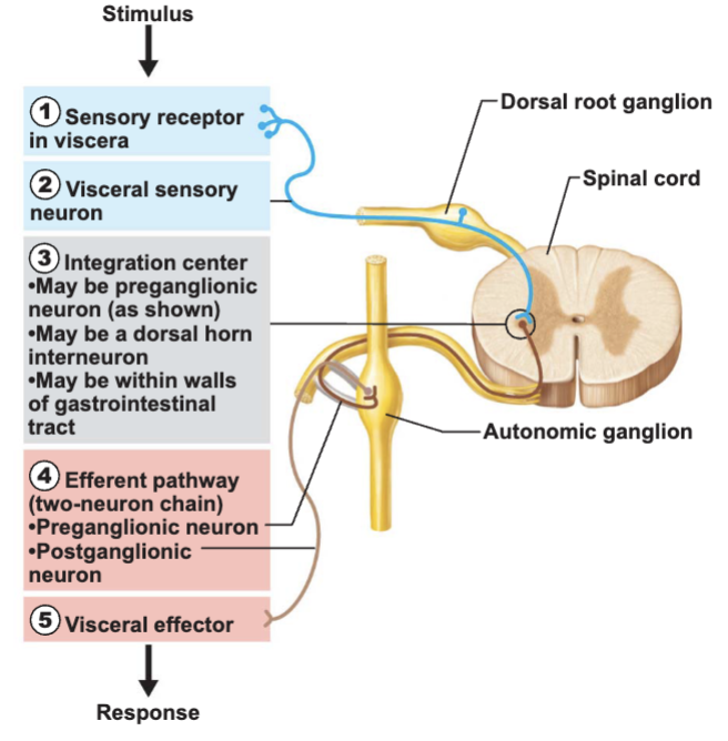
Visceral sensory and autonomic neurons
Participate in visceral reflex arcs
Defecation reflex
Micturition reflex
Some are simple spinal reflexes
Others do not involve the CNS
Strictly peripheral reflexes
Visceral Reflex Arc
Central Control of the ANS
Control by the brain stem and spinal cord
Reticular formation exerts most direct influence
Medulla oblongata
Periaqueductal gray matter
Control by the hypothalamus and amygdala
Hypothalamus - the main integration center of the ANS
Amygdala - main limbic region for emotions
Control by the cerebral cortex
Chapter 17 - The Endorcine System
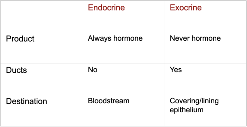
Endocrine vs Exocrine
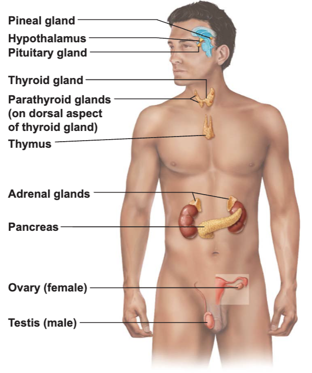
Endocrine Organs
Scattered throughout the body
Pure endocrine organs
Pituitary, pineal, thyroud, parathyroid, and adrenal glands
Organs containing endocrine cells
Pancreas, thymus, gonads, and the hypothalamus
Richly vascularized
Location of Major Endocrine Glands
Hormones
Basic hormone action
Circulate throughout the body in blood vessels
Influence only specific tissues - target cells
A hormone can have different effects on different target cells
Control of Hormones Release: 3 Mechanisms
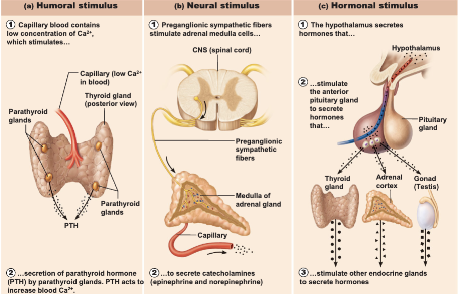
Control of Hormones Secretion
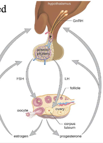
Always controlled by feedback loopes
Blood concentration declines below a minimum
More hormone is secreted
Blood concentration exceeds maximum
Hormone production is halted
The Pituitary Gland
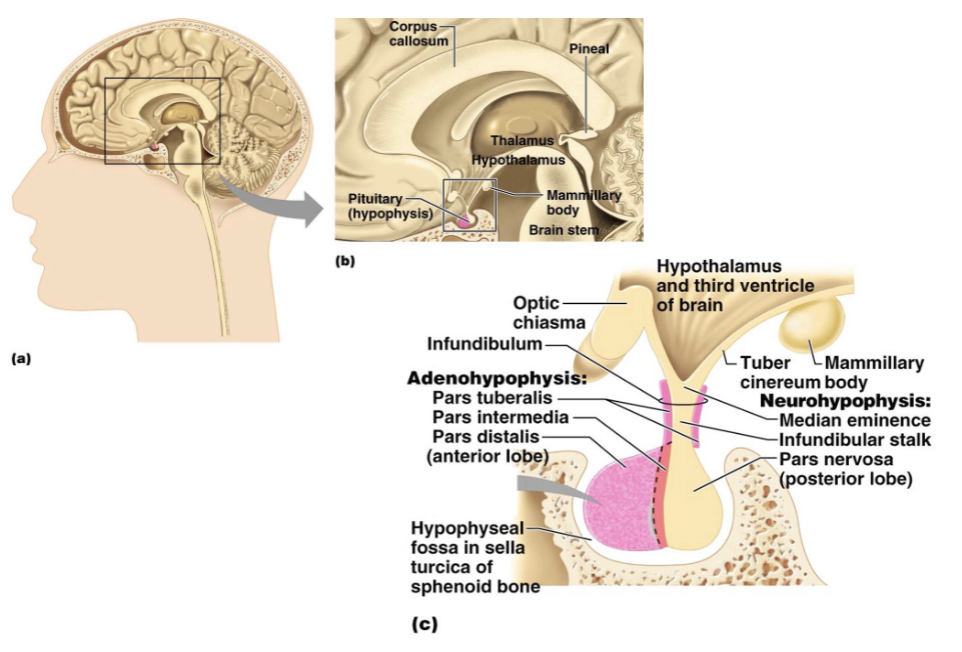
The Anterior Lobe
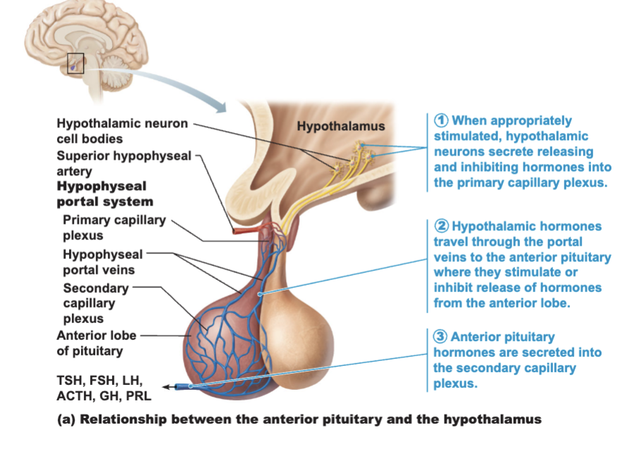
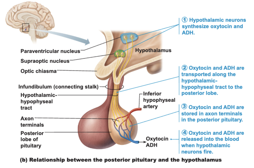
Relationship Between the Posterior Pituitary and Hypothalamus
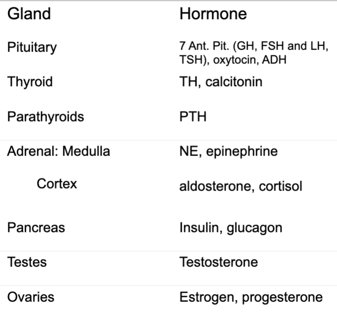
Glands and Hormones each releases
Other Endocrine Structures
Endocrine cells occur within:
Atria of heart
atrial natriuretic peptide (ANP)
The GI tract
enteroendocrine cells
The placenta
secretes several steroid protein hormones
Estrogen
Progesterone
The kidneys - cels of the juxtaglomerular apparatus secrete renin —> aldosterone (sodium reabsorption)
Endothelial cells and interstitial connective tissue - secrete erythropoietin
RBC production
The skin - modified cholesterol molecules convert to a precursor of vitamin D
A steroid hormone required for calcium metabolism/absorption
Pituitary Disorders
Gigantism - hypersecretion of GH in children
Pituitary dwarfism - hyposecretion of GH
Acromegaly - hyposecretion of GH after closure of growth plates
Diabetes insipidus - pars nervosa does not make enough ADH
Disorders of the Pancreas: Diabetes Mellitus
Caused by:
Insufficient secretion of insulin
Resistance of body cells to the effects of insulin
Type I diabetes - develops suddenly, usually before age 15
T cell-mediated autoimmune response destroys beta cells
Type II diabetes - adult onset
Usually occurs after age 40
Cells have lowered sensitivity to insulin
Controlled by dietary changes and regular exercise