Lymphatic System
Chart of Blood Pathologies
Category | Disease/Disorder | Description | Causes / Risk Factors | Key Features / Symptoms | Treatments |
|---|---|---|---|---|---|
Red Blood Cell Diseases | Anemia | Deficiency in erythrocytes or hemoglobin. | Iron deficiency, chronic disease | Fatigue, pallor, shortness of breath | Iron supplements, treat underlying cause |
Aplastic Anemia | Bone marrow failure to produce blood cells. | Idiopathic, benzene, chloramphenicol | Pancytopenia, fatigue, infections | Transfusion, antibiotics, immunosuppressants, marrow transplant | |
Hemolytic Anemia | Excessive destruction of RBCs. | Hereditary (spherocytosis) | Spherocytes, jaundice, increased reticulocytes | Splenectomy | |
Sickle Cell Anemia | Abnormal hemoglobin (HbS) leading to sickled RBCs. | Genetic (African/Hispanic ancestry) | Pain crises, stroke, poor oxygenation | Pain management, transfusions, experimental gene therapy | |
Thalassemia | Defective hemoglobin production. | Genetic (Mediterranean, Asian ancestry) | Hypochromic, microcytic anemia | Transfusions, iron chelation, stem cell transplant | |
Pernicious Anemia | Inability to absorb vitamin B12 due to lack of intrinsic factor. | Autoimmune gastritis | Macrocytic anemia, neurologic symptoms | B12 injections/oral therapy for life | |
Hemochromatosis | Excess iron accumulation in body tissues. | Hereditary or frequent transfusions | Bronze skin, fatigue, diabetes, joint pain | Phlebotomy, iron chelation therapy | |
Polycythemia Vera | Increased RBC production leading to thickened blood. | Bone marrow abnormality | Ruddy complexion, headaches, thrombosis | Phlebotomy, myelotoxic drugs | |
Clotting Disorders | Hemophilia | Deficiency of clotting factors VIII or IX. | Genetic (X-linked) | Prolonged bleeding, joint hemorrhage | Factor replacement, gene therapy in trials |
Purpura | Multiple small hemorrhages under the skin due to low platelets. | Autoimmune (ITP) | Petechiae, ecchymoses, bleeding gums | Splenectomy, immunosuppressive drugs | |
White Blood Cell Diseases | Leukemia | Malignant increase in white blood cells. | Unknown; some genetic links | Fatigue, fever, bleeding, bone pain, infections | Chemotherapy, targeted therapy, bone marrow transplant |
- AML | Immature myeloblasts dominate. | Adults | Abrupt symptoms, low platelets/RBCs | Chemotherapy, possible transplant | |
- ALL | Immature lymphoblasts dominate. | Children, young adults | Sudden onset, bone pain, fatigue | Chemotherapy; high cure rate in children | |
- CML | Mixed immature/mature granulocytes. | Adults >55 | Slow progression, fatigue | Gleevec and similar targeted therapies | |
- CLL | Mature lymphocytes dominate. | Elderly | Asymptomatic early, lymphadenopathy | Often no early treatment; monitored | |
Granulocytosis | Increased granulocytes due to infection/inflammation. | Infections, inflammation | Varies by type: eosinophilia, basophilia, neutrophilia | Treat underlying cause | |
Mononucleosis | Viral infection by Epstein-Barr virus. | EBV, saliva (kissing) | Fatigue, sore throat, swollen lymph nodes | Rest, supportive care | |
Bone Marrow Disease | Multiple Myeloma | Malignant plasma cells secrete abnormal antibodies (e.g., Bence Jones protein). | Unknown | Bone pain, fractures, anemia, renal failure, infections | Chemotherapy, pain control, bone marrow transplant |
🧪 LABORATORY TESTS
Test | Purpose | Notes |
|---|---|---|
Antiglobulin (Coombs) Test | Detects antibodies coating RBCs | Used for Rh incompatibility, autoimmune hemolytic anemia, transfusion testing |
Complete Blood Count (CBC) | Measures counts of RBCs, WBCs, platelets, Hgb, Hct, red cell indices | Fundamental screening test for many conditions |
Erythrocyte Sedimentation Rate (ESR) | Measures rate of RBC settling | ↑ in infections, inflammation, tumors; nonspecific |
Hematocrit (Hct) | Percentage of blood volume that is RBCs | Performed via centrifugation |
Hemoglobin (H, Hg, Hgb, HGB) | Measures total hemoglobin in blood | Assesses oxygen-carrying capacity |
Platelet Count | Number of platelets per mm³ or µL | Normal: 150,000–450,000 |
Prothrombin Time (PT) | Measures clotting ability | Used to monitor warfarin; INR standardizes result across labs |
Partial Thromboplastin Time (PTT) | Measures intrinsic clotting pathway | Often used alongside PT |
Red Blood Cell Count (RBC) | Number of erythrocytes per mm³ or µL | Normal: ~4–6 million/mm³ |
Red Blood Cell Morphology | Microscopic exam of RBC shape | Identifies anisocytosis, poikilocytosis, micro/macrocytosis, sickle cells |
White Blood Cell Count (WBC) | Number of leukocytes per mm³ or µL | Normal: 7,000–10,000/mm³ |
WBC Differential | Percentages of different WBC types | Includes neutrophils, lymphocytes, monocytes, eosinophils, basophils, bands |
Shift to the Left | Increase in immature neutrophils | Suggests acute infection or inflammation |
🧫 CLINICAL PROCEDURES
Procedure | Purpose | Notes |
|---|---|---|
Apheresis | Separates and removes a component from blood | Includes leukapheresis, plasmapheresis, plateletpheresis |
Blood Transfusion | Transfer of blood or components | Requires blood typing, infectious disease screening |
Autologous Transfusion | Patient’s own blood is stored and reused | Avoids transfusion reactions |
Packed Red Cells | RBCs separated from plasma for transfusion | Used to treat severe anemia |
Bone Marrow Biopsy | Core sample of marrow for microscopic exam | Used to diagnose leukemia, anemia, pancytopenia |
Bone Marrow Aspiration | Liquid marrow withdrawn via suction | Often combined with biopsy |
Hematopoietic Stem Cell Transplantation | Infusion of stem cells after marrow ablation | For leukemia, lymphoma, myeloma, autoimmune diseases |
Autologous Stem Cell Transplant | Uses patient’s own previously harvested stem cells | Less risk of GVHD |
Allogeneic Stem Cell Transplant | Uses donor cells; requires HLA match | Risk of graft-versus-host disease (GVHD); requires immunosuppressants |
GVHD (Graft-Versus-Host Disease) | Donor immune cells attack recipient tissue | Prevented with drugs like cyclosporine, methotrexate, sirolimus, mycophenolate |
🩸 Blood Tests & Components
Abbreviation | Meaning |
|---|---|
CBC | Complete blood count |
diff | Differential count (white blood cells) |
H and H | Hemoglobin and hematocrit |
Hct | Hematocrit |
Hgb, HGB | Hemoglobin |
MCH | Mean corpuscular hemoglobin |
MCHC | Mean corpuscular hemoglobin concentration |
MCV | Mean corpuscular volume |
RBC | Red blood cell; red blood cell count |
WBC | White blood cell; white blood cell count |
ESR / sed rate | Erythrocyte sedimentation rate |
PT / pro time | Prothrombin time |
PTT | Partial thromboplastin time |
INR | International normalized ratio |
SMAC | Sequential Multiple Analyzer Computer (automated chemistry analyzer) |
Fe | Iron |
g/dL | Grams per deciliter |
μL | Microliter |
mm³ | Cubic millimeter |
🧬 Cell Types & Blood Cell Terms
Abbreviation | Meaning |
|---|---|
bands | Immature white blood cells (granulocytes) |
baso | Basophils |
eos | Eosinophils |
lymphs | Lymphocytes |
mono | Monocytes |
segs | Segmented (mature) neutrophils |
polys / PMNs / PMNLs | Polymorphonuclear leukocytes (neutrophils, eosinophils, basophils) |
🧫 Diseases & Disorders
Abbreviation | Meaning |
|---|---|
ALL | Acute lymphoid leukemia |
AML | Acute myeloid leukemia |
CLL | Chronic lymphoid leukemia |
CML | Chronic myeloid leukemia |
HCL | Hairy cell leukemia |
MDS | Myelodysplastic syndrome (preleukemic condition) |
ITP | Idiopathic thrombocytopenic purpura |
DIC | Disseminated intravascular coagulation |
GVHD | Graft-versus-host disease |
EBV | Epstein-Barr virus (cause of mononucleosis) |
💉 Transplants, Therapies, & Immune Factors
Abbreviation | Meaning |
|---|---|
ABMT | Autologous bone marrow transplantation |
ASCT | Autologous stem cell transplantation |
BMT | Bone marrow transplantation |
HLA | Human leukocyte antigen (tissue typing marker) |
Ab | Antibody |
IgA, IgD, IgE, IgG, IgM | Classes of immunoglobulins (antibodies) |
G-CSF | Granulocyte colony-stimulating factor |
GM-CSF | Granulocyte-macrophage colony-stimulating factor |
EPO | Erythropoietin (stimulates RBC production) |
📊 Other Important Terms
Abbreviation | Meaning |
|---|---|
ANC | Absolute neutrophil count (WBC × % neutrophils) |
ABO | Four main blood types: A, B, AB, O |
WNL | Within normal limits |
🧬 Introduction to the Lymphatic and Immune Systems
The lymphatic system and immune system are closely connected due to their related functions.
Lymph is a clear, watery fluid that:
Surrounds body cells
Flows through thin-walled vessels of the lymphatic system throughout the body
💧 Lymph vs. Blood
Lymph:
Does not contain erythrocytes (RBCs) or platelets
Contains white blood cells (leukocytes), especially:
Lymphocytes
Monocytes
Composed of water, salts, sugars, and metabolic wastes (e.g., urea, creatinine)
Less protein than blood plasma
Originates from blood—specifically from fluid that filters out of blood capillaries into tissues (interstitial fluid)
Interstitial fluid:
Surrounds body cells
Flows into lymph capillaries → becomes lymph
Lymph flows:
From lymph capillaries in tissues
Through larger lymphatic vessels and lymph nodes
Into large lymphatic vessels in the upper chest
Finally empties into the bloodstream
🔍 Key Differences Between Lymph and Blood (Table 14-1)
🔁 Movement
Lymph: No pump; moved by muscle contractions and valves
Blood: Moved by a pump (the heart)
🩸 Contents
Lymph:
Only white blood cells (lymphocytes and monocytes)
Blood:
All blood cells: erythrocytes, leukocytes, and platelets
🧪 Fluid Composition
Lymph:
Derived from interstitial fluid
Contains water, fewer proteins, and lipids from the intestine
Blood:
Contains plasma, which includes water, proteins, salts, nutrients, lipids, and wastes
🛠 Functions of the Lymphatic System
Drainage System:
Returns proteins and fluid that have leaked from capillaries back to the bloodstream
Lipid Absorption:
Lymph vessels in intestines absorb fats and transport them to the bloodstream
Immune Defense:
Protects against foreign organisms (bacteria, viruses)
Lymphocytes and monocytes:
Originate in bone marrow, lymph nodes, spleen, and thymus
Defend by producing antibodies and attacking foreign cells
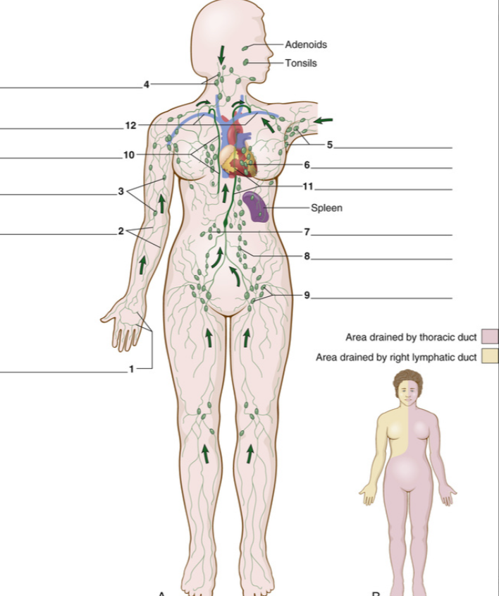
🧠 Lymphatic System – Key Points
🧭 Anatomy & Flow of Lymph
Lymph Capillaries [1]:
Begin in spaces around cells
Thin-walled, like blood capillaries
Larger Lymph Vessels [2]:
Carry lymph from lymph capillaries
Thicker walls than capillaries
Contain valves to ensure one-way flow toward the thoracic cavity
Lymph Nodes [3]:
Collections of stationary lymph tissue
Located along the path of lymph vessels
🧍♂ Major Lymph Node Regions
Cervical (neck) [4]
Axillary (armpit) [5]
Mediastinal (chest) [6]
Mesenteric (intestinal) [7]
Paraaortic (lumbar) [8]
Inguinal (groin) [9]
🌊 Lymphatic Ducts
Right Lymphatic Duct [10]:
Drains right side of head and chest
Thoracic Duct [11]:
Drains lower body and left side of the head
Both ducts empty into large veins in the neck [12] to return lymph to bloodstream
🛡 Functions of Lymph Nodes
Produce lymphocytes
Filter lymph, trapping:
Bacteria
Inflammatory substances
Cancerous cells
Contain macrophages:
Swallow (phagocytose) foreign substances
Swollen nodes:
Tenderness = more likely infection
Non-tender = more likely tumor
Contain:
B lymphocytes (B cells):
Produce antibodies
T lymphocytes (T cells):
Destroy foreign cells
Help B cells produce antibodies
❤ Special Lymphatic Organs
Spleen (Figure 14-5A)
Location: Left upper quadrant (LUQ) of abdomen, near the stomach
Functions:
Destroys old erythrocytes (RBCs) via macrophages
Filters microorganisms and foreign material
Activates B cells and T cells
Stores blood, especially RBCs and platelets
Injury risk:
Trauma (e.g., car accident) may rupture spleen
Can cause hemorrhage → may require splenectomy
After splenectomy, liver, bone marrow, and lymph nodes take over
Thymus Gland (Figure 14-5B)
Location: Upper mediastinum between lungs
Size: Large in fetus/childhood; shrinks with age
Functions:
Crucial in immunity during early life
T cells mature here
Teaches immune cells to recognize "self" antigens = tolerance
Thymectomy in infancy:
Impairs antibody production and immune cell development
Failure of tolerance = leads to autoimmune disease
Vocabulary
Term | Definition / Description |
|---|---|
Adaptive Immunity | Ability to recognize and remember specific antigens and mount an attack on them; includes humoral (B cells) and cell-mediated (T cells) immunity. |
Adenoids | Mass of lymphatic tissue in the nasopharynx. |
Antibody | Protein produced by B cells to destroy antigens. |
Antigen | Substance recognized as foreign by the body; triggers immune response. Mostly proteins on bacteria, viruses, or transplanted cells. |
Axillary Nodes | Lymph nodes located in the armpit. |
B Cell (B Lymphocyte) | Lymphocyte that matures into plasma cells to secrete antibodies; originates in bone marrow. |
Cell-Mediated Immunity | T cells respond to and destroy antigens; a type of adaptive immunity. |
Cervical Nodes | Lymph nodes in the neck region. |
Complement System | Blood proteins that help antibodies destroy their targets. |
Cytokines | Proteins secreted by cytotoxic T cells to aid antigen destruction (e.g., interferons, interleukins). |
Cytotoxic T Cell | Lymphocyte (CD8+) that directly kills antigen-bearing cells. |
Dendritic Cell | Antigen-presenting cell that shows T and B cells what to attack. |
Helper T Cell | Lymphocyte (CD4+) that aids B cells and stimulates other T cells. |
Humoral Immunity | B cells produce antibodies after exposure to specific antigens. |
Immunity | Body’s ability to resist foreign organisms and toxins; includes natural and adaptive immunity. |
Immunoglobulins | Antibodies (IgA, IgE, IgG, IgM, IgD) secreted by plasma cells. |
Immunotherapy | Use of immune cells, antibodies, or vaccines to treat disease. |
Inguinal Nodes | Lymph nodes in the groin region. |
Interferons | Cytokines secreted by T cells and other cells to regulate immune response. |
Interleukins | Cytokines that stimulate growth of B and T lymphocytes. |
Interstitial Fluid | Fluid between cells; becomes lymph when it enters lymph capillaries. |
Lymph | Clear fluid in lymphatic vessels, collected from tissues. |
Lymph Capillaries | Smallest lymphatic vessels. |
Lymphoid Organs | Lymph nodes, spleen, thymus gland, tonsils, and adenoids. |
Lymph Node | Stationary lymphatic tissue clusters along lymph vessels; contains lymphocytes and macrophages. |
Lymph Vessel | Tubes carrying lymph throughout the body; empty into veins in the upper chest. |
Macrophage | Large phagocyte in lymph nodes and tissues that engulfs foreign substances. |
Mediastinal Nodes | Lymph nodes in the chest between the lungs. |
Mesenteric Nodes | Lymph nodes in the intestinal mesentery. |
Monoclonal Antibody | Laboratory-produced antibody used to target and destroy cells; useful in immunotherapy. |
Natural Immunity | Innate, nonspecific protection present at birth; involves neutrophils, monocytes, macrophages, NK cells. |
Paraaortic Nodes | Lymph nodes near the aorta in the lumbar (waist) region. |
Plasma Cell | Mature B lymphocyte that secretes antibodies. |
Right Lymphatic Duct | Lymphatic vessel in the chest that drains lymph from the upper right body; empties into a large vein in the neck. |
Spleen | Organ in the left upper abdomen that destroys old red blood cells, activates lymphocytes, and stores blood. |
Suppressor T Cell | Lymphocyte that inhibits B and T cell activity; also called T regulatory cell (Treg). |
T Cell (T Lymphocyte) | Lymphocyte that attacks antigens directly or secretes cytokines to destroy them. |
Tolerance | Ability of T cells to recognize the body’s own antigens as "self" and not attack them; prevents autoimmune disease. |
Thoracic Duct | Large lymphatic vessel draining lower and left side of the body; empties lymph into veins in the neck. |
Thymus Gland | Lymphoid organ in the mediastinum that conditions T cells and aids immune response. |
Tonsils | Masses of lymphatic tissue in the back of the oropharynx. |
Toxin | Poison, typically a protein produced by certain bacteria, animals, or plants. |
Vaccination | Exposure to a foreign protein (antigen) that provokes immune response, providing protection. |
Vaccine | Weakened or dead antigen given to induce antibody production and adaptive immunity. |
Pathology Terminology
Term/Topic | Definition / Description |
|---|---|
Immunodeficiency | Disorders where the immune system is deficient. Can be congenital (e.g., SCID) or acquired (e.g., AIDS). |
SCID (Severe Combined Immunodeficiency Disease) | Congenital lack of B and T cells; thymus small; no immunity, leading to severe infections in infants. |
AIDS (Acquired Immunodeficiency Syndrome) | Caused by HIV, which destroys CD4+ helper T cells; leads to opportunistic infections, secondary cancers, neurologic problems. |
HIV (Human Immunodeficiency Virus) | Retrovirus that infects helper T cells (CD4+), disrupting immune response. Transmitted sexually, via blood, or mother-to-child. |
Opportunistic Infections | Infections occurring in AIDS due to weakened immunity; include candidiasis, cryptococcal infection, PCP pneumonia, toxoplasmosis, etc. |
Common Opportunistic Infections (Table 14-2) | - Candidiasis: fungal infection of mouth, skin, respiratory tract. - Cryptococcal infection: fungus from environment affecting lungs, brain. - Cryptosporidiosis: parasitic GI and CNS infection. - CMV infection: viral retinitis and enteritis. - Herpes simplex: viral blisters, encephalitis. - Histoplasmosis: fungal lung infection from bird/bat droppings. - MAI complex: bacterial systemic infection. - PCP pneumonia: fungal lung infection. - Toxoplasmosis: parasitic CNS infection. - Tuberculosis: bacterial lung and systemic infection. |
Malignancies Associated with AIDS | Kaposi sarcoma (skin tumors) and lymphoma (lymph node cancer). |
Wasting Syndrome | Weight loss, muscle weakness, decreased appetite and mental activity in AIDS patients. |
HIV Transmission Routes (Table 14-3) | Sexual contact, blood exposure (needles, transfusion), and mother to newborn (in utero or breast feeding). |
Treatment for AIDS | - Reverse Transcriptase Inhibitors (RTIs): block HIV replication enzyme (e.g., zidovudine). - Protease Inhibitors: block viral protease. - HAART: combination therapy including entry and integrase inhibitors. |
Hypersensitivity (Allergy) | Abnormal immune response to allergens; can range from mild (hay fever) to severe (anaphylaxis). |
Anaphylaxis | Severe systemic allergic reaction causing hypotension, shock, respiratory distress; requires immediate treatment (epinephrine). |
Atopy | Inherited predisposition to allergic reactions (e.g., asthma, hives, atopic dermatitis). |
Allergen Immunotherapy (Allergy Shots) | Gradual exposure to allergens to build tolerance and reduce symptoms. |
Lymphoma | Malignant tumor of lymphoid tissue; includes Hodgkin and non-Hodgkin types. |
Hodgkin Lymphoma | Malignant tumor with Reed-Sternberg cells; treated with radiation or chemotherapy; often curable. |
Non-Hodgkin Lymphomas | Diverse group mostly involving B cells; treatment varies, often chemotherapy. |
Multiple Myeloma | Malignant plasma cell tumor in bone marrow; produces large amounts of one antibody type (often IgG). |
Waldenström Macroglobulinemia | B cell malignancy producing excess IgM causing blood thickening (hyperviscosity). |
Thymoma | Tumor of thymus gland; associated with myasthenia gravis and autoimmune diseases; treated primarily by surgery. |
Quick Glossary (from Study Section)
Term | Meaning |
|---|---|
Allergen | Substance causing a hypersensitivity reaction (type of antigen). |
Anaphylaxis | Extreme allergic reaction. |
Atopy | Inherited allergic predisposition. |
CD4+ cells | Helper T cells infected by HIV. |
Hodgkin lymphoma | Lymphoid malignancy with Reed-Sternberg cells. |
HIV | Retrovirus causing AIDS. |
Kaposi sarcoma | AIDS-related skin cancer from capillary cells, caused by human herpesvirus 8. |
Non-Hodgkin lymphomas | Group of lymphoid cancers, mainly B cell types. |
Opportunistic infections | Infections common in AIDS due to weakened immunity. |
Protease inhibitor | Drug blocking viral protease to stop HIV replication. |
Reverse transcriptase inhibitor (RTI) | Drug blocking reverse transcriptase enzyme in HIV replication. |
Wasting syndrome | Weight loss and muscle weakness in AIDS patients. |
Laboratory Test and Clinical Procedures
Test/Procedure | Purpose / Description | Notes / Reference Values |
|---|---|---|
CD4+ Cell Count | Measures number of helper T cells (CD4+ T cells) in blood. | Normal: 500–1500 cells/mm³If <250-200, start anti-HIV treatment |
ELISA (Enzyme-linked Immunosorbent Assay) | Screening test for anti-HIV antibodies in blood. | Antibodies appear ~2 weeks post-infection.Positive test confirmed by Western blot. |
Immunoelectrophoresis | Separates and measures types of immunoglobulins (IgM, IgG, IgE, IgA, IgD). | Detects abnormal antibody levels in diseases like multiple myeloma and Waldenström macroglobulinemia. |
Viral Load Test | Measures amount of HIV virus in bloodstream. | Includes PCR assay and NASBA test to quantify viral RNA. |
Computed Tomography (CT) Scan | X-ray imaging producing cross-sectional views of lymphoid organs (lymph nodes, spleen, thymus). | Used to detect abnormalities in lymphoid tissue and organs. |
Abbreviation | Meaning / Description |
|---|---|
AIDS | Acquired immunodeficiency syndrome |
CAR T-cells | Chimeric antigen receptors; lab-activated to fight cancer cells |
CD4+ cell | Helper T cell |
CD8+ cell | Cytotoxic T cell |
CMV | Cytomegalovirus — causes opportunistic AIDS-related infection |
Crypto | Cryptococcus — causes opportunistic AIDS-related infection |
ELISA | Enzyme-linked immunosorbent assay — test to detect anti-HIV antibodies |
G-CSF | Granulocyte colony-stimulating factor — cytokine promoting neutrophil production |
GM-CSF | Granulocyte-macrophage colony-stimulating factor — promotes myeloid cell growth |
HAART | Highly active antiretroviral therapy — combination drug treatment for AIDS |
Histo | Histoplasmosis — fungal infection in AIDS patients |
HIV | Human immunodeficiency virus — causes AIDS |
HSV | Herpes simplex virus |
IEC | Immunoeffector cells — activated cells defending the body in immune response |
IgA, IgD, IgE, IgG, IgM | Immunoglobulins (types of antibodies) |
IL1 to IL38 | Interleukins (immune signaling molecules) |
KS | Kaposi sarcoma |
KSHV | Kaposi sarcoma herpesvirus (herpesvirus 8) |
MAI | Mycobacterium avium-intracellulare complex — causes lung/systemic disease |
MoAb | Monoclonal antibody |
NHL | Non-Hodgkin lymphoma |
PCP | Pneumocystis pneumonia — opportunistic AIDS infection |
PI | Protease inhibitor |
RTI | Reverse transcriptase inhibitor (e.g., zidovudine, lamivudine) |
SCID | Severe combined immunodeficiency disease |
Treg | Regulatory T cell (suppressor T cell) |
Toxo | Toxoplasmosis — parasitic infection associated with AIDS |