Alteration in Sensory
eye anatomy
external eye: extrinsic eye muscles, upper and lower eyelids, sclera, conjunctiva, and lacrimal structures.
eyeball → l / fluid chambers = anterior, posterior, vitreous (biggest w/ gel like liquid between lens and retina= shape of eye)
fibrous: sclera, transparent cornea
vascular: iris, choroid (retina that contains blood supply and connective cells) , ciliary body ( creates clear fluid)
neural : retina, optic nerve, and visual pathway
aqueous humor is secreted from choroid for intraocular pressure
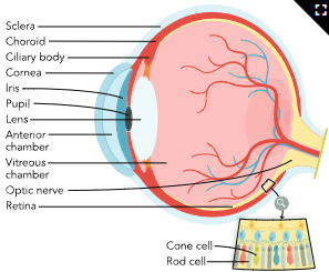
photoreceptors (rods and cones) in retina → optic nerve
rods for vision in low light and peripheral image detection
cones color differential
macular degeneration
age-related macular degeneration (AMD): blurred vision → central vision not working
build up of waste product (drusen) beneath retina
dry: outer retina to breakdown → drusen accumulation (gradual blurring = night vision, diminished central vision, diff reading)
wet: abnormal growth of blood vessel → blood leak in center retina (macula) [ rapid → sudden blurred vision/ bling spots]
>65y, genetic
→ diff w/ ADL, read, vehicle, fall
~ to have thick carotid arteries, hypercholesterolemia, hyperlipidemia, inc plaque
lab/dx:
ophthalmological examination, fundoscopy (w/ dilation eye drops)
Fluorescein angiograph: eye blood flow
nursing:
assess: Snellen chart, asler grid
fall risk
smoking cessation, dietary changes, chronic health condition management, exercise routines, and follow-up appointment schedule
dietary: antioxidants, zinc, unsaturated fats, and omega-3 fatty acids
Fruits and vegetables high in antioxidants:
Blueberries, grapes
Squash, broccoli, spinach, kale
Foods high in zinc:
Red meats: beef, pork, lamb
Dairy: milk, cheese, yogurt
Eggs
Grains: whole-grain cereal, whole-wheat bread
Unsaturated fats: olive oil, canola oil, avocado
Foods high in omega-3 fatty acids:
Salmon, soybeans, flax seed, chia seed
tx:
carotenoids lutein and zeaxanthin = slow down
wet AMD = anti-vascular endothelial growth factor (anti-VEGF) = slow abnormal vessel growth
ranibizumab, aflibercept, or brolucizumab
monthly into vitreous body of eye
photodynamic therapy (PDT) = laser beam to target abnormal blood vessel
cataracts
cloudy/ opaque eye lens by 65<
or inc risk w/
Advanced age
Diabetes mellitus
Hypertension
History of eye inflammation
Previous traumatic eye injury
Use of steroids
Previous eye surgery
Family history
Overexposure to sun or ultraviolet (UV) rays
Smoking
Alcohol use disorder
Obesity
~ w. chronic HTN, DM
pt education: stop smoking, fruits/dark green leafy vegetables (vit C/E, lutein and zeaxanthin)
wear dark UV sunglasses
assess: visual acuity (Snellen chart) , ophthalmoscope and a slit-lamp biomicroscope, fundoscopy
tx:
surgery: remove lens and implant artificial intraocular lens (IOL) → phacoemulsification (mechanically assisted extracapsular procedure)
pre op: ask for recent infection, check for anticoag/antiplatelet
post op: check for sudden changes in eye pain, purulent drainage or bleeding, floaters, other changes in vision, or swelling around the external eye.
`no water in eye or any foreign object 1 week after surgery
avoid sneezing, cough, gagging
glaucoma
progressive increase ocular pressure against optic nerve due to change in volume or decrease absorption of aqueous humor
→ loss of peripheral vision, a halo effect when looking at lights, and decreased visual acuity when in darkness.
open angle: gradual
corrective lenses don’t work
closed angle: sudden vision loss = complete obstruction of aqueous humor outflow
inc risk:
Clients older than 60
Black or Hispanic clients
Diabetes mellitus
Hypertension
Cardiac disease
Hyperlipidemia
History of eye injury or inflammation
Previous eye surgery
Family history
Severe nearsightedness (myopia)
pt education: have extensie eye exam before 40
if high risk = annual
eye drops: tilt head and look up, pull lower lid and instill w/o touching eye → close eye for 3 mins
lab/ dx:
tonometry = measure eye pressure = 11-21 normal
visual field testing (perimetry)
tx:
decrease intraocular pressure by decreasing aqueous humor or decrease fluid produced
AB: reduce humor production and inc amount of fluid drained in eye
BB/ carbonic anhydrase inhibits: decrease humor volume
Prostaglandin analog: inc drainage
Alpha agonists
|
|
Beta-blockers
|
|
Carbonic anhydrase inhibitors
|
|
Prostaglandin analogs
|
|
surgical:
laser trabeculoplasty: uses lasers on drainage angle tissue (trabecular meshwork)
→ laser thickens tissue = inc drainage ~1-2 months
trabeculectomy = drainage flap that will allow drainage
retinal detachment
retinal detaches from nourishment
from gel like humor to serous liquid → collapse and separate retina
→ dark floaters or sensation of curtain being placed in vision, blurry or distorted vision and seeing flashes of light (photopsia)
risk: consistency of the vitreous humor, diabetic retinopathy, macular degeneration, eye tumors, traumatic eye injuries (contact sports/ heavy lifitng), or inflammatory disorders, such as lupus, personal or family history of retinal detachments, previous cataract lens removal, and nearsightedness.
pt education
not preventable by annual exams but can identify risk
Wearing eye protection during sports or dangerous activities
lab/dx: ophthalmological examination, fundoscopy, slit-lamp biomicroscope
assess: confrontational visual field test (peripheral)
check intraocular pressure (<10)
surgery:
scleral buckle: silicon band sewn in place on scelate → eye indention
pneumatic retinopexy/ pars plana vitrectomy: inject absorbable gas into vitreous cavity (8 weeks to reabsorb)
post op: instructed to lie face down for up to 3 months following surgery
ear anatomy
external: auricle/ auditory canal, hair folicles
middle: tympanic and ossicle bone (malleus, incus, and stapes)
inner: cochlea, semicircular canal,
cochelea,= scala vestibuli w/ fluid perilymph (plasma like) scala media (endolymph)
ossicles transmit vibrations to endolymph fluid + hair folicles → neural impulse
hair and fluid are responsible for balance
hearing loss
Type of Hearing Loss | Causes |
|---|---|
Sensorineural ( presbycusis) → degeneration of hair follicles or low blood supply to inner ear |
|
Conductive (exteranl ear condition) |
|
Mixed |
|
risk; age-related changes to the structures of the ears, genetic predisposition, prolonged exposure to loud noise, and adverse reaction to ototoxic medications (cisplatin, furosemide).
s/s: difficulty w/ joint convos, balance, alarms, driving, dizzy,
pt education:
decrease exposure to loud or repetitive noises, regularly use ear protection (noise-canceling headphones, earbuds), and stop or decrease smoking
hearing screening, canals free of cerumen
→ erasable whiteboard for writing words, ensuring that the client can see the speaker’s mouth, or turning off obstructive noises that impede hearing.
Assistive listening devices
Hearing aids
Specialized FM radio headphones
Television amplifiers
Telephone amplifiers
Alerting devices (with flashing lights, vibration, or loud noise indicators)
Doorbell with visual indicators
Telephone with visual indicators
Alarms with visual indicators
Communication devices
Picture boards
Computerized texting
s/s: not aware of deficit until told by other pts
lab/dx:
Finger rub test | Gently rub fingers together 6 inches from client’s ear. Repeat the finger rubs six times. |
|
Whispered voice test | Stand at arm's length behind client. Ask client to cover one ear while whispering a word or number six times. |
|
audiometry: delivery of diff frequencies and sound intensity
exam w/ otoscope'
tx:
hearing aids = mild-moderate loss
improve sound quality, reduce feedback, and allow for easy volume modifications.
Do's | Don't's |
|---|---|
|
|
cochlear implants for severe SNHL
→ drilled to mastoid bone, electrode
position pt on side w/ surgery to face up
do not rotate or flex neck
no strenuous activity
soft foods to prevent jaw from closing
ear packed w/ iodoform gauze.
→ 2 weeks will become activated
tinnitus
ringing, roaring, or constant sound w/o external stim ( intermediate pr\\or continious)
Constant ringing
Loud buzzing
Pulsating hum
Low pitched roaring
Clicking
Hissing
High pitched squealing
damage to cochlear hair components, abnormar ear activity,
or metabolic disorders, temporomandibular joint disorders (TMJD), or cranial nerve abnormalities and neuro conduction
subjective =
Hearing impairment at the cochlear level or of the cochlear nerve | Otosclerosis (abnormal growth in the area surrounding the stapes bone of the middle ear) |
Eustachian tube dysfunction | Vascular disorders (atherosclerosis, arteriovenous fistula, hypertension) often present as pulsatile sounds |
Metabolic-related disorders such as diabetes mellitus, medications, autoimmune disorders, head trauma, multiple sclerosis | Temporomandibular joint disorders (TMJD); shares nerve connections with the middle ear |
Lesions of the vestibulocochlear nerve (8th cranial nerve) | Chiari malformation (a condition in which the cerebellum extends through the foramen magnum into the spinal canal) |
comorbidities: TMJD, frequent ear infections, migraines, Meniere’s disease, fibromyalgia, meningitis, excessive cerumen, thyroid disorders, Lyme disease, syphilis, and ototoxic medications (certain diuretics such as Furosemide, chemotherapy agents such as cisplatin, antibiotics such as gentamycin; acetaminophen; non-steroidal anti-inflammatory drugs).
lab/dx:
rule out: anemia, thyroid issues, or heart disease
hearing testing, otoscopic exam, CN nerve, CT/MRI
pt education:
wear headphones when in noisy or loud environments, turn down music, reduce salt intake, and decrease the use of nicotine, caffeine, and alcohol.
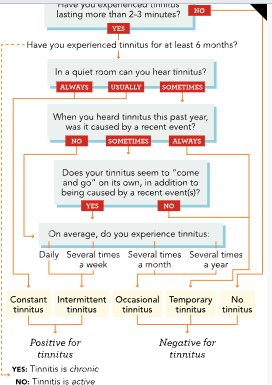
Tinnitus Screener questionnaire
tx:
TMJD, dental treatment
Discontinuation or switching of medications that cause ototoxicity
CBT
Tinnitus retraining therapy | A combination of modalities including education counseling that is specific to the causes and exacerbations of tinnitus, directive counseling which provides the client with coping mechanisms, and sound therapy which desensitizes the brain to the presence of tinnitus |
Acoustic therapy | Involves the use of external devices used to mask the sound of tinnitus. Possible devices include sound generator hearing aids, use of electric fans or other forms of white noise, and music therapy |
meniere’s disease
progressive disorder that develops from an excessive buildup of endolymphatic
in the inner ear and leads to debilitating vertigo, tinnitus, and hearing loss.
inadequate fluid drainage from the ear, a history of an autoimmune disorder, a recent viral infection, or a genetic predisposition
comorbidities: migraines, rheumatoid arthritis, lupus, and ankylosing spondylitis.
pt education: plenty of rest, avoid excessive stressful situations, and limit caffeine. Reducing the intake of dietary salt
avoid foods high in sugar and decrease the use of nicotine, caffeine, and alcohol
s/s: vertigo
Vomiting
Nausea
Blurry vision
Cold sweats
Trembling
Hearing loss
Headaches
Imbalance
Congestion in the ear
Ear fullness (ear pressure)
lab/dx: Electronystagmography (ENG) → moniotor for manifestation of impaired gait
electrocochleography (ECOG) can be used to evaluate pressure within the ears
tx:
Diuretics are beneficial in reducing edema within the endolymphatic sac.
vertigo s/s : Motion sickness (meclizine, valium) and anti-nausea (promethazine, ondansetron)
Vestibular rehabilitation for balenced