Topic 22- Respiration/Excretion
Outcomes:
Compare and contrast types of respiratory systems
Sequence of mammalian respiration
Hypothesize and diagnose the impact of variability on mammalian respiration
Compare and contrast types of osmoregulation
Compare and contrast types of nitrogenous waste
Sequence the process of excretion
Hypothesize and diagnose the impact of variability on excretion
I. Respiration
A. Overview of Respiration
Cellular respiration: Process where cells use oxygen (O₂) to generate ATP.
Organismic respiration: The physical movement of O₂ into cells and CO₂ out of cells.
In very small organisms (<1 mm thick):
Simple diffusion is sufficient, no special transport systems needed.
In larger animals:
Specialized respiratory structures are required, such as:
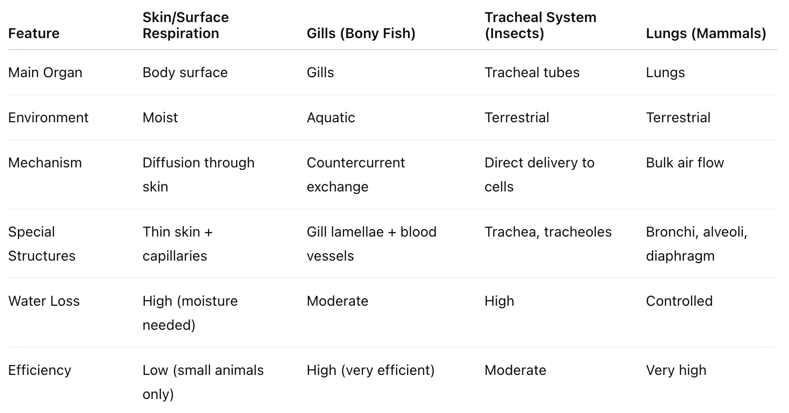
Skin/surface (cutaneous respiration)
Gills
Tracheal tubes (insects)
Lungs
These structures must meet three key conditions:
Thin walls for gas diffusion
Large surface area
Moist environment
Associated with blood vessels (except in insects)
B. Comparison of Respiratory Systems
Gills (Bony Fish):
Use countercurrent exchange to maximize O₂ absorption by maintaining a concentration gradient across the gill surface.
Tracheal System (Insects):
A network where:
Trachea → Tracheoles → direct gas exchange with cells.
Oxygen and carbon dioxide are delivered directly to each cell.
Trade-off: High water loss.
C. Mammalian Respiration
Respiratory system is a branching set of tubes ending in specialized structures:
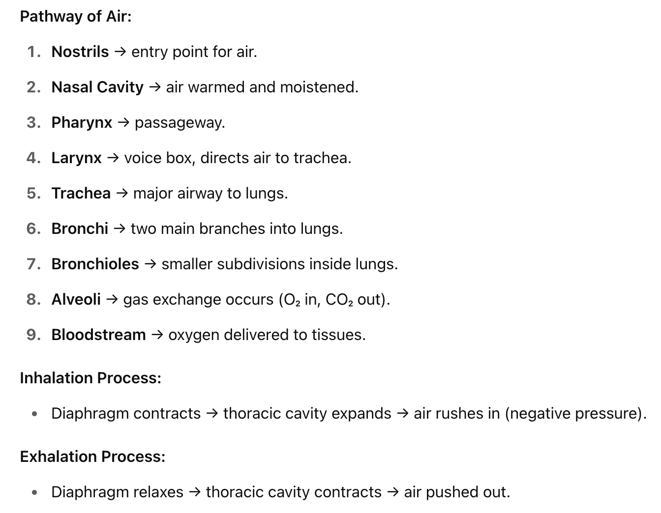
Nostrils: Air enters.
Nasal cavity: Air is warmed and moistened.
Pharynx: Passageway branching into the larynx.
Trachea: Main tube carrying air to lungs.
Bronchi: Two tubes, each leading to a lung.
Bronchioles: Smaller branches within lungs.
Lungs: Paired spongy organs (right lung larger).
Alveoli: Tiny air sacs surrounded by capillaries for gas exchange (high surface area).
Diaphragm: Muscle critical for breathing.
D. Regulation of Breathing
Mammalian breathing uses negative pressure:
Inhalation: Diaphragm contracts, thoracic cavity enlarges, air is drawn in.
Exhalation: Diaphragm relaxes, air is expelled.
Lung structure:
Each lung is enclosed by a pleural membrane to reduce friction during breathing.
Control centers:
Medulla oblongata:
Regulates depth and rhythm of breathing.
Monitors cerebrospinal fluid pH as an indicator of CO₂ concentration.
CO₂ + H₂O → H₂CO₃ (carbonic acid) → H⁺ + HCO₃⁻ (bicarbonate).
Increased acidity (H⁺) leads to increased breathing to expel more CO₂.
Pons:
Modifies the rate of breathing.
II. Osmoregulation
A. Definitions
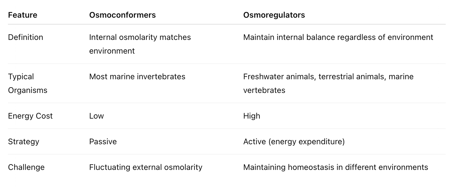
Osmoconformers:
Organisms that are isosmotic with their environment.
Most marine invertebrates.
Osmoregulators:
Organisms (like humans) that actively regulate internal osmolarity, not matching the environment.
Includes freshwater animals, terrestrial animals, and most marine vertebrates.
B. Terrestrial Vertebrate Osmoregulation
Challenge: Prevent water loss in dry environments.
Water acquisition methods:
Drinking water
Consuming moist foods
Producing metabolic water through cellular respiration
Adaptations to minimize water loss:
Protective body coverings (skin, scales)
Nocturnal behaviors to avoid daytime heat
Concentrating urine to minimize water excretion
III. Excretion
A. Introduction to Excretion
Protein and nucleic acid catabolism:
Broken down for energy or converted into fats/carbs.
Deamination (removal of amine group, NH₂) occurs in the liver.
Produces ammonia (NH₃), which is toxic and must be excreted.
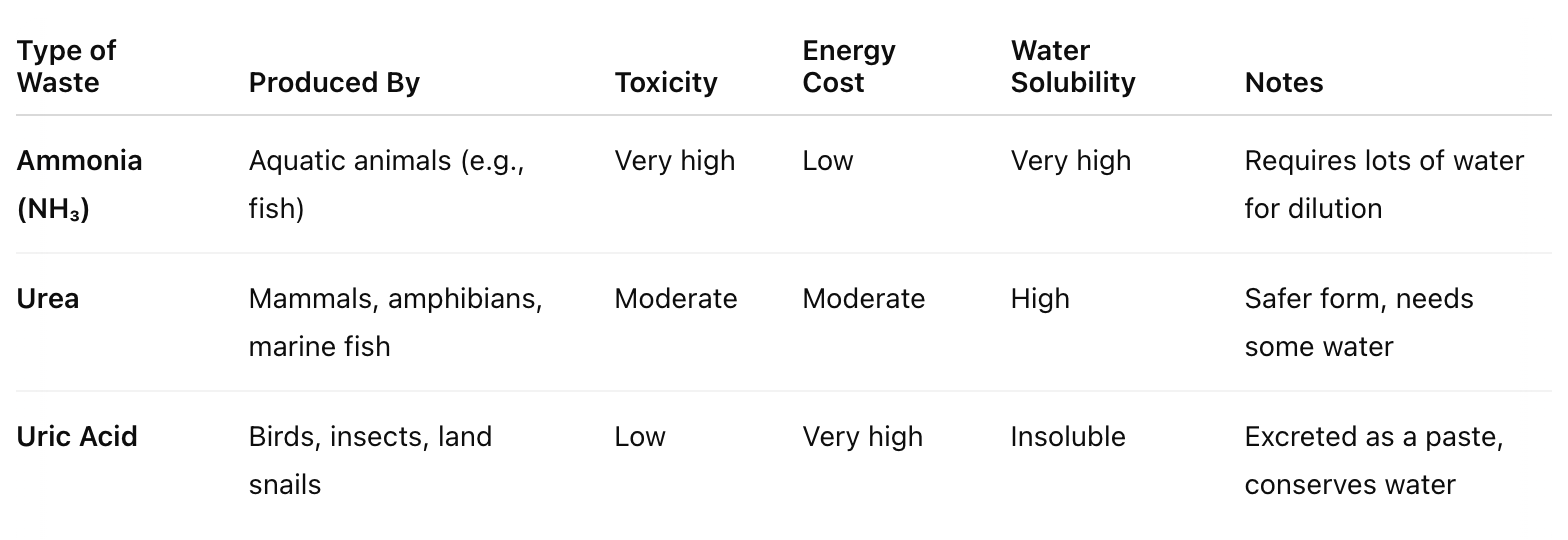
B. Types of Nitrogenous Wastes
Ammonia (NH₃):
Highly toxic.
Requires lots of water to dilute.
Low energy cost to produce.
Found in aquatic animals.
Urea:
Less toxic.
Requires moderate energy to produce.
Water soluble.
Excreted by mammals, amphibians, and marine fish.
Uric Acid:
Relatively nontoxic.
Very high energy cost to produce.
Insoluble in water (excreted as a paste).
Produced by birds, insects, and land snails.
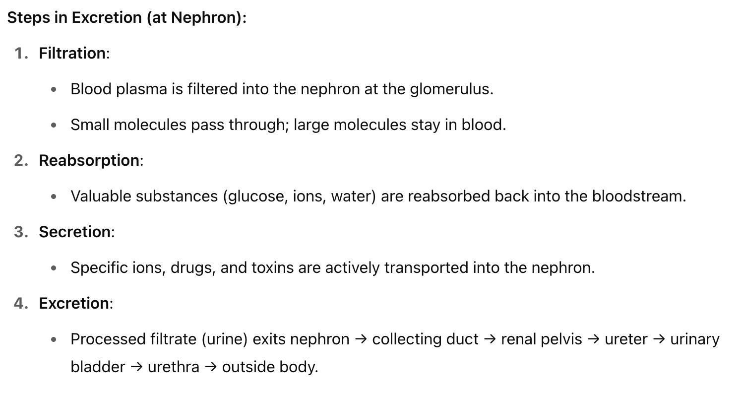
C. The Basic Excretion Process (at the Nephron)
1. Filtration
Where: In the glomerulus (capillary bed) and Bowman’s capsule of the nephron.
What happens:
Blood pressure forces plasma through a selectively permeable membrane.
Small molecules (e.g., water, glucose, salts, urea) pass through.
Large molecules (e.g., proteins, blood cells) remain in the bloodstream.
Purpose: Initial separation of waste from useful components based on size.
2. Reabsorption
Where: Mainly in the proximal tubule and loop of Henle.
What happens:
Valuable substances (like glucose, amino acids, ions, and water) are reabsorbed into the bloodstream.
This prevents the loss of essential nutrients.
Purpose: Recover materials the body still needs.
3. Secretion
Where: Primarily in the distal tubule and parts of the loop of Henle.
What happens:
Active transport of additional waste products (e.g., hydrogen ions, potassium ions, drugs) from the blood into the nephron.
Purpose: Fine-tunes chemical composition and removes excess/toxic substances.
4. Excretion
Where: Outside the kidney – urine flows from:
Collecting ducts → Renal pelvis → Ureter → Urinary bladder → Urethra.
What happens: Urine is expelled from the body.
Purpose: Eliminate final waste product from the body.
Human Excretory System Overview
Organs Involved:
Kidneys: Filter blood and regulate water, salt, and waste balance.
Ureters: Transport urine from kidneys to bladder.
Urinary Bladder: Stores urine.
Urethra: Eliminates urine from the body.
Kidney & Nephron Structure
Renal Cortex: Outer layer containing most of the nephrons.
Renal Medulla: Inner layer with loops of Henle and collecting ducts.
Renal Pelvis: Funnel-shaped structure where urine collects before entering the ureter.
Renal Artery/Vein: Supply and drain blood to/from kidneys.
Nephron Components:
Glomerulus: Capillary bed where filtration occurs.
Bowman's Capsule: Cup-like sac around the glomerulus.
Proximal Tubule: Major site of reabsorption.
Loop of Henle:
Longer in desert animals for water conservation.
Reabsorbs water and salt.
Distal Tubule: Regulates ions and pH.
Collecting Duct: Final urine processing; sends it to renal pelvis.