Chapter 10.1-10.2 Muscular Tissue
10.1 Overview of Muscle Tissue
Different types of muscle tissue:
How do they differ? microscopic anatomy, location, and the way they’re each controlled by nervous and endocrine systems
Skeletal Muscle Tissue
location: originate and insert on many bones
function: move bones of skeleton voluntarily, controlled by neurons (controlled subconsciously, i.e diaphragm and posture and stabilizing muscles)
striated - alternating light and dark protein bands, aka striations
multinucleated
Cardiac Muscle Tissue
location: heart wall
function: contract and relax heart involuntarily, internal autorhythmicity, rate can be adjusted by hormones and neurotransmitters
striated, smaller fibers, branched, cells joined by intercalated disks
uninucleate
Smooth Muscle Tissue
location: walls of internal structures like organs, blood vessels, and airways; skin, attached to hair follicle
function: move things through organs/vessels/passageways involuntarily, some muscles have autorhythmicity
non-striated - smooth
uninucleate
Functions of Muscle Tissue
Produce Body Movements
skeletal muscles, bones, and joints involved
Stabilize Body Positions
skeletal muscle contractions stabilize joints
ex: posture, standing, sitting
Store and Move Substances in the Body (example):
contractions of ringlike smooth muscles (“sphincters”) prevent outflow of contents of a hollow organ
cardiac muscles contract heart
contraction and relaxation of smooth muscle in blood vessels helps regulate blood pressure
Generate Heat
contraction produces heat via thermogenesis to maintain body temperature
ex: shivering
Properties of Muscular Tissue
Electrical Excitability
ability to respond to stimuli by producing muscles action potentials (APs)
stimuli include:
autorhythmic electrical signals that occur on muscle tissue itself (ex: heart’s natural pacemaker)
chemical stimuli like neurotransmitters released by neurons, hormones, distributed by blood, or local pH changes
Contractility
ability to contract forcefully when stimulated by a nerve impulse
skeletal muscle contractions generate tension while pulling on its attachment points
great enough tension can overcome the resistance of an object, resulting in muscle shortening and movement (doesn’t always shorten, like holding something)
Extensibility
ability of muscle tissue to stretch within limits without being damaged
connective tissue in muscle limits range of extensibility, keeping it within contractile region of the muscle cells
smooth muscle undergoes greatest amount of stretching
cardiac muscle is stretched when heart fills with blood
Elasticity
ability of muscle tissue to return to its original length and shape after contraction or extension
10.2 Structure of Skeletal Muscle Tissue
Each skeletal muscle is a separate organ made up of hundreds of cells called muscle fibers (myocytes) due to elongated shapes
muscle fibers = muscle cells = myocytes
Components:
connective tissue components: subcutaneous tissue and fascia
function: protects muscle tissue
description: surrounds muscle tissue
importance:
subcutaneous tissue (hypodermis): separates muscle from skin
composition: areolar connective tissue, adipose tissue (stores most triglycerides —> insulation and protection from trauma)
function: pathway for nerves, blood vessels, and lymphatic vessels to enter/exit muscles
fascia
location: lines body wall and limbs, surrounds muscles and organs, fills spaces between muscles
composition: dense sheet or broad band of irregular connective tissue
function: supports muscles and organs, allows free movement of muscles, carries nerves and blood and lymphatic vessels
layers that extend from fascia
function: protect and strengthen skeletal muscle
all are continuous with the connective tissue that attaches skeletal muscle to other structures (i.e to bone/another muscle)
could come together to form a tendon
could extend as a broad, flat sheet, or an aponeurosis
epimysium
location: outer layer encircling the entire muscle
composition: dense irregular connective tissue
perimysium
location: surrounds groups of 10-100+ muscle fibers, separating them into muscle fascicles
composition: dense irregular connective tissue
*application: tearing off a piece of meat, it rips apart along the fascicles
endomysium
location: penetrates interior muscle fascicles, separating individual fibers from one another
composition: mostly reticular fibers
disorder: fibromyalgia
chronic, painful, nonarticular rheumatic disorder
affects the fibrous connective tissue components of muscles, tendons, and ligaments
key signs and symptoms
pain from gentle pressure at “tender points”
muscle pain and tenderness without pressure
stiffness of muscles, tendons, and surrounding soft tissues
fatigue, poor sleep, headaches, depression, IBS, inability to do daily activities
treatment
stress reduction, regular exercise, heat, gentle massage, PT, meds for pain, low-dose antidepressant to improve sleep
nerve and blood supply
generally, 1 artery + 2 veins accompany each nerve that penetrates a skeletal muscle
nerve supply
somatic motor neurons
function: stimulate skeletal muscle contraction
composition: the axon extends from brain/spinal cord to a group of muscle fibers, and branches to other muscle fibers
blood supply
blood capillaries
description: microscopic blood vessels that are abundant in muscle tissue
function: bring in O2 and nutrients, remove heat and waste products of muscle metabolism, delivers substances to muscle fibers that are needed for contraction
each muscle fiber comes into contact with 1+ capillary
microscopic anatomy of a skeletal muscle fiber
fibers can be 4-12 in. long
arises during embryonic development from fusion of many small mesodermal cells, aka myoblasts
each fiber has 100+ nuclei
once fusion happens, the fiber can no longer do cell division
number of muscle fibers is SET BEFORE BIRTH and most last for a lifetime
components
sarcolemma
description: plasma membrane of a muscle fiber
location: above nuclei of fibers, surrounds the sarcoplasm
function: facilitate AP transmission, barrier between extra and intra cellular compartments (defines individual muscle fibers)
T tubules
description: tube-like invaginations (folded inside out) of sarcolemma, open to outside of fiber
composition: filled with interstitial fluid
location: tunnel in from surface toward center of fiber
sarcolemma and T tubule arrangement ensures that muscle APs excite all parts of muscle fiber instantly
sarcoplasm
description: cytoplasm of a muscle fiber
composition: glycogen (used to synthesize ATP), myoglobin (red-colored protein that binds oxygen molecules that traveled from interstitial fluid, releases O2 when needed by mitochondria), mitochondria (in rows throughout fiber, intentionally close to the contractile muscle proteins that use ATP)
location: within sarcolemma
disorders
muscular hypertrophy
enlargement of existing muscle fibers
microscopic cause: increased production of myofibrils, mitochondria, sarcoplasmic reticulum, other organelles
physical cause: forceful repetitive muscular activity (i.e strength training)
hypertrophied muscles have more myofibrils, so they are capable of doing more forceful contractions
testosterone promotes further enlargement of muscle fibers
GH and other hormones stimulate increase muscle fiber size
fibrosis
replacement of muscle fibers by fibrous scar tissue
muscular atrophy
decreased size of muscle fibers because of a progressive loss of myofibrils
“disuse atrophy”: atrophy from lack of use of muscles resulting in flow of impulses is reduced, i.e bedridden individuals, people with casts - REVERSIBLE condition
“denervation atrophy”: nerve supply is disrupted or cut, muscle shrinks and is irreversibly replaced by scar tissue
myofibrils
contractile organelles of skeletal muscles
sarcoplasm is stuffed with these thread-like fibers
small diameter that extends entire length of muscle fiber
prominent striations make muscle fiber look striped/striated
sarcoplasmic reticulum (SR)
fluid-filled system of membranous sacs
encircle each myofibril
terminal cisterns: dilated end sacs of SR that butt against T tubule from both sides
terminal cistern — T tubule — terminal cistern = triad
relaxed SR stores calcium, released calcium from terminal cisterns of SR, triggering muscle contraction
filaments (myofilaments)
smaller protein structures
thin filaments: made of actin
thick filaments: made of myosin
both are involved in contraction
2 thin filaments for every thick filament
arranged in compartments called sarcomeres
sarcomeres
basic functional units of a myofibril
extend from Z disc to Z disc
Z discs
plate-shaped regions of dense protein material
separate sarcomeres
A band
darker middle part of sarcomere
extends entire length of thick filaments
toward each end is a zone of overlap - where thick and thin filaments lay side by side
I band
lighter, denser area
contains the rest of the thin filaments, NO thick filaments
Z disc passes through each one
*letter I is thin - contains thin filaments
A and I bands alternate, creating striations
H band
center of each A band
contains thick, NOT thin filaments
*letter H is thick - contains thick filaments
M line
support proteins holding thick filaments together at the center of the H band
middle of sarcomere
A band is the only thing not contracting or shortening
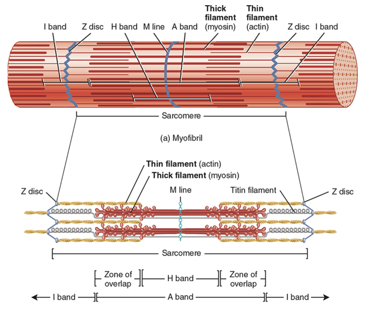
muscle proteins of myofibrils
contractile
function: generate force during contraction
motor proteins: pull cell structures to move, convert ATP to mechanical energy (motion)
myosin: main part of thick filaments, 2 golf clubs twisted together
tails: thinner end, point toward M line, neighboring tails are parallel to each other to form the shaft of the thick filament
heads: have 2 binding sites = actin-binding site and ATP binding site (also functions as ATPase), project outward from shaft in a spiral to extend to 1 of the thin filaments
actin
main part of thin filament
individual molecules join to create an actin filament, twisted in a helix
contains a myosin-binding site for a myosin head can attach
myosin bound to actin
regulatory
function: switch contraction process on/off
troponin
holds tropomyosin in place
when Ca2+ binds to troponin, it does a conformational change that moves tropomyosin away from the myosin-binding sites on actin
tropomyosin
blocks myosin from binding to actin relaxed muscle by covering the myosin-binding sites on actin
structural
function: keep thick and thin filaments aligned, give elasticity and extensibility, link myofibrils to sarcolemma and extracellular matrix
titin
most abundant of these, big, spans half of sarcomere
connects Z disc to M line to stabilize position of the thick filament
elastic - can return to normal state after extreme stretching
maintains central location of A bands
nebulin
long, non-elastic, wrapped around entire length of thin filament
anchors thin filaments to Z discs, regulates length of thin filaments during development
dystrophin
creates attachment between thin filaments of sarcomere to connective tissue matrix surrounding muscle fibers