Chapter 14
Shock and Sepsis
Cardiac Output: Cardiac output (CO) is the amount of blood ejected by the heart per minute.
Formula: CO = Stroke Volume (SV) × Heart Rate (HR).
Heart Rate (HR):
Number of ventricular contractions (heartbeats) per minute.
Influenced by:
Autonomic nervous system (sympathetic & parasympathetic stimuli).
Hormones (e.g., adrenal medulla hormones).
Baroreceptor response to blood pressure changes.
Stroke Volume (SV):
Amount of blood ejected with each ventricular contraction.
Influenced by:
Preload:
Volume of blood in ventricles at the end of diastole.
it reflects a patients fleid volume status→ filling pressures
Increased preload result in Increased CO and Decreased preload results in Decreased CO.
Afterload:
Resistance the ventricle must overcome to eject blood.
Higher afterload makes it harder for the heart to pump blood.
Contractility:
Strength of heart muscle contraction.
Poor contractility results in Decreased SV and Decreased CO.
Oxygen Delivery (DO2):
Amount of oxygen transported to tissues.
Assessed by CO and arterial oxygen content.
Arterial oxygen content depends on:
Hemoglobin levels.
Percentage of hemoglobin oxygen saturation.
amount of oxygen dissolved oxygen in plasma (PaO2).
Oxygen Consumption (VO2):
Oxygen extracted from blood at the tissue level.
Measured via venous oxygen saturation (SvO2).
Normal SvO2: 60%–75%.
Low SvO2 indicates increased oxygen extraction due to:
Low oxygen delivery (DO2).
Decreased hemoglobin, oxygen content, or CO.
Inability to meet increased oxygen demand (e.g., stress, fever, pain, lack of oxygen, Hgb, CO or high tissue demands)
If is less than 60- may indicate that the tissue is taking on too much oxyen
if is greater than 75 it means the tissue are in oxygen debt→ sign of shock
Anaerobic metabolism occurs and lactic acid builds up
Oxygen Debt:
Difference between normal VO2 and VO2 during low oxygen delivery.
Prolonged imbalance leads to greater oxygen debt, which must be repaid to maintain cellular function.
Types of Shock & Causes
Hypovolemic Shock (Loss of Circulating Volume)
Rapid fluid loss → Inadequate circulating volume
Causes:
External blood loss (trauma, GI bleeds, surgery)
Blunt trauma examples include blood loss into the abdomen due to damage to the liver, trauma to the thoracic cavity resulting in a ruptured aorta, or long bone or femur fractures
Internal bleeding (fractures, aneurysms, hemothorax, retroperitoneal bleed)
Severe vomiting, diarrhea, excessive urination, burns
Third-space fluid loss (pleural, peritoneal, joint spaces)
Cardiogenic Shock (Heart Pumping Failure)
Inadequate heart pumping → Decreased cardiac output & tissue perfusion
Causes:
Acute Myocardial Infarction (AMI)
Severe valvular dysfunction
Severe heart failure
Obstructive Shock (Mechanical Block to Circulation)
Mechanical barrier to ventricular filling or emptying → Decreased cardiac output and increase afterload.
Causes:
Impaired filling: Cardiac tamponade, tension pneumothorax
Impaired emptying/increased afterload: Severe valvular disease, pulmonary embolism
Distributive Shock (Vasodilation & Venous Pooling)
Disease states cause vasodilation → Relative hypovolemia & poor vascular tone.
resulting in increased vascular capacity and venous pooling
Causes:
Sepsis
Anaphylaxis
Neurogenic causes (brain injury, spinal injury, spinal anesthesia)
Stages
1. Initial Stage
Marked by hypoxia due to decreased oxygen delivery (DO2) to cells.
Subtle or no clinical manifestations, but cellular damage may begin.
Possible mild changes in heart rate.
Hemodynamic monitoring shows decreased cardiac output.
2. Compensatory Stage
Body activates compensatory mechanisms to maintain perfusion.
Signs & Symptoms:
Tachycardia, tachypnea
Low-normal BP with narrow pulse pressure (due to vasoconstriction)
Decreased urine output
Compensatory Mechanisms
Neural Compensation (Sympathetic Nervous System Activation)
Baroreceptors detect low BP → Sympathetic nervous system (SNS) activation.
Epinephrine & norepinephrine release → ↑HR, ↑contractility, systemic vasoconstriction.
Blood is redirected from nonessential organs (kidneys, GI tract, skin) → vital organs (heart, brain).
Endocrine Compensation (Hormonal Response)
Renin-Angiotensin-Aldosterone System (RAAS):
Low BP → Kidneys release renin → Converts angiotensinogen to angiotensin I → Converted to angiotensin II in lungs.
Angiotensin II = potent vasoconstrictor → Increases BP.
Stimulates aldosterone release → Sodium & water retention → ↑Blood volume.
ADH (Antidiuretic Hormone) Release:
Secreted by posterior pituitary → Promotes water retention.
Catecholamines (Epinephrine & Norepinephrine) Release:
Further vasoconstriction as part of fight-or-flight response.
Glucose Mobilization:
Glucocorticoids from adrenal cortex increase circulating glucose levels for energy.
Chemical Compensation (Respiratory Response)
Chemoreceptors in aorta & carotid arteries detect low oxygen.
Tachypnea (hyperventilation) → Increases oxygen levels.
Respiratory alkalosis → Constriction of carotid arteries → Risk of cerebral hypoxia & ischemia.
3. Progressive Stage
Compensatory mechanisms begin to fail → Profound hypoperfusion.
Blood shunts to vital organs → Peripheral ischemia worsens.
Metabolic acidosis develops.
Sodium-potassium pump failure → Electrolyte imbalances.
Respiratory acidosis due to CO2 retention.
4. Refractory Stage
Prolonged inadequate blood supply → Cell death & multisystem organ failure (MOF).
Aerobic metabolism stops → Only inefficient anaerobic metabolism remains.
Shock is irreversible at this stage.
1. Hypovolemic Shock
↓ Cardiac output
↓ CVP & PAOP (due to low blood volume)
↑ SVR (compensatory vasoconstriction)
↓ Venous oxygen saturation (SvO2/ScvO2) (less oxygen delivery to tissues)
Hypotension & tachycardia
2. Cardiogenic Shock
↓ Cardiac output (due to heart failure)
↑ CVP & PAOP (backed-up pressure in heart)
↑ SVR (compensatory vasoconstriction)
↓ Venous oxygen saturation (SvO2/ScvO2)
Hypotension & tachycardia
3. Obstructive Shock
↓ Cardiac output (due to obstruction in heart or vessels)
Variable CVP & PAOP (depends on obstruction type)
↑ SVR (compensatory vasoconstriction)
↓ Venous oxygen saturation (SvO2/ScvO2)
Hypotension & tachycardia
4. Distributive Shock (Vasodilation & Poor Perfusion)
A. Anaphylactic Shock
↓ Cardiac output
↓ CVP & PAOP
↓ SVR (due to massive vasodilation)
↓ Venous oxygen saturation (SvO2/ScvO2)
Hypotension & tachycardia
B. Neurogenic Shock
↓ Cardiac output
↓ CVP & PAOP
↓ SVR
↓ Venous oxygen saturation (SvO2/ScvO2)
Hypotension & bradycardia (unique feature due to loss of sympathetic tone)
C. Septic Shock
Early Septic Shock ("Warm Shock")
↑ Cardiac output (due to hyperdynamic response)
↓ CVP & PAOP
↓ SVR (due to vasodilation)
Variable or ↑ SvO2/ScvO2
Normal BP (with narrow pulse pressure) or ↓ BP, tachycardia, hyperthermia
Late Septic Shock ("Cold Shock")
↓ Cardiac output
Variable CVP & PAOP
Variable SVR
↓ Venous oxygen saturation (SvO2/ScvO2)
Hypotension, tachycardia, hypothermia
Hypovolemic Shock:
Definition: Hypovolemic shock occurs when there is inadequate intravascular volume, leading to decreased cardiac output, caused by blood or fluid loss from acute injury or illness.
Common Causes:
Blood loss (trauma, GI bleeding)
Fluid loss (vomiting, diarrhea, burns, excessive urination)
Incidence/Prevalence: Data is typically reported based on the cause (e.g., trauma) rather than hypovolemic shock directly.
Pathophysiology
Decreased Fluid Volume: Causes reduced venous return to the heart, leading to decreased stroke volume and cardiac output.
Compensatory Mechanisms:
Sympathetic Nervous System: Increases heart rate (HR), cardiac output, and induces vasoconstriction.
Endocrine System: RAAS and antidiuretic hormone (ADH) promote sodium and water retention to increase venous return.
Angiotensin II: Increases systemic vasoconstriction, boosting blood pressure.
Clinical Manifestations
Early Stage:
Normal blood pressure, increased HR
Restlessness, confusion, decreased urine output
Cool, pale, clammy skin, weak pulses, sluggish capillary refill
Hyperventilation, respiratory alkalosis
Decreased bowel sounds, hyperglycemia
Later Stages:
Lethargy, hypotension, anuria, cyanotic skin, weak/absent pulses
Increased creatinine/BUN, liver dysfunction
Severe: Coma, ischemia, multi-organ failure, death
Laboratory Tests in Shock
pH: Early alkalosis, later acidosis
PaCO2: Decreased in early shock due to hyperventilation
PaO2: Decreased in severe hypoperfusion
HCO3-: Decreased in metabolic acidosis
SvO2: Decreased, indicating poor oxygen delivery
Lactate: > 2 mmol/L, signaling anaerobic metabolism
Hemoglobin/Hematocrit: Decreased in blood loss
Glucose: Elevated as part of the stress response
BUN/Creatinine: Elevated in kidney injury due to hypoperfusion
Hemodynamic Parameters (Table 14.3)
Decreased: Cardiac output, CVP, PAOP
Increased: SVR (due to vasoconstriction)
Decreased: SvO2/ScvO2 (poor oxygen delivery)
Medical Management
Oxygenation: Maximize oxygenation by securing the airway, using 100% non-rebreather mask, or mechanical ventilation (caution with positive pressure).
Fluid Resuscitation:
Non-hemorrhagic shock: Use balanced crystalloid fluids (normal saline, lactated Ringer’s).
Hemorrhagic shock: Use a 1:1:1 ratio of blood products (PRBCs, FFP, platelets) and minimize crystalloids.
Goal: Stabilize BP, restore pulse strength, maintain urine output, lower lactate levels.
Identifying Underlying Causes:
Use ultrasound (FAST), CT, or X-rays to diagnose trauma or bleeding sources.
Surgical intervention may be necessary for bleeding control or fracture stabilization.
Nursing Management
Assessments:
Neurological: Decreased LOC due to low cardiac output and vasoconstriction.
Vital Signs: Tachycardia, hypotension (as compensation fails).
Hemodynamics: Decreased cardiac output, CVP, PAOP, and increased SVR.
Urine Output: Decreased due to compensatory mechanisms.
Skin: Cold, clammy skin (decreased perfusion).
Lab Results: ABGs, lactate, SvO2, hemoglobin, and renal/liver function tests.
Actions:
Oxygen: Apply non-rebreather mask, prepare for intubation if necessary.
IV access: Insert large-bore IV lines for fluid administration.
Fluid Replacement: Administer prescribed fluids (crystalloids, blood products).
Patient and Family Education:
Educate on hypovolemia causes and prevention.
Allow family visitation and provide updates to reduce anxiety.
Evidence-Based Practice in Fluid Resuscitation
Crystalloid Use: Excessive crystalloid use (especially normal saline) worsens coagulopathy and acidosis.
Trauma Resuscitation: Using smaller IV fluid bags (500 mL vs. 1000 mL) can reduce the total volume of saline administered, improving patient outcomes.
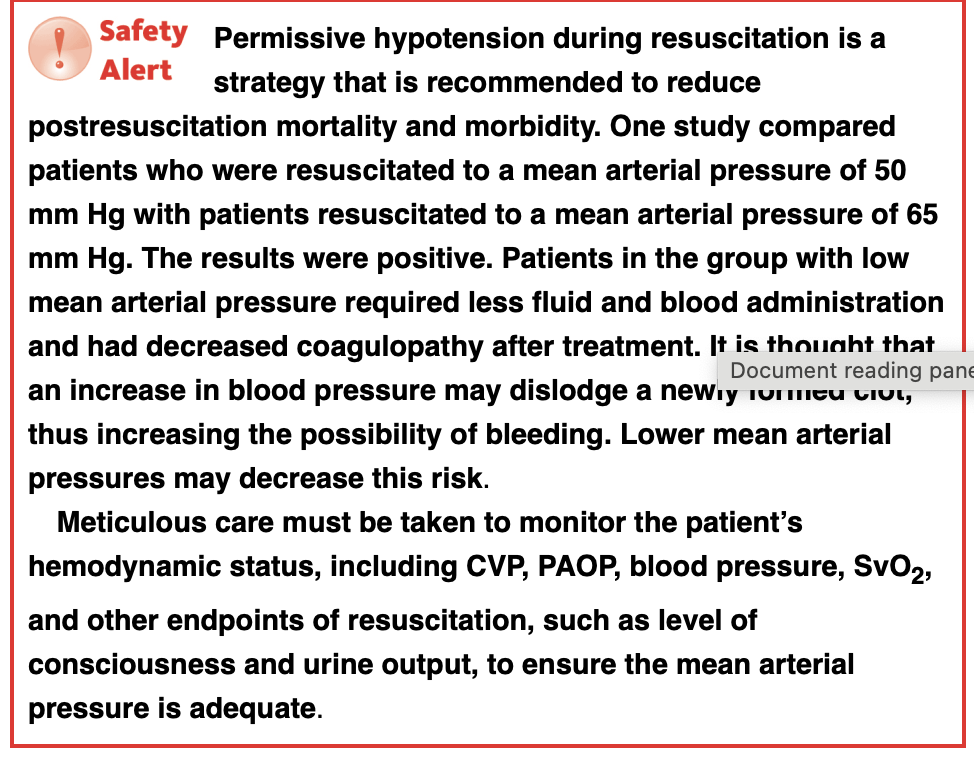
Cardiogenic Shock
The prevalence of cardiogenic shock (CS) ranges from 5-10% in patients with acute myocardial infarction (AMI).
It is more common in men due to the higher prevalence of coronary artery disease in this population.
The incidence of CS following AMI is higher in women.
Mortality rate is around 50%, with a higher rate in patients over 75 years old and a lower rate in younger patients.
Risk factors include conditions that result in acute deterioration of myocardial contraction, including:
AMI with >40% of myocardial damage.
End-stage congestive heart failure, cardiomyopathy, hypertension, diabetes, and multiple-vessel coronary artery disease.
Pathophysiology:
Cardiogenic shock is characterized by tissue hypoperfusion due to severe impairment of ventricular contraction while maintaining adequate vascular volume.
Key mechanisms include:
Reduced contractility and decreased ejection fraction, leading to low cardiac output.
Increased ventricular filling pressures and reduced cardiac output.
Venous oxygen saturation drops due to increased tissue oxygen extraction.
Sympathetic stimulation raises heart rate and myocardial workload, worsening ischemia.
Vasoconstriction raises afterload, and fluid retention worsens pulmonary edema.
A cycle of increased myocardial oxygen demand and poor tissue perfusion can lead to fatal consequences.
Clinical Manifestations:
Initial symptoms resemble those of AMI, including chest pain, diaphoresis, nausea, and vomiting.
decreased cardiac output, hypotension, and resulting compensatory mechanisms cause a decreased level of consciousness;
Other signs include:
Decreased urine output, and weak pulses.
Pale, cool skin and decreased bowel sounds.
Shortness of breath, crackles on auscultation, and low oxygen saturation due to pulmonary edema.
Laboratory findings show metabolic acidosis with elevated lactate levels.
Progressive shock leads to organ failure, bradycardia, and coma.
Laboratory and Diagnostic Tests:
12-lead ECG, cardiac enzymes, and chest radiograph are key diagnostic tools.
The ECG helps identify AMI, cardiac enzymes confirm myocardial injury, and chest radiographs rule out other causes like pneumothorax or tamponade.
Interprofessional Management:
Stabilizing Oxygenation:
Provide 100% oxygen through a nonrebreather mask.
Intubation and mechanical ventilation may be needed to support ventilation and reduce myocardial workload.
Medications:
Vasopressors (Dopamine, Norepinephrine): To support blood pressure and sustain mean arterial pressure.
Inotropic agents (Dobutamine): Increase myocardial contractility to improve cardiac output.
Nitroglycerin/Nitroprusside: Carefully used to reduce preload and afterload.
Diuretics: Used cautiously to reduce fluid overload.
Morphine/Fentanyl: For pain relief and vasodilation to reduce myocardial oxygen demand.
Emergency Revascularization:
Percutaneous coronary intervention (PCI) for balloon angioplasty or stent placement in patients with coronary obstruction.
Intra-Aortic Balloon Pump (IABP):
Used when drug therapy is inadequate. It improves myocardial oxygen supply and decreases myocardial demand by inflating during diastole to increase coronary and systemic perfusion, and deflating during systole to reduce afterload.
Ventricular Assist Device (VAD):
A mechanical pump used when other treatments are unsuccessful, assisting in pumping blood from the left ventricle to the aorta.
Nursing management
Neurological Status: Monitoring for decreased consciousness levels due to reduced cerebral perfusion.
Vital Signs: Close tracking of blood pressure, heart rate, respiratory rate, and oxygen saturation to detect early signs of shock progression.
Hemodynamic Parameters: Close monitoring of preload, afterload, and cardiac output through invasive hemodynamic monitoring.
Breath Sounds: Auscultation for crackles or rales indicating pulmonary edema, a common complication of cardiogenic shock.
Urine Output: Measuring urine output as a marker of renal perfusion and function.
Skin Color and Temperature: Observing for signs of poor perfusion, such as cold and clammy skin.
2. Laboratory Tests
ABGs: Analyzing blood gases to assess for respiratory alkalosis (early stage) or metabolic acidosis (late stage).
Venous Oxygen Saturation: Monitoring SvO2/ScvO2 to evaluate the adequacy of oxygen delivery to tissues.
Metabolic Profile: Keeping track of renal and liver function markers, such as BUN, creatinine, and liver enzymes, which can rise due to decreased perfusion.
Lactate Levels: Elevated lactate levels are a sign of inadequate perfusion, and monitoring trends in lactate can guide resuscitation efforts.
3. Actions
Oxygenation: Immediate administration of 100% oxygen to maximize tissue oxygenation.
Intubation and Ventilation: Preparing for intubation and mechanical ventilation as needed to improve oxygenation and reduce myocardial oxygen demand.
Medication Administration: Administering medications such as:
Vasoactive agents (e.g., norepinephrine, dopamine) to improve blood pressure and systemic perfusion.
Inotropic agents (e.g., dobutamine) to enhance myocardial contractility and cardiac output.
Diuretics to reduce excessive fluid if pulmonary edema is present.
Morphine to alleviate pain and decrease myocardial oxygen consumption.
Fluid Management: Carefully administering fluids to avoid exacerbating pulmonary edema while ensuring adequate circulatory volume.
Activity Restriction: Ensuring that the patient remains as inactive as possible to reduce myocardial oxygen demand.
4. Patient and Family Teaching
Educating the patient and family about the importance of rest periods to avoid exacerbating myocardial oxygen consumption.
Providing information about the causes and treatment of cardiogenic shock and myocardial infarction, emphasizing the importance of early recognition and prompt intervention to prevent complications.
Obstructive Shock: Key Points
Epidemiology
Data on incidence and prevalence are often unavailable as reports focus on the causative mechanisms.
Risk Factors: Extracardiac disorders impairing ventricular filling or emptying.
Increased afterload: Pulmonary embolism (PE).
Impaired filling: Cardiac tamponade, tension pneumothorax.
Pathophysiology
Caused by mechanical barriers to ventricular filling/emptying, resulting in decreased cardiac output.
Pulmonary Embolism (PE): Increased right heart afterload due to clot/embolus in pulmonary artery, causing ventilation–perfusion mismatch.
Cardiac Tamponade: Excessive fluid around the heart (e.g., trauma, effusion), impairing ventricular filling.
Clinical Manifestations
Common signs: Decreased cardiac output, impaired peripheral perfusion.
Decreased consciousness, poor pulses, cold/cyanotic/mottled skin, low urine output.
Chest pain, nausea, vomiting, shortness of breath.
Cardiac Tamponade: Muffled heart sounds, low BP, jugular vein distention (Beck's triad).
Hemodynamic Findings:
Variable CVP, PAOP depending on cause (impaired filling vs. emptying).
High systemic vascular resistance, low cardiac output.
Decreased venous oxygen saturation.
Interprofessional Management
Medical Management:
Oxygenation: 100% nonrebreather mask; intubation/mechanical ventilation as needed.
Definitive Treatment:
Cardiac Tamponade: Pericardiocentesis or pericardial window to drain fluid and relieve pressure.
Pulmonary Embolism (PE):
Thrombolytic therapy (dissolves clot).
Suction thrombectomy (removes clot).
Surgery (pulmonary embolectomy) if other treatments fail.
Long-term anticoagulation may be needed.
Nursing Management
Assessment and Analysis:
Monitor for signs of decreased cardiac output: hypotension, tachycardia, tachypnea, decreased consciousness.
Cold, cyanotic, mottled skin, muffled heart sounds, signs of right heart failure.
Decreased urine output, bowel sounds as shock progresses.
Nursing Diagnoses:
Altered tissue perfusion related to inadequate cardiac output.
Pain.
Shortness of breath.
Nursing Interventions
Assessments:
Neurological status: Decreased consciousness due to impaired perfusion, carotid vasoconstriction.
Vital signs: Hypotension, tachycardia, tachypnea.
Hemodynamic parameters: Elevated right/left afterloads, low cardiac output, decreased venous oxygen saturation.
Urine output: Decreased due to low cardiac output.
Skin color and temperature: Cold and clammy due to poor perfusion.
Laboratory Tests:
ABGs: May show respiratory alkalosis initially.
Venous oxygen saturation: Decreased SvO2/ScvO2 indicating inadequate oxygen delivery.
Actions:
Apply 100% nonrebreather mask to maximize oxygenation.
Prepare for intubation and mechanical ventilation to decrease VO2 and improve oxygen delivery.
Administer medications:
Vasoactive medications: Norepinephrine, dopamine (increase BP).
Definitive treatments:
Cardiac Tamponade: Pericardiocentesis, pericardial window.
Pulmonary Embolism (PE): Anticoagulation (heparin), thrombolytics, suction thrombectomy.
Prevention of Pulmonary Embolism (PE)
Avoid prolonged periods of inactivity: Encourage walking every hour, especially after surgery or periods of bed rest.
Stay hydrated: Drink plenty of fluids to help prevent clot formation.
Mobilization: Regular movement helps reduce the risk of deep vein thrombosis (DVT), which can lead to a PE.
Anticoagulation Teaching
Bleeding precautions:
Use electric razors instead of regular razors.
Use a soft toothbrush to avoid gum bleeding.
Avoid contact sports to minimize injury risk.
If on Coumadin (Warfarin):
Regular lab testing: Monitor the International Normalized Ratio (INR) to ensure proper anticoagulant dosing.
Dietary considerations: Limit foods high in vitamin K (e.g., green leafy vegetables), as they can reduce the effectiveness of anticoagulants.
Neurogenic Shock
Epidemiology and Pathophysiology
Cause: Disruption of the sympathetic nervous system, often due to:
Spinal cord injury
Regional spinal anesthesia
Brain injury
Incidence:
Approximately 20% in patients with cervical spinal cord injury
Rare in lower spinal cord injuries
Effects on the Body:
Vascular tone: Significantly decreased, causing vessels to relax and dilate (lack of vasoconstriction)
Vascular volume: Increased in peripheral vessels
Venous return: Decreased to the right heart, resulting in decreased cardiac output (relative hypovolemia)
Heart response:
Unable to compensate with tachycardia
Unopposed parasympathetic activity may lead to bradycardia
Systemic hypoperfusion: Results in anaerobic metabolism and metabolic acidosis
Susceptibility: Patients are particularly susceptible to orthostatic hypotension
Clinical Manifestations
Skin: Warm, dry, and flushed due to systemic vasodilation
Hemodynamics:
Decreased cardiac output
Decreased right and left filling volumes
Decreased systemic vascular resistance (due to vasodilation)
Interprofessional Management
Medical Management
Focus: Provide cardiovascular support while resolving the primary cause of shock
Treatment Approaches:
Fluid resuscitation: Cautious administration to increase vascular volume
Vasoactive medications: Dopamine, epinephrine, norepinephrine, phenylephrine
Bradycardia treatment: Atropine (to increase heart rate)
Pacing: Transcutaneous or transvenous pacing for recurrent bradycardia
Ventilatory support: Intubation and mechanical ventilation if required
Nursing Management
Assessment and Analysis
Cardiovascular Effects:
Decreased cardiac output due to reduced vascular tone and venous return
Bradycardia may occur instead of tachycardia due to sympathetic disruption
Pink and flushed skin due to vasodilation
Other Vital Signs:
Hypotension due to decreased venous return
Low heart rate (bradycardia)
Low respiratory rate due to cervical spinal injury
Hemodynamic Parameters:
Low cardiac output, CVP, and PAOP
Low systemic vascular resistance due to vasodilation
Nursing Diagnoses/Problem List
Altered tissue perfusion: Related to decreased cardiac output
Bradycardia: Due to disruption of sympathetic nervous system activity
Nursing Interventions
Assessments:
Vital signs: Monitor for hypotension and bradycardia
Hemodynamic parameters: Monitor for low cardiac output and low systemic vascular resistance
Respiratory rate and SpO2: Watch for low respiratory rate due to cervical spinal injury
Actions:
Administer IV fluids cautiously to increase vascular volume
Administer vasoactive drips to increase vasomotor tone and systemic vascular resistance
Administer atropine for bradycardia
Prepare for transcutaneous or transvenous pacing if bradycardia recurs
Raise the head of the patient’s bed slowly to prevent orthostatic hypotension
Apply venous thromboembolism prophylaxis (e.g., sequential compression devices, heparin) due to high risk of venous thromboembolism
Teaching:
Spinal cord injury teaching: Focus on long-term healthcare and life impacts of spinal cord injury
Psychosocial support: Essential for both the patient and their family (social workers, clergy)
Anaphylactic Shock
Epidemiology and Pathophysiology
Definition: Anaphylactic shock is a severe, life-threatening systemic hypersensitivity reaction.
Cause: Triggered by an allergic reaction leading to:
Histamine release causing:
Widespread venous dilation
Increased capillary permeability
Smooth muscle contraction
Incidence:
Approximately 30 to 950 per 100,000 people per year
Common Triggers:
Food: Seafood, peanuts
Medications: Antibiotics, etc.
Pathophysiology:
Airway compromise: Due to airway muscle contraction and throat/tongue swelling
Circulatory collapse: Due to widespread vasodilation and capillary leak, reducing venous return and cardiac output.
Clinical Manifestations
Airway Compromise:
Shortness of breath
Tachypnea
Wheezing
Stridor
Cyanosis
Confusion (due to hypoxia)
Untreated: Can progress to respiratory arrest.
Circulatory Problems:
Tachycardia
Hypotension
Cool, pale, clammy skin
Weak pulses
Edema
Skin/Mucosal Changes:
Flushing
Urticaria: Itchy, raised red rash
Angioedema: Swelling deeper in the skin (around eyes and mouth)
Hemodynamic Changes:
Hypotension
Tachycardia
Decreased cardiac output
Decreased filling pressures
Decreased systemic vascular resistance
Interprofessional Management
Medical Management
Treatment Priorities:
Remove trigger (if possible):
Stop medications (antibiotics, blood transfusions)
Remove bee stinger, etc.
If trigger removal is impossible, do not delay definitive treatment.
First treatment: Intramuscular (IM) epinephrine is the preferred method (repeat 2–3 times if needed).
IM epinephrine raises therapeutic concentration rapidly and reduces cardiovascular complications (compared to IV).
Epinephrine Effects:
Bronchodilation: Relieves respiratory distress.
Vasoconstriction: Restores blood pressure.
If necessary, administer IV epinephrine and IV vasopressin for refractory cases.
Second priority: Assess airway and oxygenation.
Start oxygen via 100% nonrebreather mask.
Be prepared for intubation if airway is compromised.
Circulatory Support:
IV fluids for volume expansion.
Adjunctive Medications:
Antihistamines: Counter histamine-mediated vasodilation and bronchoconstriction.
Corticosteroids: Reduce inflammation, shorten the anaphylactic reaction.
Inhaled bronchodilators: Relieve bronchoconstriction.
Nursing Management
Assessment and Analysis
Characteristics of Anaphylactic Shock:
Sudden and severe hypersensitivity reaction.
Histamine release causes smooth muscle contraction, vasodilation, increased capillary permeability, and decreased venous return.
Manifestations:
Respiratory: Wheezing, stridor, shortness of breath.
Skin: Urticaria (raised, red, itchy rash), angioedema (deep swelling).
Circulatory: Tachycardia, hypotension, weak pulses, decreased filling pressures, decreased cardiac output.
Nursing Diagnoses/Problem List
Impaired gas exchange: Related to airway compromise.
Impaired tissue perfusion: Related to decreased cardiac output.
Shortness of breath: Due to airway compromise.
Rash/itching: Due to histamine release.
Edema: Due to capillary leak and angioedema.
Nursing Interventions
Assessments:
Vital signs & Hemodynamics:
Hypotension and low filling pressures due to vasodilation.
Tachycardia as compensatory mechanism.
Low systemic vascular resistance due to vasodilation.
Respiratory:
Shortness of breath, wheezing, decreased oxygenation due to airway smooth muscle contraction and edema.
Skin/Peripheral Perfusion:
Urticaria and angioedema indicate an allergic reaction.
Cool, clammy skin suggests poor peripheral perfusion.
Actions:
Trigger removal: Immediate removal of the cause (if possible).
Administer IM epinephrine: To relieve respiratory distress and restore blood pressure.
Administer Oxygen via 100% nonrebreather mask or prepare for intubation if needed.
Insert IV line and administer IV fluids: To counteract fluid shifts from increased vascular permeability.
Administer adjunctive medications:
Antihistamines: Counter histamine-mediated effects.
Corticosteroids: Reduce inflammation.
Inhaled bronchodilators: Relieve bronchoconstriction.
Teaching
Teach Cause of Anaphylaxis: Understanding the trigger to avoid future anaphylactic reactions.
EpiPen Usage: Teach patient and family how to use an EpiPen for immediate treatment if anaphylaxis occurs. This is critical as allergic reactions often happen outside of the hospital setting.
Sepsis shock
1.7 million adults in the U.S. develop sepsis annually.
270,000 deaths in the U.S. annually from sepsis.
Gram-positive infections are more common but gram-negative and fungal infections are also significant.
Sepsis incidence is increasing due to:
Aging population.
More invasive medical procedures and devices.
Increased use of chemotherapy and immunosuppressive drugs.
Antimicrobial resistance.
Mortality has decreased due to advances in treatment.
Pathophysiology
Innate immune response triggered by pathogen invasion:
Mobilization of macrophages and neutrophils.
Activation of pro-inflammatory cytokines, complement proteins, and coagulation system.
Sepsis occurs when inflammation becomes uncontrolled and systemic.
Excessive pro-inflammatory cytokines damage endothelial cells, causing:
Vasodilation, hypotension, and capillary permeability.
Microvascular thrombosis and organ dysfunction.
Impaired fibrinolysis due to low levels of protein C and antithrombin III.
Clinical Manifestations
Early Sepsis (Hyperdynamic):
Tachycardia, bounding pulses, warm, flushed skin, and fever.
Normal blood pressure initially.
Decreased organ perfusion (confusion, low urine output).
Increased cardiac output and low systemic vascular resistance.
Late Sepsis (Hypodynamic):
Cool, pale skin, weak pulses, hypothermia.
Persistent tachycardia, low blood pressure.
Severe organ hypoperfusion (lethargy, coma, anuria).
Decreased cardiac output and variable filling pressures.
Interprofessional Management
Diagnosis
Early identification is crucial; involves:
Assessing infection signs (fever, increased WBC).
Specific symptoms (e.g., pneumonia, UTI, peritonitis).
Laboratory tests (CBC, metabolic profile, blood cultures).
Imaging (X-ray, CT scan, MRI).
Severity assessed using NEWS, MEWS, and SOFA scores.
Medical Management
Prevention:
Hand hygiene, aseptic techniques, and removal of invasive devices.
Fluid Resuscitation:
30 mL/kg of balanced crystalloid within 3 hours for hypoperfusion.
Norepinephrine is the first-line vasopressor if hypotension persists.
Antibiotics:
Administer within 1 hour of sepsis identification.
Blood cultures should be taken before antibiotics.
Corticosteroid Therapy:
Used only if unresponsive to fluid and vasopressors.
Bundle of Care (HOUR 1)
Fluid Resuscitation: 30 mL/kg balanced crystalloid.
Lactate Measurement: Check levels and reassess if ≥2 mmol/L.
Blood Cultures: Obtain before antibiotic administration.
Antibiotic Administration: Broad-spectrum antibiotics within 1 hour.
Vasopressors: Start if hypotension persists and maintain MAP ≥65 mm Hg.
Ongoing Monitoring
Frequent assessment of vital signs, organ perfusion, and lab results.
Use of dynamic measures like echocardiography to assess fluid responsiveness.
Regular evaluation of capillary refill time to monitor resuscitation.
Stress Ulcers
Common in ICU patients due to critical illness and impaired mucosal protection.
Causes:
Increased bile salts and toxins.
Poor gastric perfusion in shock states.
Increased acid secretion in head trauma.
Location: Typically in the fundus or body of the stomach, but can also occur in the antrum, duodenum, or distal esophagus.
Presentation:
Asymptomatic in 75% of ICU patients not receiving prophylaxis.
May present with occult or overt bleeding or clinically significant bleeding.
Perforation is rare.
Primary risk factors for bleeding:
Mechanical ventilation.
Coagulopathy.
Other risk factors: shock, sepsis, trauma, head injury, upper GI bleeding history, liver disease, NSAID use.
Prophylaxis:
Indicated for patients at high risk of bleeding.
Medications: proton pump inhibitors (e.g., pantoprazole), H2 blockers (e.g., famotidine), or sucralfate.
For patients unable to receive enteral meds, intravenous PPIs or H2 blockers can be used.
Treatment of bleeding:
Similar to management of hemodynamically unstable patients.
Include oxygen, fluid resuscitation, blood products, and acid suppression therapy.
Patient remains NPO (nothing by mouth).
Disseminated Intravascular Coagulopathy (DIC)
Cause: Most commonly caused by sepsis; results from enhanced coagulation and release of procoagulant factors.
Phases:
Thrombotic phase:
Excessive clotting due to thrombin production.
Clots lodge in microvasculature causing ischemia, necrosis, and organ ischemia.
Patients may present with cyanosis and ischemia in extremities (e.g., fingers, toes, nose).
Bleeding phase:
Breakdown of clots due to fibrinolysis, which leads to bleeding.
Fibrin degradation products act as anticoagulants, impairing clot formation.
Results in excessive bleeding due to the inability to form clots.
Diagnosis:
Lab results: Decreased fibrinogen, increased fibrin degradation products, elevated D-dimer, low platelets, prolonged clotting times, decreased antithrombin III levels.
Management:
Treat underlying disorder (e.g., sepsis).
Supportive treatments: volume replacement (crystalloids), blood products (FFP, platelets).
Anticoagulation is typically not recommended routinely.
Multiple Organ Dysfunction Syndrome (MODS)
Cause: Results from excessive inflammation due to severe injury or sepsis, leading to impaired tissue perfusion (DO2).
Factors contributing to MODS:
Apoptosis (programmed cell death) is accelerated in sepsis.
Widespread damage to vascular endothelium causing increased capillary permeability and vasodilation.
Microvascular dysfunction leads to uneven blood flow to organ systems.
Enhanced coagulation forms clots that obstruct microcirculation.
Hypermetabolism: Increased glucose production leads to cellular oxygen demand.
Mitochondrial toxicity reduces the ability to use oxygen effectively.
Progression:
Initial organ failure typically begins with the lungs (e.g., ARDS).
Sequential failure: Renal, hepatic, and GI systems may follow.
Mortality rates:
ARDS: ~40% mortality.
Three or more systems fail: 80-90% mortality.
Cardiovascular and neurological failure can lead to 100% mortality.
Management:
Supportive care to maximize oxygenation and restore intravascular volume.
Treat underlying infections (e.g., antibiotics).
Maintain blood volume and hemoglobin levels.
Mechanical ventilation often required for respiratory support.
Nursing Management
Early Clinical Manifestations of Septic Shock:
Increased cardiac output, tachycardia, and decreased systemic vascular resistance.
Presenting as warm, flushed skin, bounding pulses.
Later Manifestations (due to poor tissue perfusion):
Hypotension, tachycardia, hyperventilation.
Decreased consciousness, weak pulses, cold, cyanotic, and mottled skin.
Reduced urine output and bowel sounds.
DIC manifestations: Necrosis in extremities and excessive bleeding from puncture sites.
Nursing Diagnoses:
Altered tissue perfusion related to inadequate cardiac output.
Excessive clotting and bleeding.
Nursing Interventions:
Assessments:
Neurological status: Decreased consciousness due to decreased cerebral blood flow.
Vital signs: Hypotension (vasodilation), tachycardia, fever (early), hypothermia (later).
Hemodynamic readings: Initially increased cardiac output, but later decreases.
Urine output: Decreased due to poor perfusion.
Skin: Initially warm, later cold, clammy; monitor for tissue necrosis.
Bleeding: Monitor for excessive bleeding due to clotting factor consumption.
Laboratory Tests:
ABGs: Respiratory alkalosis progressing to metabolic acidosis.
Venous oxygen saturation: Decreased due to poor oxygen delivery.
Metabolic profile: Indicators of renal and liver failure.
Lactate levels: Elevated in poor perfusion; normalization indicates recovery.
Clotting studies: Decreased fibrinogen, increased fibrin degradation products, prolonged clotting times suggest DIC.
Actions in Sepsis Management
Hand Washing and Aseptic Technique: Prevent infection by ensuring all procedures are performed with sterile techniques.
Oxygen Administration: Maximizing oxygen delivery is essential to improve tissue oxygenation.
Intubation Preparation: Prepare for mechanical ventilation if respiratory failure is anticipated.
Fluid Replacement: Administer fluids to restore intravascular volume and stabilize blood pressure in septic shock.
Lactate Level Monitoring: Lactate levels help assess the adequacy of tissue perfusion; elevated levels signal anaerobic metabolism.
Cultures: Obtain blood, urine, sputum, and wound cultures to identify the pathogen.
Antibiotics: Administer appropriate antibiotics after cultures are taken to target the identified microorganism.
Vasoactive Medications: Administer norepinephrine or other vasoactive agents if fluid therapy does not restore blood pressure.
Supportive Care: Ensure nutrition, DVT prophylaxis, stress ulcer prophylaxis, and early mobilization, as well as managing delirium and providing mouth care.
Teaching Points
Patient and Family Education: Educate about the importance of hand washing and infection prevention.
Family Support: Allow family visitation to reduce anxiety for both the patient and the famil