Class 2 Ruth Part 1 Development and Differentiation of the Gonads and Sexual Duct Systems
Origin and Development of the Urogenital System
The development of the gonadds is the second step of sex determination from a bipotential gonads—ones that have the potential to be differentiated into male and female depending on the proliferation of certain signals.
Developmental Source: The urogenital system arises from the intermediate mesenchyme. This is the center of the mesoderm. If the embryo was cut laterally, it would be in the middle. The cells of the intermediate mesochyme at 19d develop into the urogenital ridge.
Constituent Components:
Kidneys. There are three stages of the kidney development in the embryo. The second stage, the temporary mesonephros, is the most pertinent to the stage of development of the urogenital ridge and migration of the PGCs within weeks 4 and 5.
The Wolffian duct is the drain that leads to the mesonephros and then the glomerus as a temporary waste disposal system. We don’t need to know any of this.
Gonads.
Adrenal cortex.
Urinary and reproductive ducts.
Regional Specialization:
Nephrogenic Cord: This specific area gives rise to the urinary system.
Gonadal Ridge: This area gives rise to the genital system.
Formation of the Urogenital Ridge: A longitudinal elevation of mesoderm known as the urogenital ridge () forms on either side of the dorsal aorta.
Genital Ridge Emergence: These ridges appear due to the proliferation of coelomic epithelial cells along the medial border of the mesonephros (the temporary kidney).
Human Timeline: This occurs around day to day .
The Indifferent Gonad
Definition: The bipotential or indifferent gonad is the common precursor from which both male and female gonads develop. At this stage, the embryo carries the potential for two reproductive systems.
Human Timeline:
Week 4: The bipotential/indifferent gonad is the genital ridge formed along the medial aspect of the mesonephros, which is characterized by a thickening of mesodermal tissue. The PGCs will migrate from the umbilical chord/yolk sac inwards.
Week 5 - 6: Migrating primordial germ cells (PGCs) arrive at the gonadal ridge.
Week 7: The gonad remain undifferentiated until this point. Until the chromosomal determination and proliferation signals (genetic determination and expression of those specific genes and cell specific signals) will there be differentiation
Structure and Formation:
Indifferent State: Consists of an external cortex and an internal medulla.
Gonadal Cords: Once PGCs arrive at the ridge, the coelomic epithelium sends short epithelial finger-like projections called gonadal cords toward the interior. They move inward, creating a more organized structure that eventually develops into either testes or ovaries depending on the chromosomal and hormonal influences present.
Primordial Germ Cell (PGC) Migration
Mouse Timeline ( = Embryonic Day):
(Epiblast): First appearance of PGCs.
: Approximate cell count is .
: Approximate cell count is .
Identification: These cells are stained/identified by Alkaline phosphatase.
Modes of Migration: Will migrate to the genital ridge. begins passive, moves into active
Passive Migration (): Cells move from the base of the allantois () to the hindgut endoderm. This is a result of embryonic tissue remodeling rather than directed movement. Key players include adhesion molecules, extracellular matrix (ECM) receptors, and regulators of morphogenesis.
Active Migration ():
(): Cells exhibit active ameboid movement to the dorsal mesentery.
(): Cells enter the gonadal ridge.
Regulation: Active migration is directed by chemotactic signals, survival factors, and cell-matrix interactions. A specific chemokine gradient provides guidance. signaling ensures the survival of the cells, while integrin-ECM interactions facilitate movement.
Gonad Differentiation and Fate Determination
Timeline After Entry: Once PGCs enter the genital ridges, their fate is determined by their environment. If it is going to be a boy, the PGCs will migrate to the primodial chords and begin to develop there around 8 weeks of development. In contrast, if the developing embryo is female, the PGCs will not enter the primordial chords, allowing the cortex to develop further into ovarian follicles. This happens around the medulla. males develop testes before females develop ovaries (16 weeks vs 20 weeks)
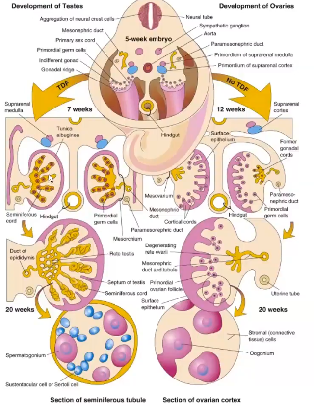
TDF is SRY. Also, in the initial migration of the PGCs, divide and reproduce themselves (mitosis). When they get to the gonads there are a lot of them. However the males ones will stop to divide. They will begin to develop and divide again during puberty. In a female, the PGCs undergo meiosis. The primordial cysts in females develop very peripherally, whereas the male gonads develop more centrally.
Male PGCs: Undergo mitotic arrest.
Female PGCs: Enter meiosis.
Environmental Cues:
Males: Cytochrome catabolizes retinoic acid. The medulla differentiates into a testis, while the cortex regresses.
Females: Retinoic acid produced in the mesonephros tubules stimulates meiosis. The cortex differentiates into an ovary, and the medulla regresses.
Primary Sex Determination: This is dictated solely by the organism’s karyotype ( or ).
Morphological Structures of the Embryonic Gonads
Embryonic Testis (Example: Mouse):
Testis Cords (TC): Composed of Sertoli cells and germ cells.
The sertoli are the male parallel to the granulosa cells in the development of female gonads and cysts.
Leydig Cells: Located in the interstitial area (IC). These are a specific type of תאי ביניים that are responsible for the hormonal development (androgens)
Interstitial cells (תאי ביניים): will develop into blood vessels, but are avariety of different supporting cells.
Embryonic Ovary (Example: Mouse):
Morphology: Unlike the testis, the ovary does not undergo major morphological changes until after birth.
Internal Organization: Composed of a cortex and a medulla.
Germ Cell Distribution: Cells accumulate in the cortex; those remaining in the medulla die via apoptosis.
Ovarian Reserve (Maatouk et al., 2008): Primarily established via germ cell nests that later break down to form primordial follicles containing a single oocyte and pregranulosa cells.
Development of the Sexual Duct Systems
Indifferent Sexual Duct System (Week 5 - 6 in Humans):
Two pairs of genital ducts are present in all embryos.
Mesonephric Ducts (Wolffian Ducts): Future male system. (deferens)
Paramesonephric Ducts (Müllerian Ducts): Future female system.
We need to know here: week 5-6 we are still talking about indifferent. The wolffian is primary in the development in the male system, and the Mullerian plays a primary role in the development of the female system.
Male Sexual Duct Development:
AMH Secretion: Sertoli cells secrete Müllerian inhibiting substance (AMH; Anti-Müllerian Hormone). Sertoli support the sex cells (may be referred to as תאי ביניים at this stage). In fertility tests, they test the AMH levels.
Regression: Paramesonephric ducts undergo apoptosis, leaving only small remnants at the cranial and caudal ends.
Testosterone Secretion: Sertoli cells secrete growth factors to stimulate Leydig cell differentiation, which then secrete testosterone. The female parallel of Leydig cells are Theca cells.
Differentiation: Testosterone influences the mesonephric ducts to develop into the paired ductus deferens () and epididymis ().
Female Sexual Duct Development:
Absence of Testosterone: Mesonephric (Wolffian) ducts regress, leaving only rudimentary structures.
Absence of AMH: Paramesonephric (Mullerian) ducts develop into the major female structures.
Uterine Tubes: Formed from the cranial portions of the paramesonephric ducts.
Uterus and Vagina: Toward the caudal ends, the ducts fuse at the midline to form the uterus and upper vagina. Epithelial tissue from the Müllerian tubercle hollows out to form the lower portion of the vagina.
Hormonal Influence: Full development depends on estrogenic hormones secreted by the ovaries.
Comparison of Indifferent Structures and Adult Derivatives
Indifferent Structure | Male Derivative | Female Derivative |
|---|---|---|
Genital Ridge | Testis | Ovary |
Primordial Germ Cells | Spermatozoa | Ova |
Sex Cords | Seminiferous tubules (Sertoli cells) | Follicular (granulosa) cells |
Mesonephric Tubules | Efferent ductules; Paradidymis | Oöphoron; Paroöphoron |
Mesonephric (Wolffian) Ducts | Appendix of epididymis; Epididymal duct; Ductus deferens; Ejaculatory duct; Seminal vesicles | Appendix of ovary; Gartner’s duct |
Mesonephric Ligaments | Gubernaculum testis | Round ligament of ovary |
Paramesonephric (Müllerian) Ducts | Appendix of testis; Prostate utricle | Uterine tubes; Uterus; Upper vagina |
Definitive Urogenital Sinus (lower) | Penile urethra; Bulbourethral glands | Lower vagina; Vaginal vestibule |
Early Urogenital Sinus (upper) | Urinary bladder; Prostatic urethra; Prostate gland | Urinary bladder; Urethra; Glands of Skene |
Labioscrotal Folds | Scrotum | Labia majora |
Genital Tubercle | Penis | Clitoris |
Progenitor/Precursor | Sertoli cells; Leydig cells | Granulosa cells; Theca cells |
Germ Cell Location | Inside testis cords (Medulla) | Inside follicles (Cortex) |