PROTOZOA
Kingdom Protista: Protozoa
General Characteristics of Kingdom Protista
Traditionally includes single-celled and colonial eukaryotic microbes lacking tissue organization.
'Protista' - Greek word "protistos," meaning "the very first"
Typically unicellular with a nucleus bound to organelles.
Flagella or cilia for locomotion.
First used the term by Ernst Haeckel in 1866.
Protists are a polyphyletic grouping of independent clades evolved from the last eukaryotic common ancestor, not a natural group (clade).
Clade: a monophyletic group with a common ancestor.
Polyphyletic group: organisms with mixed evolutionary origin, excluding their most recent common ancestor.
Inhabit almost any environment with liquid water.
Sub-Kingdoms of Protista
Protozoa ('animal like')
Algae ('plant like')
Slime & Water Molds ('fungus like')
Protozoa
General Information
Considered the most engrossing and vivid group of microorganisms.
Greek origin meaning 'first animals'.
About 65,000 species exist.
Single-celled creatures with movement, feeding, and behavior properties.
Cells contain eukaryotic organelles, except chloroplasts.
Classified by cell architecture, motility structure, and hosts.
Unicellular, higher protists, Eukaryote, and binary fission replication.
Chemoheterotrophic like animals, do not photosynthesize.
Are 10 times more diverse than bacteria and viruses.
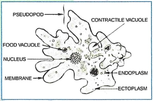
Protozoan Form & Function
Organelles specialized for feeding, reproduction, and locomotion.
Cytoplasm divided into:
Ectoplasm: clear outer layer involved in locomotion, feeding, and protection.
Endoplasm: granular inner region housing the nucleus, mitochondria, and vacuoles.
Outer boundary - is a cell membrane (not cell wall) that regulates the movement of food, wastes, and secretions.
Most ciliates have constant shapes; some protozoa change constantly (e.g., ameba).
Feeding
All protozoa are Heterotrophic: they derive nutrients from other organisms through phagocytosis or osmotrophy.
Phagocytosis: proccess of cell engulfing large particles using its plasma membrane, creating a phagosome.
Osmotrophy: feeing mechanism involving the movement of dissolved organic compounds by osmosis.
Osmotrophs - Organisms that use osmotrophy
Protozoan Locomotion
Except for Apicomplexa, protozoa are motile via:
Pseudopods: 'false foot'
Used for locomotion, food capture, and endocytosis.
Involves actin filament disassembly and assembly.
Actin: fine protein fibers contributing to structure and support.
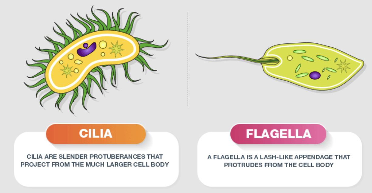
Flagella: long whiplike structure
Longer and fewer than cilia, but structurally identical.
Undulate in a whip-like fashion for movement.
Require ATP to move.
Cilia: shorter than flagella
Numerous, short projections that beat in an oar-like fashion for movement.
Consist of microtubule core (axoneme) enclosed in a cell membrane.
Ring of 9 doublets, plus two singlets, centrally located pattern.
Anchored by a basal body pattern.
Life Cycle & Reproduction
General life cycle includes a trophozoite (motile feeding stage); not all produce cysts.
Life cycle dictates transmission mode to another host.
T. vaginalis does not form cysts; transmitted by intimate contact.
Cysts can be dispersed by air currents.
E. hystolitica and G. lamblia are transmitted via contaminated water and food.
Many form a resistant, dormant cyst structure.
Trophozoite: active feeding stage of parasitic protozoa.
Both cyst and trophozoite stages may be found in feces.
Encystment and Excystment
Encystment: Cell rounds up, loses motility, early cyst wall formation, mature cyst (dormant, resting stage) forms during adverse conditions.
Excystment: Cyst wall breaks open, Trophozoite is reactivated when moisture and nutrients are restored, release of active cellular form.
Taxonomic Classification of Protozoa
Sarcodina (Rhizopoda)
Motion: by Pseudopodia (Pseudopodes)
Replication: Binary Fission
Example: Amoeba / Ameba
Shape: Pleomorphic
Pathogenic species for man: Entamoeba hystolytica.
Disease: Amoebiasis or amoebic disentery
Three forms:
forma manga
forma minuta
cyst
Non-pathogenic species:
Entamoeba gingivalis
Entamoeba coli
Have a complex cytoskeleton
Marine, freshwater and terrestrial species
Diversity of pseudopodia
Lobopodia - wide and rounded
Filopodia - slender and may be branched
Life Cycle of Entamoeba histolytica
Host: Homo sapiens
Transmission: Fecal-oral route (alimentary)
Infective stage: mature cyst
Localization: Large Intestine
Pathogenicity:
Intestinal amoebiasis: formation of ulcerus of the wall of the intestine, acute or chronic diarrhea, stool containing blood and mucus; may be asymptomatic infection.
Extra-intestinal amoebiasis: abscess of liver, lung, brain, skin.
Laboratory diagnosis: Fresh stools are examined under the microscope. E. histolytica (forma magna and cysts with 4 nuclei) can be demonstrated in the stools.
Prophylaxis: Treatment of patients and asymptomatic cyst carriers; protection of foodstuffs and water from flies and contamination with feces, the staff of catering establishments must be examined for cysts carriage, health education of the population.
Shelled Amebas
Foraminefera or forams (Phylum Foraminifera) and Radiolarians (Phylum Radiolaria) are shelled amebas. They are the responsible for the chalk deposits in the oceans
Foraminefera (forams)
mostly marine organisms.
May be benthic or planktonic.
Planktonic - have spines to increase surface area, and thus they are buoyant.
Forams create natural wonders in the form of chalk and limestone
Pink sands of Bermuda
White cliffs of Dover in England
Block of Pyramids
Radiolaria
Mostly planktonic
Siliceous, spherical test.
Up to 20 cm in some colonial species
Sporozoa (Apicomplexa)
Lack locomotor organelles
All members of Phylum Apicomplexa are endoparasites.
Sporozoites (Gr. sporos - seed, and zoon - animal) - special sporelike cells produced by sporozoa following sexual reproduction which are important in infection transmission.
Possess an apical complex
Attaches or penetrates host cell
Cone contains disgestve enzymes
It includes the ff:
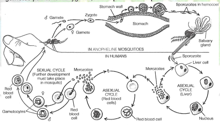
Malaria
Anopheles mosquito is the vector carrier of plasmodium.
Four species of Plasmodium that cause malaria
Plasmodium falciparum
Diseases: Tropical malaria or malignant tertian (Most fatal form of malaria)
Incubation Period: 12 Days
Plasmodium vivax
Benign Tertian / Tertian Malaria
Can cause death to young and old patients
Incubation period: 14 days
Plasmodium malariae
Quartan Malaria
Not lethal
Incubation Period: 30 days
Plasmodium ovale
Tertian ovale
Very seldom in Phils.
14 days IP
MALARIA PARASITES OF MAN
Intermediate host: Homo sapiens (Man)
Definitive host: Anopheles mosquito
Transmission: by bite of female Anopheles mosquito
Infective stage for man: sporozoite
Infective stage for mosquito: gametocyte
Localisation: blood, liver
Clinical manifestations: fever, anemia, splenomegaly, hepatomegaly
Laboratory diagnosis: Microscopy of thin and thick films blood smears. Different stages of the parasite (trophozoites, schizonts, and gametocytes) can be demonstrated in the blood.
Prophylaxis: Malaria may be prevented by chemoprophylaxis and personal protective measures against the mosquito vector (Anopheles).
Complex life Cycle of Plasmodium
Sporozoite - motile, infective stage possessing apical complex
Injected into blood by mosquito.
Attack liver cells.
Merozoite - motile, reinfective stage, also has apical complex.
Reinfect liver cells or move to RBC
Cyclic merozoite release correlates w/ chills, fever, fatigue due to hemoglobin loss and capillaries blockage.
Gametocytes - reproductive stage. Usually, male and female gametocytes pair up and release gametes (sexual reproduction)
Spore - gametes pair to form zygotes, and a protective capsule is secreted
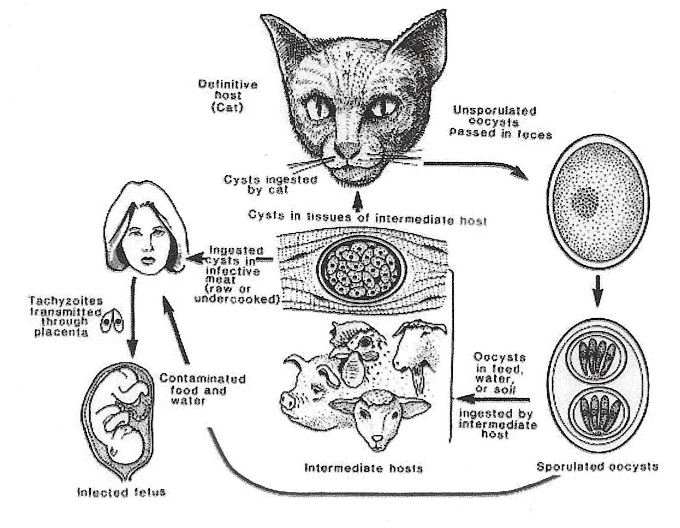
Toxoplasmosis
Toxoplasma gondi: transmitted by the ingestion of oocysts from cat feces. Infection can lead to ocular problems and is also a cause of neonatal toxoplasmosis
Geographical distribution: Cosmopolitan
4 forms of Morphology: pseudocysts; trophozoites; cysts and oocysts.
Definitive hosts: cats and other Felidae
Intermediate hosts: birds and mammals, including humans
Localisation: brain, eyes, skeletal and cardiac muscles, liver, and lungs
Transmitted to humans by:
1) ingestion of undercooked infected meat (cysts and pseudocysts);
2) contamination of food or drink with infected cat feces (oocyts);
3) transplacental (congenital)
Life Cycle of Toxoplasma gondi
Oocysts pass from cat intestine to cat feces.
Oocysts sporulate in soil and are viable for longer than one year
Humans ingest oocysts either from soil or cat raw tissue infected with cysts. The alimentary route of infection takes place on ingestion of meat, milk, and dairy products of animals sick with toxoplasmosis, uncooked eggs of affected birds, and water contaminated by sick animals.
Transmitted via placenta when mother develops infection during gestation-congenital infection.
Invade intestinal wall after entering host (usually orally) and disseminate via lymphatics and bloodstream forming trophozoites. Toxoplasma gondii can spread to many host cells.
Mastigophora (Zoomastigophora)
Flagellates
Motion: by flagella. Flagellum arises posteriorly and can be connected to other parts of body, pulls animal through the blood.
Reproduction: longitudinal binary fission.
Complex life cycles include alternation of hosts.
Intermediate hosts commonly serve as vectors, which transport developing parasites from one definitive host to another.
Parasitical species parasites of tissues and blood(Their transmission requires a biological vector.):
Trypanosoma
Leishmania
Species living in the digestive tract and genitals (ransmission does not require a biological vector):
Lamblia intestinalis
Trichomonas vaginalis
Trichomonas hominis
Mastigophora Species includes:
Trypanosoma brucei gambriense
Trypanosoma brucei rhodesiense
Trypanosoma cruzi
Leishmania donovani
Leishmania tropica
Lamblia intestinalis
Trichonomas vaginalis
Trichonomas intestinalis
Trichonomas buccalis
Trypanosoma brucei gambiense and Trypanosoma brucei rhodesiense
Parasites: Trypanosoma brucei gambiense and Trypanosoma brucei rhodesiense
Disease: African trypanosomiasis, (sleeping sickness)
Geographical distribution: West and Central Africa
Morphology: spindle-shaped cells with an undulatory membrane and pointed flagella at the ends. The organisms are motile, 25-40 micron in length. Transmission: by bite of infected tsetse flies (Glossina palpalis)
Reservoir hosts (T.b.gambiense): man, domestic pig, cattle, dog, antelope.
Reservoir hosts (T.b.rhodesiense): hartebeest, lion, hyena. Localisation: blood, lymph nodes, cerebrospinal fluid, brain, muscles.
Trypanosomiasis
Pathogenicity
From the site of bite trypanosomes reach the blood and lymphatics where they multiply.
There is perivascular infiltration with chronic inflammation, leading to meningoencephalitis.
The patient suffers from fever, rash, headache, lymphadenopathy, oedema of the brain. There are alternating periods of fever and apparent recovery. This is followed by depression and progressive lethargy.
Rhodesien form develops within weeks to months, Gambian form develops within years. The disease becomes chronic and persists for months and even years.
Labarotatory Diagnosis:
microscopic examination of blood and of material obtained by puncture of the enlarged lymph nodes;
examination of the cerebrospinal fluid (availability of trypanosomes).
Prophylaxis:
treatment of patients;
protection of the population from the bites of tsetse flies (Glossina palpalis);
the use of insect repellents, extermination of vector flies.
Chaga's Disease
Parasite: Trypanosoma cruzi
Disease: American trypanosomiasis, or Chagas' disease
American trypanosomiasis (Chagas' Disease) was discovered in 1909 by C. Chagas in Brazil.
Geographical distribution: South and Central America
Morphology: typical, small (20 micron) trypomastigotes (with flagella) are found in peripheral blood and amastigotes (intracellular without flagella) - in tissues.
Transmission:
1) by bite of infected bug species of the family Triatomidae;
2) congenital;
3) by blood transfusion.Reservoir hosts: armadillos, opossums, rodents, monkeys, dogs, cats.
Localisation: blood (in acute phase), cells of lymph nodes, spleen, liver, brain, muscles.
Clinical manifestation: fever, edema of the face, and enlargement of the thyroid gland, lymph nodes, spleen, and liver, heart alterations.
Laboratory diagnosis:
1) examination of patient's blood;
2) guinea pig inoculation with 5-10 ml of patient's blood;
3) serologic tests.Prophylaxis:
1) extermination of bugs;
2) chemoprophylaxis with special preparations in endemic areas.
Leishmaniasis
Parasite: Leishmania tropica
Disease: Cutaneus leishmaniasis
Geographical distribution: Asia, Africa, Europe
Morphology: Intracellular amastigotes (without flagellum) 3 to 6 micron long by 1.5 to 3 micron in diameter live in men. Promastigotes (with flagellum) develop in the intestine of the sand fly.
Transmission: by sand fly vector
Phlebotomus sergenti (in Iran, Iraq, and India); Phlebotomus papatasi (in southern France, Italy, and certain Mediterranean islands).
Reservoir hosts: man, dogs, wild rodents.
Localisation: cells of skin.
Clinical manifestation: development of a cutaneous papule that evolves into a nodule, breaks down to form an indolent ulcer, and heals, leaving a depressed scar.
Laboratory diagnosis: detection of the Leishmania parasites in cells of skin.
Prophylaxis: early diagnosis, extermination of sandflies and dogs and rodents infected with leishmaniasis, and vaccination
Disease Table
Disease | Parasite | Geeographical Distribution | Transmission | Reservoir Host |
|---|---|---|---|---|
African sleeping sickness / African trypanosomiasis | Trypanosoma brucei gambiense and Trypanosoma brucei rhodesiense | West and Central Africa | By bite of infected tsetse fly (Glossina palpalis) | T.b.gambiense are: man, domestic pig, cattle, dog, antelope. T.b.rhodesiense are: hartebeest, lion, hyena. |
Chaga's Disease or American trypanosomiasis | Trypanosoma cruzi | South and Central America | 1) by bite of infected bug species of the family Triatomidae; 2) congenital; 3) by blood transfusion. | armadillos, opossums, rodents, monkeys, dogs, cats. |
Cutaneus leishmaniasis | Leishmania tropica | Asia, Africa and Europe | sand fly vector - Phlebotomus sergenti (in Iran, Iraq, and India); Phlebotomus papatasi (in southern France, Italy, and certain Mediterranean islands). | Man, dogs and wild rodents |
Visceral leishmaniasis, or kala-azar | Leishmania donovani | India, Pakistan, China, Central Africa and Central America | sand fly vector Phlebotomus | man, dogs (except in India), cats, rodents. |
Lambliosis
Parasite: Lamblia intestinalis
Disease: lambliosis
Geographical distribution: cosmopolitan.
Morphology: Trophozoites are bilateral, symmetrical, pear-shaped organisms with an elongated posterior and two symmetrically placed nuclei. The body of the parasite is from 10 to 18 micron long with four pairs of flagella. Cysts are oval-shaped which are 10-14 micron and have four nuclei.
Host: man
Transmission: fecal-oral (alimentary)
Infective stage: cyst
Localisation: the small intestine (duodenum) and gall-bladder.
Trichomoniasis
Parasite: Trichomonas vaginalis
Disease: Urogenital trichomoniasis
Geographical distribution: cosmopolitan.
Morphology: Trophozoite is a pear-shaped (7 to 23 micron long) with four anterior flagella and a fifth forming the edge of an undularing membrane. The axostyle extends the length of the body.
Host: man
Transmission: by sexual contact; otherwise (through contact with toilet seats and towels, for example).
Localization: vagina, urethra, prostate.
Clinical Manifestations: vaginitis in women, more commonly asymptomatic in men, but may lead to prostatitis or urethritis. The main symptoms are dysuria, pruritis, yellow and frothy discharge.
Laboratory diagnosis: microscopic examination of the vaginal fluid, scrapings, or washing.
Giardiasis
Giardia lamblia - Parasite lives in small intestine
Ciliophora
Or Ciliates are sophisticated protozoans
Trophozoites are motile by cilia
Most have definite mouth and feeding organelle.
Reproduce by transverse binary fission and sometimes by conjugation.
Ciliates have both macronuclei and micronuclei.
Macronucleus - genes are actively transcribed
Micronucleus - master copy of genome; inactive except during cell division
Shapes and numbers of these nuclei varies across genera
Bean shaped in Paramecium
String of beads in Stentor
Most ciliates are free-living and harmless.
Function of alveoli is to store
Release of causes changes in ciliary beat, and discharge of extrusomes
Trichocysts - long shafts that are thought to defend against predators
Toxicysts - longs shafts with toxin that are used for prey capture
Mucocysts - release mucus and creates sticky surface for prey capture or protective cysts
Balantidiasis
Parasite: Balantidium coli
a large motile ciliated parasite that lives in the colon of pigs, humans and rodents and can lead to colonic ulceration
Disease: Balantidiasis
Morphology: The trophozoite is from 75 to 200 micrometer, in length, asymmetrical, oval, with cilia, a cytostome, anal pore, the macronucleus, the micronucleus, two contractile vacuoli. Cyst with a double-layer membrane, from 30 to 60 micron in diameter.
Hosts: man, domestic swine
Transmission: fecal-oral (alimentary)
Localization: large intestine
Clinical Manifestations: colitis, ulcers and abscesses of colon, diarrhoea, blood and mucus in the stool.
Laboratory diagnosis: microscopic examination of the feces.
Prophylaxis: protection of foodstuffs and water from contamination with swine feces and observation of individual hygiene when talking care of the animals (domestic swine).
Microspora
Unique feature: lack mitochondria
Intracellular parasites (live inside host cells)
The term microsporidia is also used as a general nomenclature for the obligate intracellular parasites belonging to the phylum Microsporidia.
Microsporidia, are characterized by the production of resistant spores that vary in size, depending on the species. They possess a unique organelle, the polar tubule or polar filament, which is coiled inside the spore as demonstrated by its ultrastructure. The microsporidia spores of species associated with human infection measure from 1 to 4 μm and that is a useful diagnostic feature.
There are at least 15 microsporidian species that have been identified as human pathogens.
Enterocytozoon bienusi: a microsporidian that parasitises the small intestine. Also more common in the immunocompromised.
Keratoconjunctivitis, skin and deep muscle infection
Clinical manifestations of Microsporidian Species
Microsporidian species | Clinical manifestation |
|---|---|
Anncaliia algerae | Diarrhea, acalculous cholecystitis |
Enterocytozoon bieneusi | Keratoconjunctivitis, infection of respiratory and genitourinary tract, disseminated infection |
Encephalitozoon cuniculi and Encephalitozoon hellem | Keratoconjunctivitis, infection of respiratory and genitourinary tract, disseminated infection |
Encephalitozoon intestinalis (syn. Septata intestinalis) | Infection of the GI tract causing diarrhea, and dissemination to ocular, genitourinary and respiratory tracts |
Microsporidium (M. ceylonensis and M. africanum) | Infection of the cornea |
Nosema sp. (N. ocularum), Anncallia connor | Ocular infection |
Pleistophora sp. | Muscular infection |
Trachipleistophora anthropophthera | Disseminated infection |
Trachipleistophora hominis | Muscular infection, stromal keratitis, (probably disseminated infection) |
Tubulinosema acridophagus | Disseminated infection |
Vittaforma corneae (syn. Nosema corneum) | Ocular infection, urinary tract infection |