COVID 19 histopathology
Overview of COVID-19
COVID-19 is a significant public health crisis, first identified in December 2019.
The disease is caused by the SARS-CoV-2 virus, leading to over 481 million infections and more than 6 million deaths globally by the end of the study.
Affects all age groups, with older individuals and those with pre-existing conditions facing higher risks.
Characteristics of the Disease
Clinical presentation ranges from asymptomatic to multiorgan failure and death.
Common symptoms include:
Fever
Cough
Fatigue
Possible nasal discharge, headaches, diarrhea, and gastrointestinal issues.
Virology and Pathogenesis
Coronaviruses belong to the Coronavirinae subfamily and can impact both humans and animals.
Viral Particle:
Size: 80 to 220 nm, with a positive-strand RNA genome.
Genome structure: 5’-RdRp-S-E-M-N-3’, involved in encoding structural proteins (N, E, M, S) and replication proteins (RhRp).
Entry Mechanism:
The virus enters host cells by binding its spike (S) protein to the ACE2 receptor, influencing viral pathogenesis.
Histopathological Findings
Common findings include:
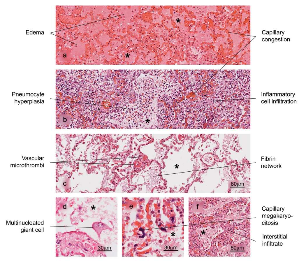
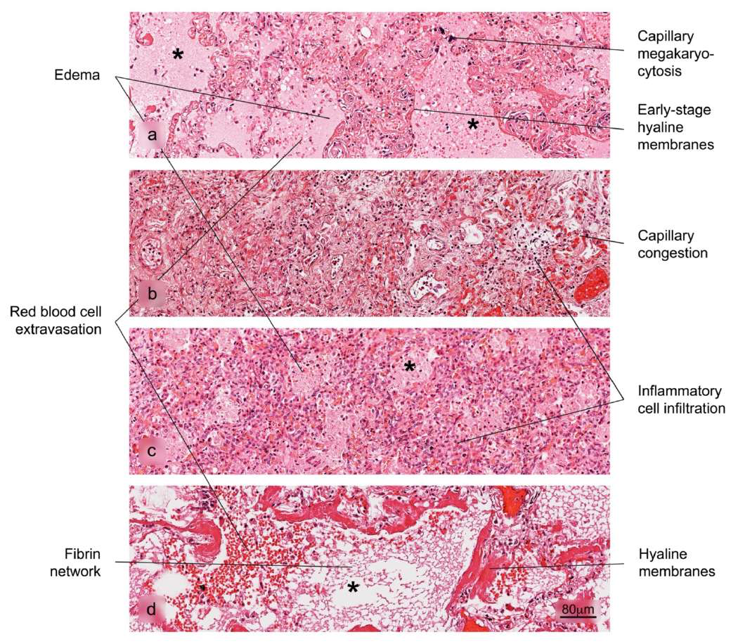
Diffuse alveolar damage, vascular congestion, edema.
Pulmonary hemorrhage, cardiomegaly, and ventricular dilation observed in fatal cases.
Histology indicates shared patterns with other respiratory viral illnesses, but COVID-19 displays unique vascular issues such as thrombosis and microthrombosis.
Damage to Organs
Lung
Predominantly affected, with findings of:
Edema, desquamation of pneumocytes.
Hyaline membranes and signs of acute respiratory distress syndrome.
Other Organs
Liver
Alterations include cirrhosis, microvesicular steatosis, and lobular inflammation.
Kidney
Chronic kidney disease and acute duct lesions.
Heart
Myocardial fibrosis and inflammatory infiltrates.
Study Limitations in Colombia
Limited necropsy and postmortem tissue sampling during COVID-19 restrictions hinder comprehensive studies.
The research is based on 50 fatal cases confirmed through various diagnostic methods, providing insights into the histopathology of COVID-19.
Figures Overview
Lung Histology: Normal alveolar structure and bronchiole sections.
Lung Damage: Different stages of Diffuse Alveolar Damage detailed.
Additional Alterations: Various forms of edema, reactive hyperplasia, and vascular changes.
Spleen & Liver Histology: Comparisons of normal and affected structures in these organs.
Summary of Findings: Frequencies of identified histopathological changes in various organs, evidencing the extensive impact of COVID-19.
Conclusion
The histopathological changes provide valuable insight into the disease's progression and complications due to SARS-CoV-2.
Ongoing evaluations and further studies are essential for understanding the long-term effects and pathological mechanisms involved.