L3 - Isolation and analysis of cell organelles and molecules
Fluorescent antibodies or stains:
- labeling %%live%% cells,
- Proportion of staining = proportion of DNA,
- Fluorophore = fluorochrome.
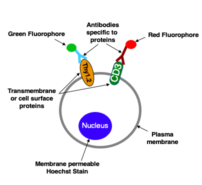
- Antibodies:
- Fluorophores bounds to antibodies against specific cell surface proteins
- Membrane Permeable Dyes:
- Membrane permeable fluorescent dyes labels intracellular structures
- (i.e. Hoechst stain binds DNA in nucleus).
- Cells with bound antibodies or that have taken up the dyes can then be sorted and counted.
Fluorescent Activated Cell Sorting (FACS):
- Cells pass through a laser light beam in a thin tube,
- Both fluorescent light emitted and scattered are measured by detectors,
- Individual cells are forced through a nozzle and given a ,
- Cells with different electric charges are and collected.
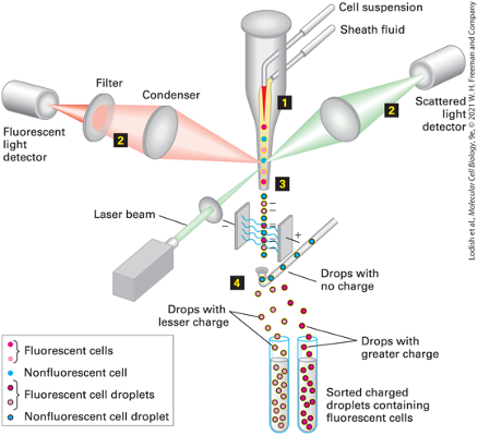
- Many different types fluorophores can be used to differentiate different cells
Quantification of Cells Expressing 2 Different Cell Surface Markers by FACS:
- As the cells pass through the FACS machine, the intensity of the green and red fluorescence emitted by each cell is recorded
- Each dot represents a single cell
- The proportion of each cell population can be calculated.
\n 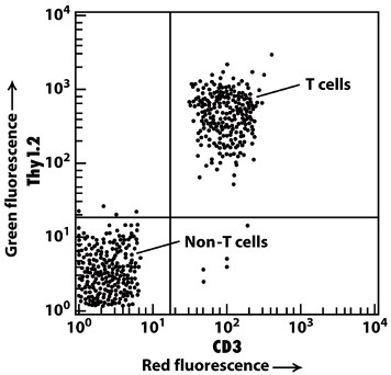 \n
\n 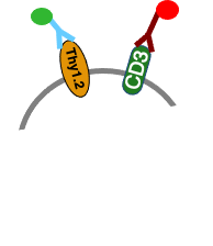
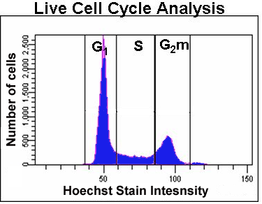
Cell Cycle Analysis by FACS:
- Cells that have replicated their DNA but not fully divided (G2) will have twice the Hoechst stain fluorescence intensity of non-dividing cells (G1).
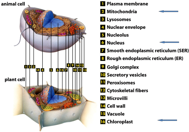
Organelles of Cells:
 Cell Organelle Isolation:
- STEP ONE: DISRUPTION of CELL PLASMA MEMBRANE
- i) mechanical homogenization
- ii) sonication (ultrasound) (destroys the membrane)
- iii) pressure (cells are forced through a very narrow valve) (cause the cell to rupture)
- iv) non-ionic detergents i.e., Triton X-100
- Agent that causes cell membranes to dissipate
- disperses phospholipid bilayer
- v) placing cells in hypotonic solution
- Osmotic effect that makes cell blow up (swollen)
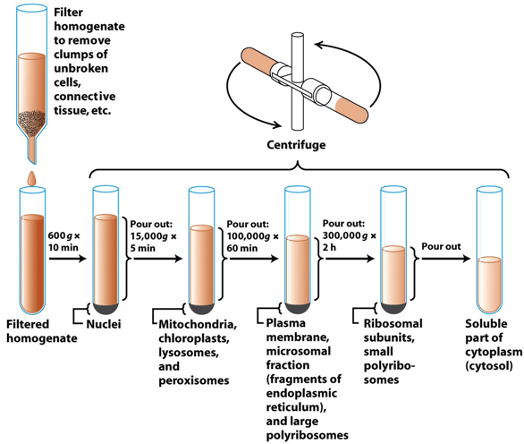
- STEP TWO: CENTRIFUGATION of CELL HOMOGENATE
- i) differential
- ii) equilibrium density-gradient.
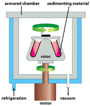
Centrifugation:
- Refrigeration:
- Cools down the system to prevent the samples from overheating
- Vacuum
- Prevents heat conduction
- Armoured chamber:
- Prevents the materials from leaving the system in a projectile motion
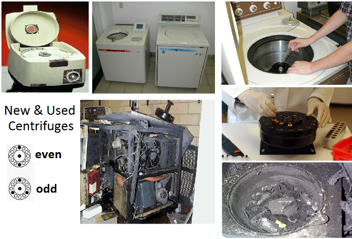
- Distribution
- equal weight, even distribution, balance the system to prevent shaking \n
- equal weight, even distribution, balance the system to prevent shaking \n
- Differential Centrifugation:
- spinning homogenate yields pellet & supernatant
- increasing centrifugal force (gravity) to isolate organelles based on mass
- larger organelles = easy to separate, low force required
- smaller organelles = need high speed to separate
- important to do it sequentially
- big, medium, small.
- g = Relative centrifugal force (RCF).
- Relative g required to separate organelles:
- 600g * 10 min
- Nuclei
- 15,000g * 5 min
- mitochondria, chloroplasts, lysosomes, and peroxisomes
- 100,000g * 60 min
- plasma membrane, microsomal fraction(framents of endoplasmic reticulum) and large polyribosomes
- 300,000g * 2h
- ribosomal subunits, small polyribosomes
- Pour out
- soluble part of cytoplasm (cytocol)
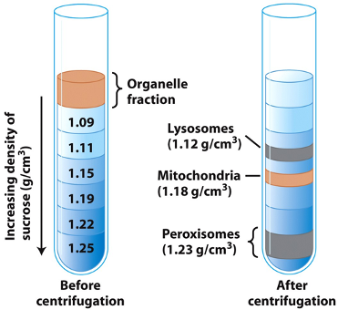
- Equilibrium Density-Gradient Centrifugation:
- separation based on density
- homogenate is applied to a gradient of sucrose
- at high speed/several hours, organelles migrate to sucrose layer equal their own density and remain there.
- each layer then can be suck up for later use
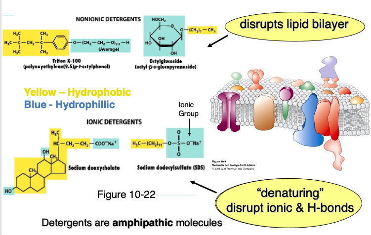
Separating Protein from Organelles:
- Detergents:
- molecules.
- non-ionic detergents:
- only disrupts lipid bilayer.
- ionic detergents:
- by denaturing ionic & H-bonds that breaks down proteins.
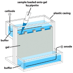
SDS-PAGE:
- Electrophoretic separation of proteins is most commonly performed in polyacrylamide gels.
- usually carried out in the presence of the ^^negatively charged detergent SDS^^ and called
- SDS binds to and destabilizes the hydrophobic side chains within the core of proteins
- All polypeptide chains are forced into extended negatively charged conformations with a @@similar charge-to-mass ratio@@.
- The mobility of the SDS-protein complexes are influenced primarily by molecular size, i.e MW=daltons
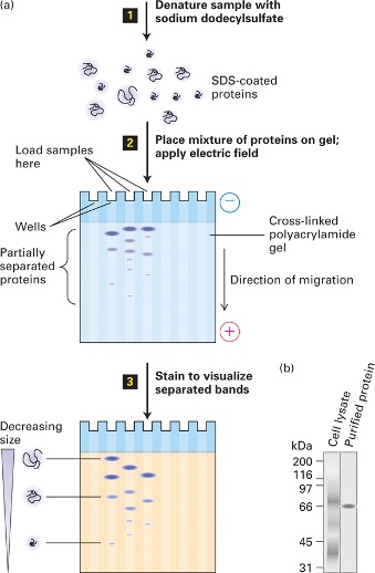
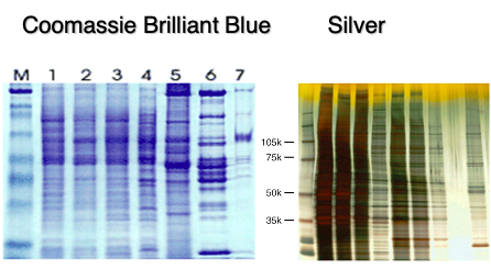
Quantification and Observation of Protein:
- Chemical dyes allow the visualization of protein gradients
\n
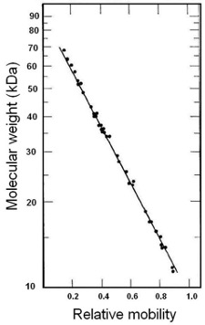
- {{There is a linear relationship between the @@log MW@@ and the electrophoretic mobility.{{
- Low mass proteins have greater mobility than high mass proteins
Western Blotting (Immunoblotting):
- Very useful in separating proteins as many proteins have similar/same molecular mass
Procedure:
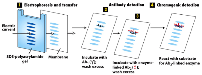
- Electrophoresis and transfer
- Electric current helps SDS-polyacrylamide gel to separate the proteins
- Antibody detections
- Incubate with antibody1 that recognizes the desired protein
- Ab 1 recognizes the desired protein
- Incubate with enzyme linked antibody2
- Ab2 recognizes Ab1
- Enzyme-labelled antibody2 recognizes the antibody1
- wash excess after procedure
- Chromogenic detection
- React with substrate for antibody2 -linked enzymes
- Enhanced chemiluminescence substrate reacts with enzyme in Ab2 (glows)
- luminal reacts with the enzyme product and emits light
- imaging with x-ray detector or very sensitive camera
- forms a bright band
- sometimes inverted to be a dark band
Immunoblotting to detect changes in levels of a specific protein:
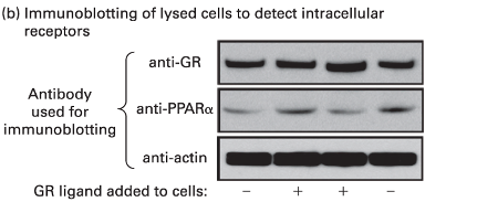
- Loading control: actin
- Does not change by the amount of reactant
- Tests if the gel was applied properly
- if actin band changes width/intensity, then there’s excess gel
\n