Unit 10 Restrictive & Lung Perfusion Disorders
Chronic Interstitial (Restrictive) Lung Diseases
Interstitial Lung Diseases (ILDs)
Definition of ILDs:
Diffuse interstitial lung diseases (ILDs) are a diverse group of lung disorders characterized by similar inflammatory and fibrotic changes in the lungs’ interstitium or interalveolar septa.
The presence of ILDs results in a stiff and noncompliant lung, leading to their classification as restrictive lung disorders.
Unlike obstructive lung diseases where the physiologically functioning airways are primarily affected, ILDs result in stiffness making it difficult for the lungs to expand.
Etiology and Pathogenesis of Interstitial Lung Diseases
Onset of ILDs:
ILDs may exhibit either an insidious or abrupt onset.
The course can be rapidly progressive, slowly progressive, or remain static.
Etiologies of ILDs:
Occupational and Environmental Inhalants:
Pneumoconiosis (including coal miner’s pneumoconiosis, silicosis, asbestosis)
Hypersensitivity pneumonitis (e.g., farmer’s lung, pigeon breeder’s lung)
Drugs and Therapeutic Agents:
Cancer drugs (e.g., bleomycin, busulfan, cyclophosphamide, methotrexate)
Amiodarone
Immunologic Lung Diseases:
Sarcoidosis
Collagen vascular diseases (e.g., systemic lupus erythematosus, rheumatoid arthritis, scleroderma)
Pathophysiology of ILDs:
ILDs result from inflammatory conditions that affect the interalveolar structures, leading to lung fibrosis and a stiff lung.
Unlike obstructive lung diseases that primarily involve the airways, ILDs exert effects on the collagen and elastic connective tissues located in the alveolar walls.
Common patterns of lung dysfunction in ILDs include:
Diminished lung volumes.
Reduced diffusing capacity of the lung.
Varying degrees of hypoxemia.
Effects of ILDs:
Characteristically, ILDs are defined by the following:
Reduced compliance of lung tissue leading to increased effort needed to expand the lung.
Increased work of breathing for affected individuals, often leading to dyspnea, an increased respiratory rate, and decreased tidal volume.
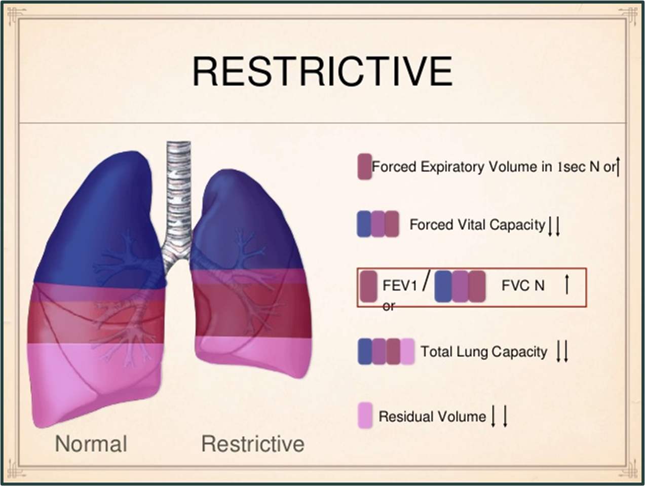
Illustrated in pulmonary function tests showing reduced Forced Vital Capacity (FVC).
Ventilation and perfusion mismatch may occur, affecting the alveolocapillary membrane, leading to decreased diffusion of O2 from the alveoli to the blood, resulting in hypoxemia.
Common ILDs affecting adults include: aspiration, atelectasis, bronchiolitis, sarcoidosis, pulmonary fibrosis, inhalation disorders, pneumoconiosis, allergic alveolitis, pulmonary edema, and acute respiratory distress syndrome.
Clinical Manifestations of Interstitial Lung Diseases
General Clinical Manifestations:
Clinical manifestations typically reflect restrictive changes rather than obstructive changes; however, some individuals may exhibit both forms.
Key Symptoms:
Dyspnea (breathlessness) is the most common manifestation, eventually leading to cyanosis, without wheezing or other signs of airway obstruction.
Initial breathlessness commonly exacerbates with physical activity before progressing to incapacitation.
Breathing patterns are characterized by:
An increased respiratory rate with diminished tidal volume, an attempt to maintain minute volume while reducing the effort.
Non-productive cough may develop, especially with ongoing exposure to inhaled irritants.
Physical changes include clubbing of the fingers and toes, along with a significant decrease in lung volumes such as vital capacity and total lung capacity (TLC).
Diagnosis of Interstitial Lung Diseases
Diagnostic Approach:
Diagnosis involves a comprehensive personal and familial history, focused on exposure to environmental, occupational, and injurious agents.
Initial diagnosis may utilize chest radiographs, with serial chest films tracking disease progression over time.
Surgical lung biopsy remains the preferred and most definitive diagnostic method, allowing for histological examination and culture.
Occupational and Environmental Interstitial Lung Diseases
Categories:
Occupational and environmental ILDs can be classified into pneumoconiosis, drug-induced ILD, and hypersensitivity diseases.
Pneumoconiosis:
Resulting from the inhalation of inorganic dust and particulate matter.
Types include:
Silicosis
Asbestosis
Coal miner’s pneumoconiosis
Talcosis
Berylliosis.
Hypersensitivity Diseases:
Result from inhalation of organic dusts and occupational antigens.
An example includes byssinosis, a disease affecting cotton workers that shares characteristics of both pneumoconiosis and hypersensitivity diseases.
Sarcoidosis
Definition:
Sarcoidosis is a systemic disorder characterized by the presence of noncaseating(no necrosis → caseation) granulomas in affected tissues and organs, most commonly the lung and lymphatic system.
Granulomas develop absently of known exogenous agents that cause granulomatous inflammation.
Predominantly affects individuals between ages 10-40, with a significantly higher prevalence in individuals of Black descent compared to White descent.
Its etiology remains unclear but may involve environmentally-triggered processes in genetically predisposed individuals, sometimes showing familial clustering.
Clinical Manifestations of Sarcoidosis:
Symptoms vary significantly; one of the most common presentations includes lung involvement, often found incidentally through imaging or in response to severe respiratory symptoms.
Clinical signs include:
Dyspnea, nonproductive cough, and chest pain
Constitutional symptoms can also appear: fever, night sweats, anorexia, weight loss, fatigue, and myalgia.
Involvement of the eyes (e.g., anterior uveitis) and skin (e.g., skin papules and plaques) is common.
The disease follows an unpredictable course, marked by either chronic progression or periods of activity and remission influenced by corticosteroid treatment.
Diagnosis of Sarcoidosis:
Diagnosis is based on comprehensive history and examination, excluding other disease processes, chest radiography, and biopsy to confirm granulomas.
CT and MRI use in diagnosis is controversial.
Approximately 65-75% of affected individuals recover with minimal clinical and radiographic abnormalities.
Disorders of the Pulmonary Circulation
Physiological Role:
Blood circulation through the pulmonary capillaries facilitates gas exchange, with simultaneous increases in oxygen content and decreases in carbon dioxide content.
Efficient gas exchange relies on the matching of ventilation and perfusion in the pulmonary circulation.
Mismatched Ventilation and Perfusion:
If ventilation and perfusion are mismatched, gas exchange efficiency is compromised, potentially leading to hypoxemia and other pulmonary circulation pathologies.
Key issues include pulmonary embolism and pulmonary hypertension.
Pulmonary Embolism
Definition:
Pulmonary embolism occurs when material (emboli) lodges in a branch of the pulmonary artery, obstructing blood flow.
Types of emboli include:
Thrombus (blood clot)
Air (accidental injection during intravenous infusion)
Fat (mobilized from bone marrow or fat depots post-injury)
Amniotic fluid (entered the maternal circulation accidentally)
Etiology and Pathogenesis of Pulmonary Embolism:
An embolus results from a blood clot that detaches from its point of origin and travels within the vascular system.
Most pulmonary emboli arise from thrombi in deep vein thrombosis (DVT) located in the upper or lower extremities.
Risk factors contributing to the development of DVT include:
Venous Stasis:
Risk increases due to immobilization or heart failure.
Endothelial Injury:
Trauma or caustic intravenous transfusions can damage the vessel lining.
Hypercoagulability:
Conditions such as inherited coagulation defects, malignancy, hormone replacement therapy, oral contraceptives, pregnancy.
Genetic risk factors include mutations like factor V Leiden, deficiencies in antithrombin II, protein S or C, and mutations in the prothrombin gene.
The effects of an embolus depend on its size and location, leading to complications such as hypoxic vasoconstriction, pulmonary edema, atelectasis, pulmonary hypertension, shock, or death.
Clinical Manifestations of Pulmonary Embolism:
Symptoms vary based on embolus size and location:
Commonly reported symptoms include chest pain, dyspnea, and increased respiratory rate.
Small emboli may be clinically silent and overlooked, especially in the elderly or acutely ill.
Moderate-sized emboli can manifest with:
Dyspnea accompanied by pleuritic pain, apprehension, slight fever, and blood-streaked sputum.
Tachycardia as a compensatory response to reduced oxygenation, with rapid and shallow breathing.
Massive emboli may cause:
Sudden collapse, crushing substernal chest pain, shock, and loss of consciousness.
Signs include rapid and weak pulse, low blood pressure, distended neck veins, cyanotic and diaphoretic skin.
Massive pulmonary emboli can be fatal.
Diagnosis of Pulmonary Embolism:
Diagnosis relies on clinical signs/symptoms, blood gas tests, venous thrombosis studies, troponin and D-dimer tests, lung scans, and helical CT scans.
Ancillary tests help exclude conditions presenting similar symptoms.
An elevated pulmonary vascular resistance necessitates monitoring right heart strain through ECG.
Preventive measures include the use of leg compression devices and promoting active range-of-motion exercises.
Pulmonary Hypertension
Pulmonary Circulation:
Low-pressure system built to accommodate variations in blood flow from the right heart and to facilitate gas exchange.
The main pulmonary artery and its major branches are thin-walled and compliant.
Distal pulmonary arterioles can adjust their diameter based on various physiological factors including blood flow, vasoactive substances, and oxygen tension.
Pulmonary Hypertension:
Defined by an elevation of pressure in the pulmonary arterial system, which can be acute or chronic depending on etiology.
Etiological Factors:
Changes in pulmonary arterial walls contribute to pulmonary arterial hypertension (PAH), a serious condition.
Secondary pulmonary hypertension can arise from:
Occlusion due to pulmonary embolism or
Disruption caused by heart or lung disease.
Etiology and Pathogenesis of Pulmonary Hypertension:
Factors contributing to PAH include:
Decreased cross-sectional area of pulmonary arteries.
Loss of blood vessels due to scarring or destruction adversely affecting alveolar walls.
Vasoconstriction responses due to hypoxia.
The need to accommodate increased blood flow without corresponding arterial changes.
Occlusion of pulmonary outflow at elevated left atrial or ventricular pressures.
Pulmonary Arterial Hypertension (PAH)
Definition and Classification:
PAH emerges specifically within pulmonary arteries.
According to the World Health Organization, PAH categorizes into five groups based on the underlying mechanisms:
Group I: Idiopathic pulmonary arterial hypertension
Group II: Pulmonary venous hypertension
Group III: Hypoxemia-associated pulmonary hypertension
Group IV: Secondary to chronic thrombotic or embolic disease
Group V: Miscellaneous conditions leading to PAH. (Brown-Richards, 2024, pp. 974-975)
Clinical Features of PAH:
PAH, while rare, demonstrates debilitating characteristics including abnormal proliferation, vascular smooth muscle contraction, coagulation issues, and severe intimal fibrosis, leading to artery obstructions.
Elevated pressure in untreated PAH can culminate in progressive right heart failure, lowered cardiac output, and eventual death.
Advances in PAH treatment recently have arisen, though the condition remains life-threatening.
Etiology and Pathogenesis of PAH:
PAH may follow an autosomal dominant familial inheritance pattern, often with low penetrance.
Many pathologies associated with PAH include collagen vascular disorders (e.g., scleroderma), drug exposure, HIV, portal hypertension, and persistent pulmonary hypertension in newborns.
Proposed mechanisms of vascular changes in PAH remain unclear but could involve:
Increased serotonin transporter expression.
Decreased nitric oxide and prostacyclin levels.
Elevated levels of growth factors including endothelin, vascular endothelial growth factor, and platelet-derived growth factor.
Clinical Manifestations of PAH:
Defined by persistent elevations in pulmonary artery pressure while left ventricular pressures remain normal, distinguishing it from left-sided heart failure.
Typical symptoms range from dyspnea and decreased exercise tolerance to right heart failure characterized by peripheral edema and functional limitations.
Other common symptoms encompass fatigue, angina, and syncope (fainting or near-syncope).
Diagnosis is based on ruling out secondary hypertension and requires mean pulmonary artery pressures above 25 mm Hg at rest or 30 mm Hg with exercise.
Secondary Pulmonary Hypertension
Overview:
Most pulmonary hypertension cases are secondary, arising from other conditions, including chronic hypoxemia from COPD, ILD, or sleep-disordered breathing, and increased resistance due to diastolic dysfunction of the left heart or chronic thromboembolic disorders.
Etiology and Pathogenesis:
Prolonged exposure to hypoxemia frequently causes pulmonary vessel constriction as a response to elevated carbon dioxide levels.
Marked hypoxemia during sleep conditions (e.g., sleep apnea) contributes to rising pulmonary arterial pressures.
Clinical Manifestations and Diagnosis:
Symptoms reflect elevated arterial pressure combined with the underlying heart or lung pathology.
Diagnosis includes radiographic findings and echocardiography, along with Doppler ultrasonography data.
Cor Pulmonale→ Right sided heart failure d/t lung disease
Definition:
Cor pulmonale refers specifically to right-sided heart failure resulting from primary lung disease or pulmonary hypertension.
Elevated pressures induced by pulmonary conditions contribute to hypertrophy and eventual right ventricular failure.
Clinical Manifestations of Cor Pulmonale:
Symptoms include manifestations from the primary lung disease, along with typical signs of right-sided heart failure.
Common features include:
Venous congestion, peripheral edema, dyspnea, and a productive cough, worsening during heart failure episodes.
Other signs like plethora (redness), cyanosis, warm and moist skin due to compensatory polycythemia, and altered consciousness due to carbon dioxide retention may manifest.
Acute Respiratory Disorders
Function of the Respiratory System:
The primary role is the addition of oxygen to and removal of carbon dioxide from the blood.
Disruptions in this physiologic function can occur in acute lung injury (ALI)/respiratory distress syndrome (RDS) and acute respiratory failure.
Different mechanisms impair gas exchange in ALI/RDS leading to serious, life-threatening conditions with substantial morbidity and mortality risk.
Acute Respiratory Distress Syndrome (ARDS)
Definition:
ARDS is the most severe manifestation of acute lung injury (ALI).
Early Clinical Manifestations of ARDS:
Characterized by diffuse crackles, dyspnea, cyanosis, tachypnea, tachycardia, and diaphoresis.
Predisposing Factors:
Common causative factors include:
Genetic predispositions, septicemia, near-drowning, aspiration, chest trauma, multitrauma (especially with blood transfusion history).
Other contributors include pneumonia, cardiopulmonary bypass surgeries, pancreatitis, drug overdose, burns, fat embolism, radiation therapy, oxygen toxicity, toxic gas inhalation, and disseminated intravascular coagulation (DIC).
Severe and prolonged shock can lead to ARDS due to ischemic lung tissue damage.
Onset and Clinical Course of ARDS:
The onset generally occurs within 12-18 hours post-injury or negligence-event.
Initial signs may be obscured by primary pathology effects.
ARDS often correlates with multiple organ dysfunction or failure due to severe body insults.
Typical indicators include restlessness, dyspnea, rapid and shallow respirations, and increased heart rate.
Chest X-ray typically reveals diffuse bilateral lung tissue infiltrates with no evidence of myocardial dysfunction per the ECG. → know this
Clinical Progression in ARDS:
Notable blood gas measurements usually show a significant drop in arterial PO2 levels.
As lung congestion escalates, accessory muscle use is apparent, rales may be audible, productive cough yielding frothy sputum occurs, and cyanosis with lethargy or confusion may arise.
A combination of respiratory and metabolic acidoses develops as gas diffusion is impaired, leading to anaerobic metabolism activation.
Acute Respiratory Failure
Definition:
Acute respiratory failure is characterized by impaired gas exchange secondary to heart and/or lung failure, impacting one or both of its functions (oxygenation and carbon dioxide elimination).
It isn’t a specific disease but can stem from various conditions hampering ventilation, ventilation-perfusion matching, or gas diffusion.
Causes and Types:
Acute respiratory failure may occur in healthy individuals due to sudden disease or trauma affecting the respiratory system.
It may also evolve in chronic lung or neuromuscular diseases.
Generally divided into two types:
Hypoxemic respiratory failure (due to impairment of gas exchange)
Hypercapnic or hypoxemic respiratory failure (due to ventilatory failure).
Hypoxemic Respiratory Failure
Factors Contributing to Hypoxemic Respiratory Failure:
Severity in lowering arterial PO2 is influenced primarily by:
Ventilation-Perfusion Mismatching: Occurs when some lung areas are ventilated but not perfused or vice versa; this is commonly observed in advanced COPD cases.
Impaired Diffusion: Gas exchange between alveolar air and pulmonary blood can be hindered due to increased diffusion distances or decreased permeability/surface area of respiratory membranes.
Conditions primarily associated with impaired diffusion include ILD, ARDS, pulmonary edema, and pneumonia.
Hypercapnic/Hypoxemic Respiratory Failure
Characteristics of Hypercapnic/Hypoxemic Respiratory Failure:
Individuals cannot sustain adequate ventilation, resulting in carbon dioxide retention (hypercapnia) and potential hypoxemia.
Often occurs due to external conditions impacting lung function (like depression of respiratory centers, neuromuscular disorders, or chronic lung disease exacerbation).
Resulting hypoventilation typically produces two critical arterial blood gas changes:
Increased PCO2 levels
Possible hypoxemia, which can usually be rectified with supplemental oxygen.
Clinical Manifestations and Diagnosis of Hypercapnic/Hypoxic Respiratory Failure
Symptoms and Diagnosis:
This type of respiratory failure presents with varying degrees of hypoxemia and hypercapnia.
No universally accepted PO2/PCO2 levels dictate respiratory failure (PO2 < 50 mm Hg, PCO2 > 50 mm Hg, or both).
Generally, these thresholds serve as guidelines, to interpret alongside patient history and physical assessment findings.