Chapter 24: The Respiratory System
Overview of the Respiratory System
The respiratory system comprises:
Nose
Nasal cavity and sinuses
Pharynx
Larynx
Trachea
Conducting passageways leading to lungs
It is important to understand and know that the respiratory system is divided into the Upper & Lower Respiratory Tracts
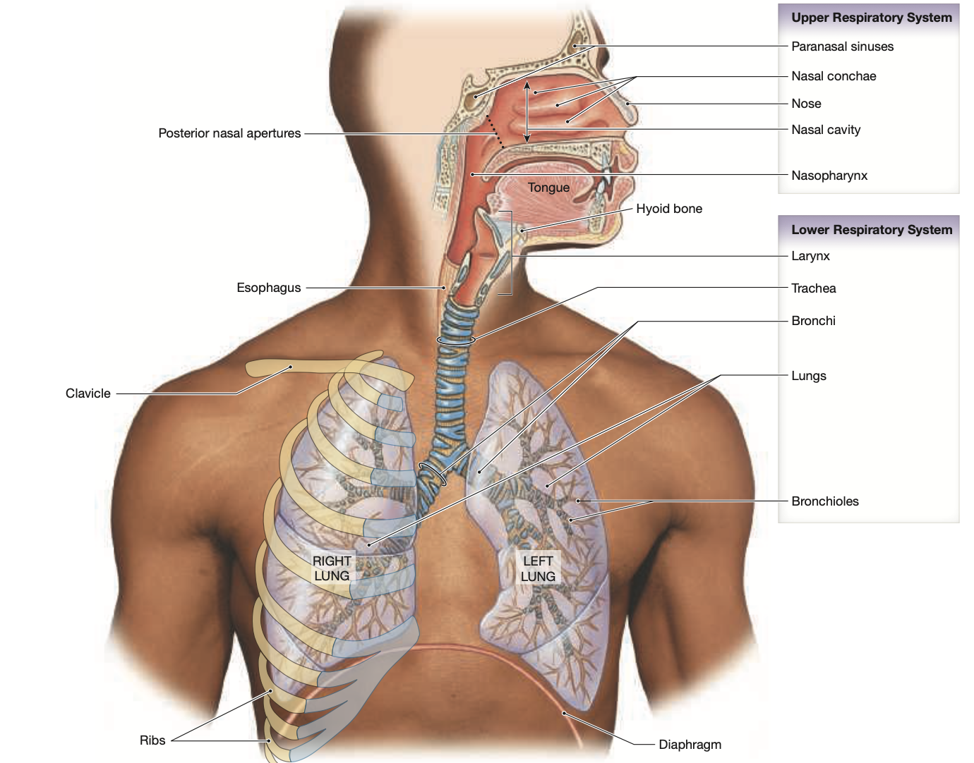
Structure of the Respiratory Tract
Upper Respiratory System:
Paranasal sinuses
Nasal conchae
Nose
Nasal cavity
Nasopharynx
Lower Respiratory System:
Larynx
Trachea
Lungs
Bronchi
Bronchioles
Alveoli
Respiratory Tract Portions:
Conducting Portion: The portion of the Respiratory System involved air transportation— all of the respiratory organs from the Nasal cavity to bronchioles
Respiratory Portion: The portion of the Respiratory System involved in gas exchange— Bronchioles and Alveoli
Functions of the Respiratory System
Gas exchange between air and blood
Cells obtain O2 and eliminate CO2
Moves air to/from exchange surfaces
Protects respiratory surfaces
Defends against pathogens
Permits vocal communication
Regulates blood volume, pressure, and pH
Respiratory Epithelium
Lines conducting portions down to terminal bronchioles
Structure: Pseudostratified, ciliated, columnar epithelium with mucous cells
Produces mucus to trap particles
Lamina propria (connective tissue layer) beneath epithelium forms a mucosa
Stratified squamous surrounds the pharynx
Defense Mechanism:
Mucous glands
Alveolar macrophages
Hairs and cilia
Upper Respiratory System
Nose and Nasal Cavity
Air enters via nostrils (nares)
Nasal vestibule is guarded by hairs (screen large particles)
Air flows through meatuses and conchal surfaces
Hard and soft palate divide oral and nasal cavities
Pharynx
Common chamber for digestive and respiratory systems— serves as a passageway connecting the nose to the mouth and throat
Parts:
Nasopharynx— The back of the nose that extends down to the soft palate, allowing airflow from the nasal cavities to the throat.
Oropharynx— The back of the mouth that lies behind the oral cavity, it serves as a passageway for both air and food, facilitating respiration and digestion.
Laryngopharynx— The most inferior portion of the pharynx, it connects with the Layrnx (respiratory) & the Esophagus (digestive)
Lower Respiratory System
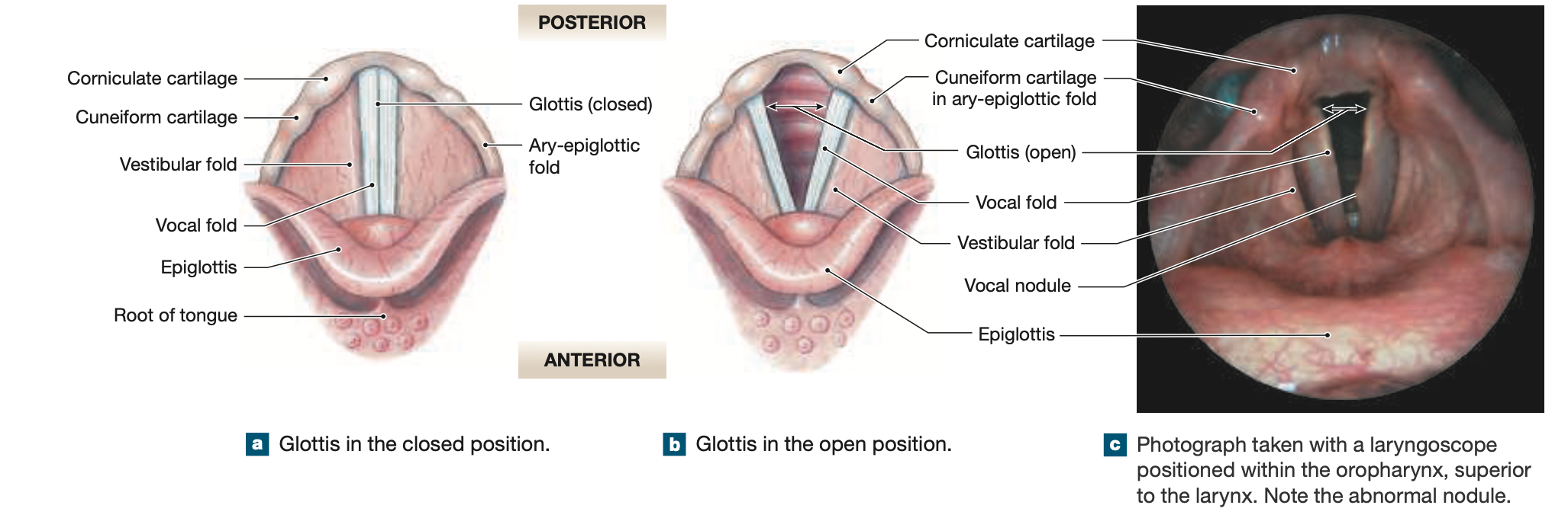
Larynx
Protects glottis; involved in sound production
Epiglottis— A flap of cartilage located at the root of the tongue, it acts as a valve to cover the larynx during swallowing, preventing food or fluid from entering the airway.
Contains vocal folds and vestibular folds
Regulated by intrinsic and extrinsic laryngeal muscles
Laryngeal Muscles—
Laryngeal Ligaments—
Trachea
Extends from cervical to thoracic vertebrae
Contains C-shaped tracheal cartilages for support
Allows for flexibility during swallowing
Trachea Glands— produce mucus to trap foreign particles and keep the airway moist., located in the submucosa of the trachea,.
The trachea branches at the Carina, forming the L & R Primary Bronchi—
The diameter of the R primary bronchi is larger than the L
The R Bronchi is shorter than the L
The R Bronchi decesnds toward the lung at a steeper angle than the L
If food is asperated down the trachea it will likely end up in the R lung
Lobes and Fissures of the Lungs
Right Lung: 3 lobes (superior, middle, inferior) & 2 fissures (horizontal, oblique)
Left Lung: 2 lobes (superior, inferior) & 1 fissure (oblique)
The R Lung is slightly larger than the L lung
Lobes separated by fissures
Main Bronchi and Lungs
Trachea branches into left and right primary bronchi
Each bronchus enters lung at the hilum (root includes bronchus, vessels, nerves)
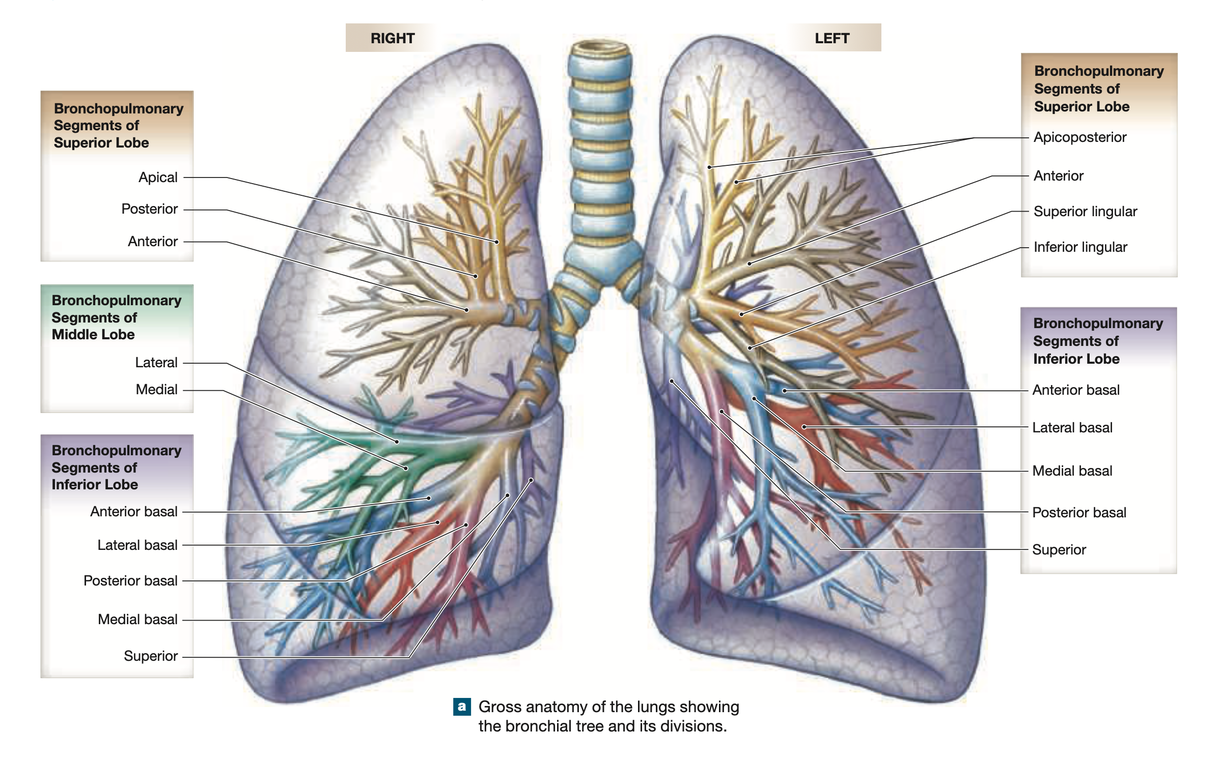
The bronchi further divide into secondary & tertiary bronchi, which decrease in diameter and increase in the number of branches
Bronchopulmonary Segments— are the smallest functionally independent regions of the lungs. Consists of tertiary bronchi, bronchioles, & alveolar structures.
Each lung is divided into segments (10 right, 8-9 left)
Bronchioles— Smaller branches of the bronchi that lead to the alveoli, where gas exchange occurs. They are mainly composed of smooth muscle and elastic fibers, allowing for the regulation of airflow and resistance.
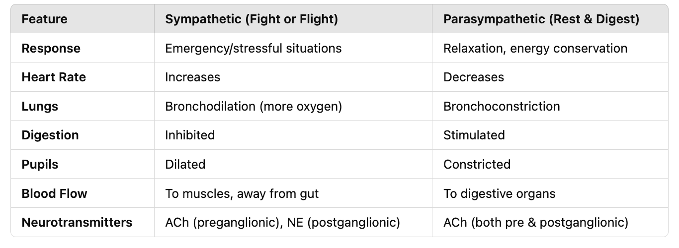
The autonomic nervous system contorols—
Bronchodilation— (sympathetic) Increased airflow
Bronchoconstriction—(parasympathetic) Decreased airflow
Alveolar Structures
Alveolar Ducts: Smallest airways leading to clusters of alveoli.
Alveolar Sacs: Groups of alveoli connected to a common duct.
Alveoli: Tiny air sacs with thin walls— simple squamous epithelium where oxygen enters the blood and CO₂ is expelled (gas exchange occurs).
Oxygen Diffusion into Blood—
Oxygen moves from the alveoli (high O₂ concentration) → through the respiratory membrane → into the capillary blood (low O₂ concentration).
Oxygen binds to Hb in RBCs, which is then transported to tissues.
Carbon Dioxide Exchange
Carbon dioxide (CO₂) from the tissues is carried in the blood (dissolved in plasma, bound to hemoglobin, or as bicarbonate).
In the capillaries, CO₂ diffuses from blood (high CO₂ concentration) → into the alveoli (low CO₂ concentration) → exhaled out through the airway.
Blood-air barrier:
Type 1 Cells— Simple squamous epithelium that facilitates gas exchange due to their thin structure, allowing oxygen and carbon dioxide to diffuse easily between the alveoli and blood.
Has to be a thin membrane in order to facilitate quick gas exchange
Type II Cells— Cuboidal epithelial cells that secrete pulmonary Surfactant, which reduces surface tension in the alveoli and prevents their collapse during exhalation.
Alveolar macrophages protect the epithelium
Pleural Cavities and Membranes
Each lung in a separate pleural cavity lined by pleura (serous membrane)
Two types:
Parietal pleura— Inner membrane (thoracic wall)
Visceral pleura— Outer membrane (lungs)
Respiratory Muscles and Pulmonary Ventilation
Major Muscles:
Diaphragm— Inhalation & exhalation
External intercostals— Elevate the ribs to assist in inhalation
Diaphragm contraction increases thoracic cavity volume
Accessory muscles assist during heavy breathing (e.g., scalene, pectoralis minor)
Respiratory Changes at Birth
Fetal lungs are fluid-filled; inflate upon first breath
Brain Control of Respiration
Controlled by the Pons & Medulla Oblongata
Respiratory rhythmicity center: Sets pace
Apneustic center: Sustains inspiration
Pneumotaxic center: Inhibits deep breaths
Receptors/Reflexes involved:
Mechanoreceptors (lung volume changes)
Chemoreceptors (CO2, pH changes)
Protective reflexes (injury/irritation)
Aging and the Respiratory System
Efficiency declines with age due to:
Deterioration of elastic tissue
Chest cage movement restrictions
Reduced lung volume and capacity
Development of emphysema in older age