Chapter 11: Embryo and Fetal Development
Study Notes
PLACENTAL FUNCTIONS
Functions
Responsible for the transmission of nutrients to fetus and excretion of wastes
Also produces hormones that affect pregnancy and lactation
Structure
Fetal membranes give rise to the placenta.
All farm species have a Chorio-allantoic type of placenta. The outer layer of allantois fuses with the chorion.
This brings fetal blood vessels in allantois close to umbilical vessels, which are in the connective tissue between allantois and chorion.
Chorionic Villi
Project into uterine endometrium to increase the area for nutrient and waste exchange.
Present in all farm species
Villi are diffusely spread over uterus in pig and early on in mares.
Later, mares develop microcotyledons.
Ruminants: cotyledons form connected to maternal caruncles.
Membranes of Placenta First Functions
Chorion Trophoblast
Encloses other membranes and associates with the uterine lining.
Allantois
Hindgut Blood vessels connect fetus and placenta
Amnion
Innermost membrane
Encloses fetus in mass fluid
Umbilical cord amnion Encloses allantoic wraps around blood vessels
Yolk sac
Endoderm
Vestigial
Some nutrients
In monotocous species, the horn of the uterus with the pregnancy is called the Gravid horn, and the other is the Nongravid horn.
Chorion often extends into the nongravid side.
In pigs, chorions often come in contact but with no vascular anastomosis. True also of sheep with multiple pregnancies.
In cows, vascular anastomosis does occur, resulting in freemartins if opposite sexes connect.
Placental and Maternal circulations do not mix in farm species. Their placentas are classified as epitheliochorial: uterine epithelium contacts chorion.
Maternal blood flows to the placenta by uterine arteries and is carried away by uterine veins.
Blood flow to uterus increases 2-3 times at maternal recognition. A further increase is seen at implantation.
About 80% of the blood goes to the placenta.
Umbilical arteries carry blood from the fetus to the placenta, and umbilical veins carry blood from the placenta to the fetus.
In species with cotyledons, maternal capillaries surround the cotyledons which have fetal capillaries within.
Exchange of Gases
Oxygen from maternal blood passes to fetus and CO2 goes from fetus to mother
Fetal Hemoglobin has a higher affinity for oxygen and a lower affinity for CO2 than does an adult.
Nutrient exchange
All nutrients for a fetus come from mother.
Some flow across due to concentration differences, and others use a carrier.
Glucose, amino acids, vitamins have specific carrier systems.
Placenta converts glucose from mother to fructose.
Amino acids cross quickly, but larger proteins do not cross well.
Free fatty acids cross by diffusion.
Immunoglobulins do not cross the placenta in farm species.
Fetal Growth and Development
The fetus has priority over mother for nutrients, and if maternal nutrition is inadequate, this leads to problems such as Pregnancy Toxemia in ewes.
The fetus can pull calcium from the maternal skeleton to supply its needs.
Fetal growth is most rapid early in terms of percent change in size.
But the greatest increase in weight occurs late in gestation.
Calves gain over half of the weight in the last two months.
This is the time when maternal nutrition can affect fetal growth.
Twins are smaller at birth than singles in monotocous species.
Maternal parity and size can affect birth weight.
Primiparous animals usually have smaller young.
Heat stress in late pregnancy can decrease birth weight by decreasing uterine blood flow.
Hormones of Pregnancy
Progesterone is required for maintenance of gestation.
The source of P4 is not always the CL in some stages of pregnancy.
In the sow, cow, and doe, the CL is required throughout pregnancy because it is the only source of progesterone.
In ewes the CL provides progesterone for the first half of gestation, and the placenta produces P4 for the last half.
In mares, the CL produces P4 for about 40 days, at which time accessory CL's are formed
The formation of accessory corpora lutea is due to the secretion of PMSG, also called Equine Chorionic Gonadotropin (eCG).
PMSG acts like FSH in other species, but in horses acts more like LH.
It causes development and ovulation of follicles about day 35 of gestation.
PMSG is produced by Endometrial Cups.
Endometrial cups develop from trophoblast cells called chorionic girdle cells. They detach from chorion and invade endometrium.
PMSG is produced from day 40 to 130 of gestation.
Genotype of fetuses can affect PMSG production. Accessory CL's regress about day 150 and placenta takes overproduction of progesterone.
Progesterone levels decrease when accessory CL regress, but the placenta produces enough to maintain pregnancy.
Estrogen increases in mares about day 90 of gestation and remains elevated through pregnancy.
Estrogen produced by the Feto-Placental unit. Neither placenta nor fetus has all the enzymes needed to make estrogen.
Progesterone is made by the placenta. Some of it goes to the fetus to be converted to androgens.
Then androgens come back to the placenta to be aromatized to estrogen.
This is true for all of the farm species.
High levels of estrogen cause the increased blood flow to the uterus during late gestation.
Elevated estrogen and P4 during pregnancy stimulate mammary gland development.
Relaxin is produced by the placenta in mares and in CL in sows and cows. Its function is to relax or soften connective tissue. This allows the uterus to expand to accommodate a growing fetus.
Relaxin also dilates the cervix and relaxes pelvic ligaments near parturition.
Placental Lactogen or Chorionic Somatomammotropin
Resembles Growth hormone and Prolactin structurally and functionally.
Stimulates mammary gland development.
It also spares glucose from mother for fetus. (Growth regulator)
Mother can use other energy sources, but the fetus needs glucose.
Stimulates fetal growth.
The human placenta also makes a placental lactogen.
hCG: human chorionic gonadotropin
Produced by the embryo as early as day 7 of pregnancy
Maintains CL
Often substituted for LH in research or in treating reproductive problems.
Other hormones produced by the placenta.
Pregnancy Specific Protein B cows and ewes
Found about day 25 of gestation in cows and increases throughout gestation.
Function still unknown.
Can be used a pregnancy test
Beta-endorphin, Corticotropin Releasing Hormone GnRH.
Functions of these are not known.
Placenta protects fetus from immunological rejection by mother.
Not sure how the placenta is protected from attack.
Some of these hormones are likely to have a role in regulating the immune system.
Section 1. It Starts in the Oviduct
the developing embryo reaching the blastocyst stage of development and during this time period, the embryo is still encased in the zona pellucida and making its way from the ampullary–isthmic junction (AIJ) to the uterus.
The trip through oviduct will take the embryo from 5 to 7 days (depending on species)
Once the trip is complete, the embryo will exit the zona and attach itself in a process called Hatching
Hatching: the process of the developing embryo exiting the zona pellucida
After hatching, the embryo has exited the zona; it must take up resident on the endometrium (form an attachment, so the mother’s physiology can supply everything it will need to grow and survive until the time of Parturition
Parturition: delivery of the fetus
This will be the longest period of pregnancy, and it and the few days it takes the embryo to travel to the uterus will constitute what is termed the length of Gestation
Gestation: length of pregnancy
Section 2. Early Pregnancy in the Uterus
Section 2.1 Before Implantation
these cellular divisions are taking place as the embryo travels from the AIJ to the uterus.
In general, the transport time of the embryo from the ovary to the AIJ and, because after ovulation there is less estrogen to cause contractions, it takes between 4 and 5 additional days to reach the uterus.
Days after ovulation
Species | Develop to 2-cell | Develop to 8-cell | Reach the uterus |
Bovine | 1 | 3 | 3.5 |
Equine | 1 | 3 | 5 |
Ovine | 1 | 2.5 | 3 |
Porcine | <1 | 2 | 2 |
During their transport, the embryos must signal their presence to their mother in what is called Maternal Recognition
Maternal Recognition: physiological recognition of pregnancy by the maternal reproductive tract
Maternal recognition is not a conscious event. It is the mother’s physiology being signaled there is an embryo in the reproductive tract.
Cows and sheep have a specific protein, call a trophoblastic protein (TP), which signals the mother the embryo is there. In cattle, it would be called bovine trophoblastic protein 1 or bTP1, in sheep ovine trophoblastic protein, or oTP1.
Table 2 shows these signals and when they occur. Note that, in estrus species, this signal is occurring during diestrus, which if you think about it make perfect sense. If the CL is not supported in late diestrus, it begins to regress and stop producing progesterone. However, pregnancy is dependent on progesterone to keep the uterine lining producing things the embryo needs and the myometrium from contracting. So if the CL is lost, pregnancy will be over before it begins (hence the reason PGF2a can be given to abort estrus animals).
Pig embryos appear to use a different system where the embryos elongated and physically touch the uterine wall telling the mother’s track they are there. This touch may include the embryo secreting placental estrogen that it “paints” onto the walls of the endometrium. Horse use a similar system to pigs but it is unclear whether it includes the estrogen stimulus. Each of these varied mechanisms is designed to stop the action of PGF2α, which would cause regression of the corpus luteum (CL). Humans use a hormonal signal, using human chorionic gonadotropin (hCG) to signal the mother’s reproductive tract. Remember hCG is luteinizing hormone (LH) like, so here the mechanism is to directly support the CL. Regardless of the exact signaling pathway, the signal transforms the track and more specifically the CL from a cyclic system into a system that is designed to support a pregnancy.
Species | days | |
Bovine | 16-17 | |
Ovine | 12-13 | |
Porcine | 11-12 | |
Equine | 14-16 |
The last major event that must occur prior to implantation is hatching. When we hear hatching, most of us think of a chick hatching out of an egg by chipping through the shell. Embryos, on the other hand, use a combination of enzymes to weaken the zona wall and pressure in the blastocoel cavity to force its way out of the zona Blastocyst Development - Day 3 to Day 5
Section 2.2 Implantation and Early Growth
Once the embryo has exited the zona pellucida, the cells can grow to normal size. Further, the removal of the zona allows more nutrients to enter the embryo, and cell duplication speeds up. The inner cell mass will begin to develop three distinct tissue layers called the ectoderm, mesoderm, and endoderm (Figure 1), which will eventually form all tissues of the body
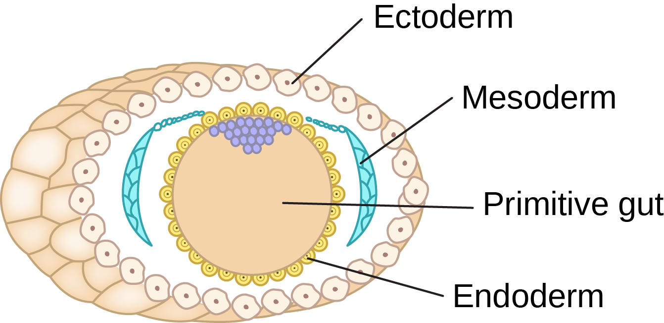
Ectoderm | Mesoderm | Endoderm |
Skin Hair, hooves, and horns Sweat glands (including mammary glands) | Heart and blood vessels Renal system | Digestive system (Including liver and pancreas) |
Nervous system | Skeleton (including muscle) | Pulmonary system |
Oral and nasal cavity Outer reproductive tract (vulva, vestibule, clitoris, glans penis) | Internal reproductive tract (Gonads, Female including ovaries oviduct, uterus, cervix, and vagina Male including epididymis, vas deferens, and accessory glands) | Major glands |
The embryo sets inside the three membrane layers of the placenta, which will serve as both the “lifeline” to mom’s uterine lining and protection for the embryo. These membranes, the amnion, allantois, and chorion, provide distinct functions necessary for the embryo’s survival (Figure 2). The amnion immediately surrounds the developing embryo and baths it in a pool of fluid, amnion fluid. The fluid will serve two major functions as the embryo grows and transforms into a fetus. First, it is like an airbag in a car in that it protects the embryo from any potential trauma as the mother moved through the environment. However, unlike an airbag, the amniotic fluid keeps growth conditions for the embryo at a constant, perfect temperature, an incubator for growth. The second thing the amnion does is allow space for growth. Walk over to the wall, put your hand on the wall and try and push it through. While you might push away from the wall, the wall did not give. Now think of an embryo trying to grow against the solid walls of the uterus surrounding it on all sides. Without the fluid giving it space, the embryo would have a difficult time growing and would never form correctly.
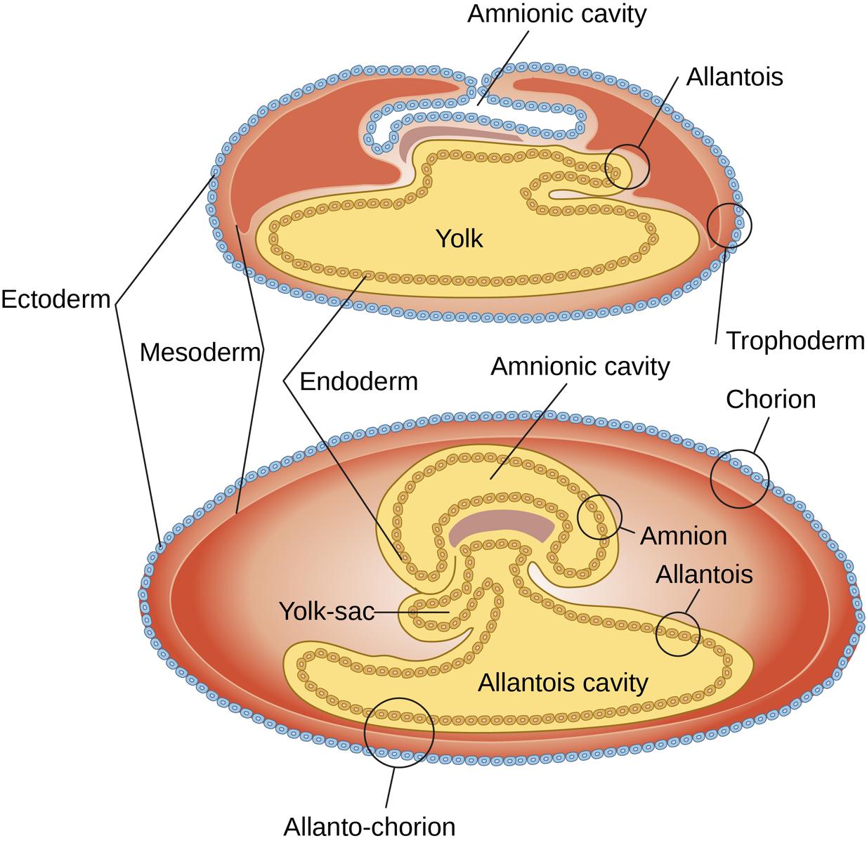
The allantois sac is connected directly to the hindgut and will serve as the embryo/fetus’ “garbage can” for the disposal of metabolic waste. Initially, the chorion will be the point of connection of the endometrium and the conceptus. Over time, the chorion and allantoic membranes may fuse to form a single allanto–chorionic membrane, or the chorion may be eroded placing the allantoic membrane in direct contact with the endometrium and making it easier for the waste materials to be transported out of the placenta into the mother’s blood and disposed of.
While there are significant differences in how the various species attach their placentas, the concept is the same, to place placental (fetal) blood supplies as close to material blood supplies without the two actually mixing. This relationship will maximize the exchange of nutrients and waste between the two and allow rapid conceptus growth. Watch the following animation which will demonstrate this process The Placenta: Its Development and Function.
Section 2.3 Issues in Early Pregnancy
Normal development during early pregnancy is crucial to a successful pregnancy. However, early pregnancy is also a period of tremendous loss. Termed Embryonic Mortality or “Embryo Wastage”, a significant number of pregnancies will be lost due to the death of fertilized ova and embryos up to end of implantation (Table 4). These losses are considered a normal process to correct abnormal genetics or relieve overcrowding in the uterus and may be related to such things as: progesterone deficiency, inbreeding, increasing maternal age, overfeeding, high environmental temperature, multiple embryos, overcrowding in utero, chromosomal aberrations, lactation, defective semen, immunologic incompatibility, or other factors.
Species | Days of Gestation | Embryos Lost % |
bovine | 8-16 | 25-40 |
ovine | 9-15 | 25-40 |
porcine | 8-16 | 25-40 |
equine | 30-36 | 30-40 |
Another significant issue during early pregnancy is the creation of twins or higher-order multiple pregnancies. While multiple gestations are the norm in litter-bearing animals, only a small percentage of non-litter bearing animal pregnancies result in multiple births (Table 5). Twins can be the result of a single fertilization event (i.e., one egg one sperm) or two separate eggs each being fertilized by its own sperm cell. Twins from a single fertilization event are referred to as Monozygotic, Identical, or Paternal twins; mono—one, so monozygotic—one zygote.
Monozygotic, Identical, or Paternal twins: twins resulting from one sperm fertilizing one egg
Alternatively, we can have twins that form as a result of multiple fertilization events, which are referred to as Dizygotic of Fraternal twins
Dizygotic of Fraternal twins: twins results from two separate fertilization events
Dizygotic twins are the result of the body ovulating two egg cells and each egg cell being fertilized by a separate sperm cell. Genetically they are no closer related than siblings born during different pregnancies. However, they share the same birth date.
This type of twin is rare, accounting for 10% of all twins in cattle and 30% in humans. There are a number of theories as to how this type of twin forms. These include (1) separation of cells after the first cleavage with each cell developing independently, (2) formation of two inner cell masses within the blastocyst, (3) division of inner cell mass at the time of hatching, or (4) after implantation, the inner cell mass differentiates into two primitive streaks. These type twins are chromosomally identical, which mean they always have to be the same sex.
Section 3. Early Development and Transitioning from Embryo to Fetus
Section 3.1 Early Organ Development
Obviously, the embryo begins as the one cell zygote.
The first differentiation step is as the cells of the blastocyst separate into the true embryo (the inner cell mass) and the placenta.
As discussed above the inner cell mass develops the three tissue layers of the embryo, which in turn gives rise to all of the body organs.
There is a set pattern of development.
The first tissue of the body to start the differentiation and development process is the nervous system.
It starts as a ridge at the top of the embryo; what is termed the neural crest.
The second organ system to start forming and the first to become functional is the heart.
Teleologically speaking, it is absolutely necessary for the heart to become functional first.
Teleologically: defining how somethings development in terms of its function
By the time the heart starts beating, the tissue is approximately 2 cm3.
At this point, the tissue is too large for simple diffusion to supply oxygen and nutrients, so without its “pump,” the embryo would die.
From these beginnings, the embryo will begin to enlarge and begin to develop limbs and shape more like an adult animal.
In fact, an embryo is said to transform into a fetus when you can tell the animal species.
At that point, its outer differentiation is complete, but it is still growing and developing its organ system
While the nervous tissue is the first to form, it is among the last tissues to become fully functional and finishes its development after birth.
Section 4. Fetal Development
While pregnancy is a continuous process from conception to Parturition, it can be seen of consisting of three separate stages of development.
Parturition: delivery of the fetus
In humans, we would call these trimesters as each is roughly three months long.
The first phase was discussed above and consisted of growth from conception (the single cell zygote) until the embryo is identifiable as a fetus
This phase of development is mainly concerned with getting the structure of the fetus in place (establishing all limb structures externally and the beginnings of all organs internally).
This phase of development will start with the single-cell zygote and ends with a fetus that in the cow would weigh approximately 500 grams.
1st - 3rd | 2nd - 3rd | 3rd - 3rd |
Maternal Recognition | Completion of Differentiation | Most Rapid Increase in Weight (%) |
Implantation | ||
Embryo to Fetus | Sexual Differentiation | Maturation of Lungs |
Most Rapid increase in Cell number (%) | Organ Maturation | Parturition |
If the first third of pregnancy can be thought of as getting the structure right, the second third of pregnancy can be thought of as being the period of biochemical maturation.
All but one of the organ systems will become functional, meaning that the biochemistry to run the organ begins to function.
As stated above, sexual differentiation will occur during this second period, but the biochemical signal occurs earlier in development
Interestingly, all mammals have two different duct systems as they develop.
The Mullerian duct that at maturity, would become the oviduct, uterus, cervix, and vagina, and the Wolffian duct that supplies the epididymis, vas deferens, and accessory glands.
You might have noticed there was no mention of the gonads.
There is only one set of gonads, which will become ovaries or testis depending on which of these ducts develops.
In mammals, the presence of an XX or XY chromosomes in the developing fetus will signal the duct systems as to which duct should develop.
If the chromosome arrangement is XY, there is a cascade of events that leads a burst of androgens and the Wolffian duct develops and the Mullerian duct regresses; the fetus is male, and the gonads become testis, and the gametes become sperm.
In the absence of this signal, that is, XX chromosomes, the gonads become ovaries, the gametes develop into oocytes, the Mullerian duct develops, and the fetus is female.
This will include the reproductive system, which we will discuss below.
The one system that will not at least be partially functional by the end of the second third of pregnancy are the lungs. Why?
They are not needed at this point as mom is still doing all of the breathing at this point, and it appears to be the fetus conserving energy to do other things.
While there is growth since most of the energy is going into establishing biochemical pathways, physical growth is restricted, and in the cow, the fetus only increases in size from that 500 grams at the start of this period to approximately 2.5 kilograms (2500 grams) when it is completed.
Species | day | |
bovine | 45 | |
ovine | 35 | |
porcine | 30 | |
equine | 45 |
However, from time to time things do not go as planned.
Two good examples of developmental problems are Freemartins and Hermaphrodites.
Freemartins general occurs in cattle (but can occur in other species) and are always the result of a twin pregnancy where one twin is female, and the other one is male.
During the period of sexual differentiation, the male fetus has the burst of androgens, which tells him to develop his Wolffian duct.
Unfortunately, because in cattle the placental membranes of the twins fuse together, little sister also gets part of the androgen signal.
This partial signal is not enough to trigger her Wolffian duct to develop, but is enough to prevent her Mullerian duct from developing normally.
Generally, these animals are born with a blind pouch (an incomplete vagina that does not connect to a uterus) where their vagina should be.
Further, because the developing ovaries received mixed signals, they fail to develop, and the animal is generally sterile.
Hermaphrodites, or what are sometimes called individuals with ambiguous genitalia, results in partial development of both tracts.
Hermaphrodites are not generally associated with a twin pregnancy, and it is unclear how they received mixed signals to cause both ducts to develop.
However, it is known that most are sterile, although there are reports of reproductively sound individuals in all species.
The last part of pregnancy is a period of rapid growth. For example, our 2.5 kg cow will grow rapidly up to 45 kg at parturition.
This rapid weight gain requires good nutrient, meaning this is the time when nutrient restrictions in the mother could have the most impact on the fetus.
Nutrient restrictions may not only impact the weight of the fetus at delivery, but it might also have long-term consequences on the future development of the animal outside the womb Pregnant Cow Nutrition: Effect Carcass on Characteristics.
Section 5. The Placenta in Pregnancy
The placenta serves several roles that are essential to the fetus’ growth, development, and survival.
First, it provides the connection link to the mother who supplies the nutrients for growth and development.
Second, it provides protection for development.
Finally, it is a major hormone producer, making hormones that maintain the pregnancy and regulate growth.
Let’s look first at the placenta’s role in connecting the fetus to the mother’s endometrium.
The objective here is to position the mother’s and fetus’ blood supplies as close together as possible without them mixing the two sources of blood.
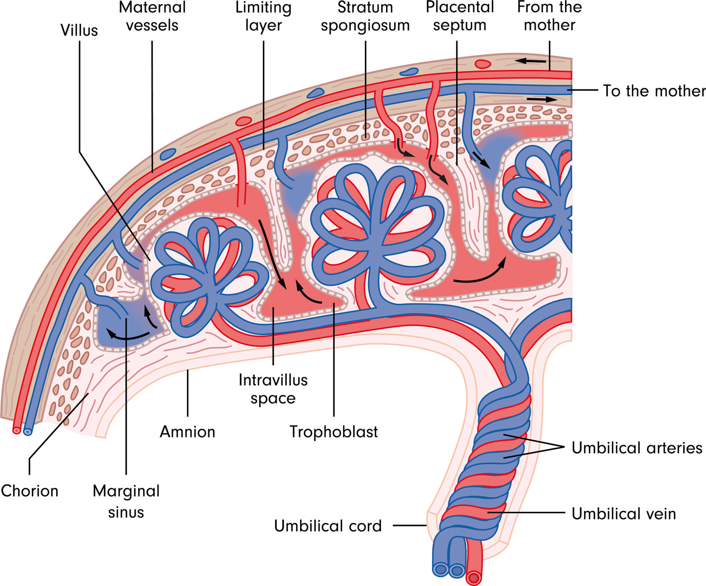
While the exact arrangement varies from species to species, Figure 4 demonstrates the complexities of this arrangement.
Because it supplies all nutrients and carrying away waste, maternal blood flow is essential for normal fetal development.
In larger species, the maternal blood flow to the uterus will increase from 500 to 1000 mL per minute toward the end of pregnancy.
About 80% of this “new” blood will end up right over the top of the placenta.
This does not happen by chance, the placenta releases factors, which will grow new maternal blood vessels right where the placenta is growing.
the placenta provides protection and space for normal development by surrounding the developing fetus with the fluid-filled amnion.
In extreme cases, loss of the fluid can lead to malformation or total absence of limbs
In fact, the placental hormones are critical to maintaining pregnancy, priming the mother’s body to synthesize milk and to regulate regulating fetal growth.
The placenta is the major producer of progesterone for the majority of pregnancy.
Since progesterone maintains the uterine environment necessary for pregnancy, this means the placenta must function, or pregnancy will be lost.
The combination of progesterone and estrogen coming from the placenta are necessary to trigger growth and organization of the mammary tissue to allow for milk synthesis.
The placenta also makes relaxin to help create the space for fetal growth.
Finally, the placenta makes placental lactogen.
From the name, it should be obvious that this hormone helps regulate milk synthesis.
However, placental lactogen is also a growth regulator for the fetus.
Placental lactogen will make the mother somewhat insulin resistant.
Because she is insulin resistant, her blood sugars are higher.
Higher blood sugars in mom translated to more nutrients available to the fetus, which causes it to grow at faster rates.
This can be both a good thing and a bad thing.
Adding growth is good if within a control range.
However, if the fetus grows too fast, it will get too large and have trouble at delivery (to be discussed in Chapter 12), so it is important that placental lactogen controls fetal growth within the right rates.
Section 6. Why Should the Producer Care?
Hopefully, the section title is a rhetorical question.
Everything we do in animal science begins with the recombination of genetics at conception.
The quality of meat, the quantity of milk, the overall appearance of our animals are all influenced by the genes that formed them.
However, it is not just this reset of the genes that matter when determining how an animal will start and how it will finish.
It is well known that to “catch an animal up” that is born small requires both time and usually money (in the form of higher feed bills).
So anything we can do to make sure our animals hit the ground at the right size and weight will affect our profitability.
Further, knowing the critical periods to add additional nutrition or limit exposure to pesticides might prevent avoidable disasters in our operations.