Lab Exam 2
Integumentary System (Skin Model)

Epidermis: Superficial, avascular layer composed of keratinized stratified squamous epithelium.
Dermis: Deeper, vascular connective tissue layer beneath the epidermis; contains nerve endings, hair follicles, and glands.
Dermal Papilla: Finger-like upward projections of the dermis that interlock with the epidermis to form skin ridges (fingerprints).
Hair Follicle: Tubular invagination of the epidermis surrounding the hair root.
Hair Root: The portion of the hair embedded below the skin surface within the follicle.
Hair Shaft: The visible portion of the hair extending above the skin surface.
Sebaceous Gland: Multi-lobed, oil-producing gland connected directly to hair follicles; secretes sebum.
Eccrine Sweat Gland: Coiled tubular gland located in the dermis with a duct leading directly to a surface pore for thermoregulation.
Pacinian Corpuscle (Lamellar): Large, onion-like layered sensory receptor located deep in the dermis or hypodermis; detects deep pressure and vibration.
Meissner’s Corpuscle (Tactile): Small, oval sensory receptor situated high in the dermal papillae; detects light touch.
Arrector Pili Muscle: Smooth muscle band extending from the upper dermis to the hair follicle; contracts to pull hair upright ("goosebumps").
Axial Skeletal System
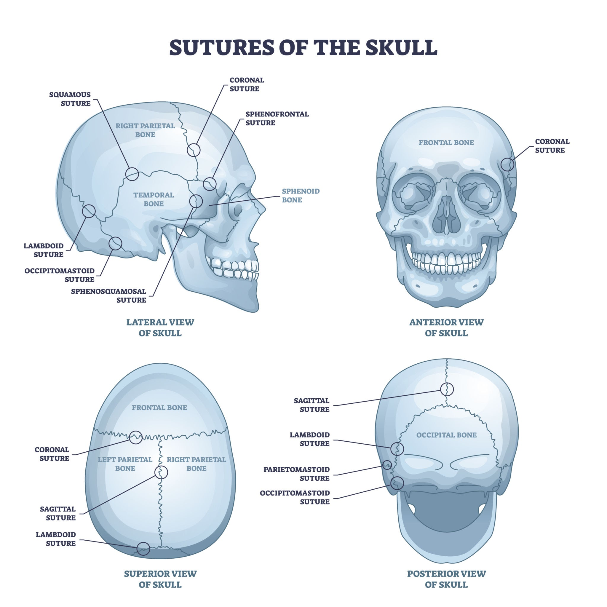
Skull Sutures
Coronal: Connects frontal bone with parietal bones across the anterior skull.
Sagittal: Connects the right and left parietal bones along the midline top of the skull.
Lambdoidal: Connects parietal bones with the occipital bone along the posterior skull.
Squamosal: Connects parietal bone with temporal bone on the lateral side of the skull.
Cranial Bones & Features
Parietal Bones: Pair forming superior and lateral walls of the cranium.
Temporal Bones: Pair on inferior sides of cranium.
Zygomatic Process: Posterior projection forming part of the cheekbone.
Zygomatic Arch: Formed by zygomatic process of temporal bone and temporal process of zygomatic bone.
External Acoustic Meatus/Canal: Outer canal opening leading to the eardrum.
Mastoid Process: Rounded projection posterior to the ear for muscle attachment.
Styloid Process: Needle-like downward projection for muscle and ligament attachment.
Carotid Canal: Anteromedial opening on inferior surface for internal carotid artery.
Jugular Foramen: Large posterior opening on inferior surface for internal jugular vein and cranial nerves IX, X, XI.
Frontal Bone: Anterior skull bone forming forehead and upper orbits.
Supraorbital Foramen/Notch: Small opening above orbit for nerves and vessels.
Occipital Bone: Posterior and inferior base of skull.
Foramen Magnum: Large central opening allowing brainstem to connect with spinal cord.
Occipital Condyles: Smooth rounded processes flanking foramen magnum that articulate with atlas (C1).
Sphenoid Bone: Butterfly-shaped bone spanning the floor of the central cranium.
Greater & Lesser Wings: Lateral extensions forming parts of cranial floor and orbits.
Sella Turcica: Saddle-shaped depression holding the pituitary gland.
Optic Canal: Anterior canal for optic nerve (CN II).
Superior Orbital Fissure: Slit-like opening in orbit for CN III, IV, V1, VI.
Foramen Ovale: Oval opening for mandibular nerve (CN V3).
Foramen Spinosum: Small posterolateral opening for middle meningeal artery.
Foramen Lacerum: Irregular jagged opening at junction of sphenoid, temporal, and occipital bones.
Ethmoid Bone: Spongy bone between orbits forming nasal roof and septum.
Perpendicular Plate: Superior vertical plate forming upper nasal septum.
Crista Galli: Superior median ridge anchoring dura mater.
Cribriform Plate: Horizontal perforated plate flanking crista galli for olfactory nerve fibers (CN I).
Facial Bones & Features
Maxilla: Upper jawbone forming central face, hard palate, and floor of orbits.
Mandible: Lower jawbone (only movable skull bone).
Mental Foramen: Opening on anterior lateral body for mental nerve and vessels.
Zygomatic Bones: Cheekbones.
Temporal Process: Posterior process joining temporal bone.
Zygomatic Arch: Arch formed with temporal bone.
Lacrimal Bones: Small bones in medial orbital wall.
Nasolacrimal Canal: Groove housing tear duct into nasal cavity.
Nasal Bones: Small paired bones forming bridge of nose.
Vomer: Inferior plow-shaped bone forming lower nasal septum.
Hyoid Bone: U-shaped floating bone in anterior neck; anchors tongue muscles.
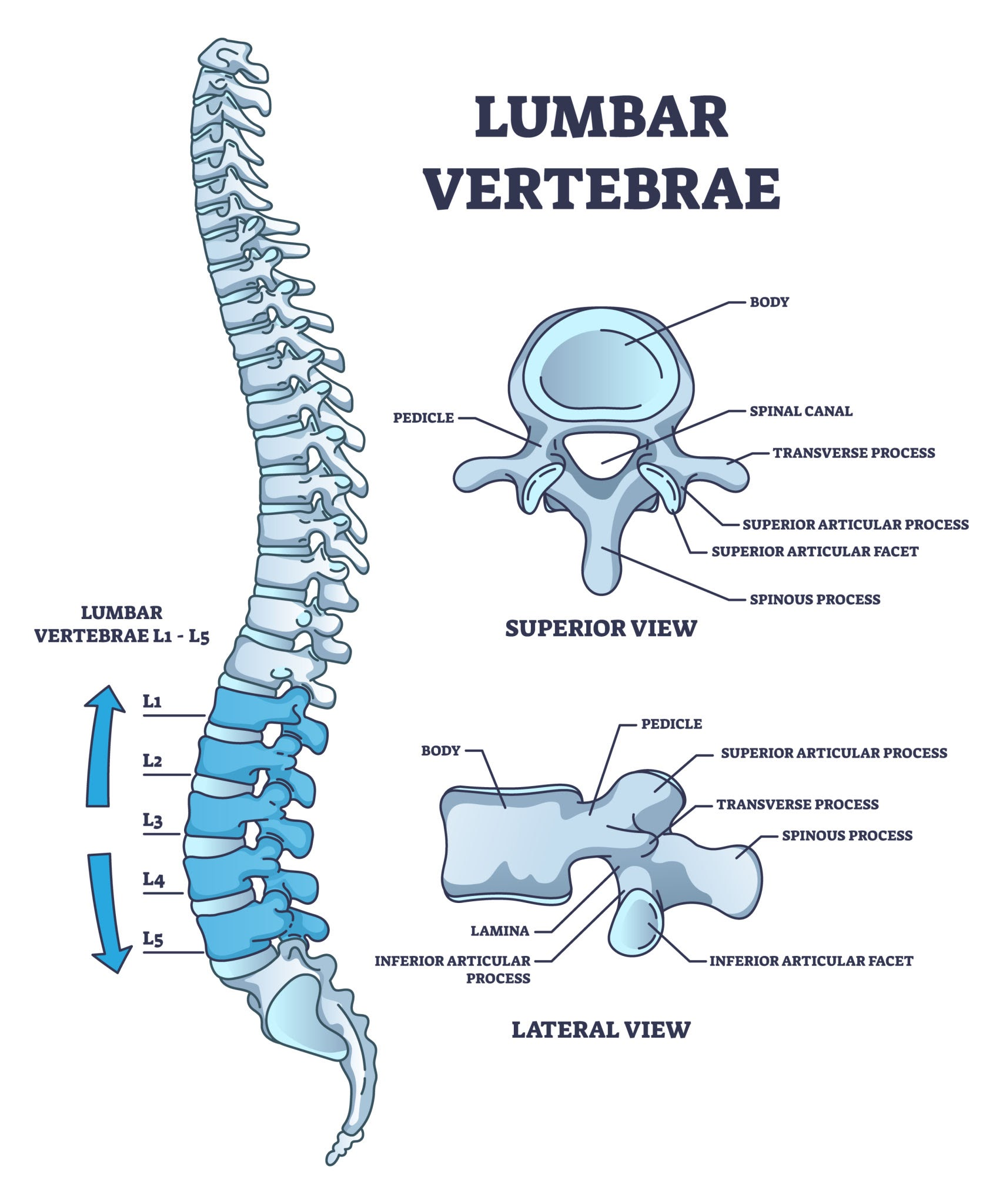
Vertebrae
Universal Structures:
Body: Anterior solid weight-bearing block.
Vertebral Foramen: Central canal housing spinal cord.
Spinous Process: Posterior projection felt along spine.
Transverse Process: Lateral projections for muscle attachment.
Pedicle: Bony bridge connecting body to transverse process.
Lamina: Bony roof connecting transverse process to spinous process.
Cervical Vertebrae (C1–C7): Smallest vertebrae; feature transverse foramina for vertebral arteries.
Atlas (C1): Ring-like bone without body or spinous process; has superior facets articulating with occipital condyles and facet for dens anteriorly.
Axis (C2): Has vertical peg (dens/odontoid process) acting as pivot for atlas.
Thoracic Vertebrae (T1–T12): Heart-shaped bodies with costal facets for rib articulation; long downward-sloping spinous processes.
Lumbar Vertebrae (L1–L5): Massive blocky bodies; short, hatchet-shaped spinous processes.
Sacrum: Triangular bone formed by 5 fused vertebrae.
Coccyx: Tailbone formed by 3–5 fused vestigial vertebrae.
Sternum & Ribs
Sternum: Breastbone consisting of superior Manubrium, middle Body (gladiolus), and inferior Xiphoid Process.
True Ribs (1–7): Direct cartilage attachment to sternum.
False Ribs (8–12): Indirect cartilage attachment (8–10) or no anterior attachment (11–12).
Floating Ribs (11–12): Subgroup of false ribs with unattached anterior ends.
Appendicular Skeletal System

Pectoral Girdle & Upper Limb
Clavicle: S-shaped collarbone.
Scapula: Shoulder blade.
Spine: Posterior prominent ridge.
Supraspinous Fossa: Depression superior to spine.
Infraspinous Fossa: Depression inferior to spine.
Acromion Process: Lateral apex of spine articulating with clavicle.
Glenoid Cavity: Lateral socket articulating with humerus head.
Coracoid Process: Anterior hook-like projection for muscle attachments.
Subscapular Fossa: Smooth anterior concave surface facing ribs.
Humerus: Arm bone.
Anatomical Neck: Smooth groove surrounding head; Surgical Neck: Narrowing distal to tubercles (frequent fracture site).
Deltoid Tuberosity: Lateral mid-shaft roughened ridge for deltoid attachment.
Trochlea: Medial spool-shaped distal condyle for ulna.
Capitulum: Lateral rounded distal condyle for radius.
Olecranon Fossa: Deep posterior distal depression for ulnar olecranon process.
Ulna: Medial forearm bone.
Trochlear (Semilunar) Notch: C-shaped wrench-like groove grasping humerus trochlea.
Coronoid Process: Anterior lip of trochlear notch.
Olecranon Process: Posterior prominent elbow tip.
Styloid Process: Distal medial point at wrist.
Radius: Lateral forearm bone.
Head: Disc-shaped proximal end; Radial Tuberosity: Medial bump for biceps insertion; Styloid Process: Distal lateral point at wrist.
Carpals (Wrist - 8 bones): Proximal row (lateral to medial): Scaphoid, Lunate, Triquetral, Pisiform. Distal row (lateral to medial): Trapezium, Trapezoid, Capitate, Hamate.
Metacarpals: Palm bones numbered I–V starting at thumb.
Phalanges (Hand): Finger bones (Proximal, Middle, Distal); Pollex (thumb) has only proximal and distal.
Pelvic Girdle & Lower Limb
Os Coxae (Hip Bone):
Ilium: Superior region featuring Iliac Crest (upper rim) and Greater Sciatic Notch (posterior cutout for sciatic nerve).
Ischium: Inferior posterior region featuring Lesser Sciatic Notch, Ischial Tuberosity (sit-bone weight bearer), Acetabulum (deep socket for femur head), and Obturator Foramen (large hole between ischium and pubis).
Pubis: Inferior anterior region joining at Pubic Symphysis (cartilage pad).
Femur: Thigh bone.
Head: Proximal sphere articulating with acetabulum; Neck & Shaft: Angle connecting head to long body.
Linea Aspera: Posterior longitudinal ridge on shaft.
Medial & Lateral Condyles: Distal rounded joint surfaces.
Intercondylar Fossa: Deep posterior notch between condyles.
Patellar Groove/Surface: Smooth anterior distal surface for kneecap.
Patella: Sesamoid kneecap bone.
Tibia: Medial shin bone.
Tibial Tuberosity: Anterior proximal bump for patellar ligament insertion.
Anterior Crest: Sharp shin ridge.
Medial Malleolus: Distal inner ankle bump.
Fibula: Lateral slender leg bone.
Head: Proximal enlargement; Lateral Malleolus: Distal outer ankle bump.
Tarsals (Ankle - 7 bones): Talus (articulates with tibia), Calcaneus (heel bone), Navicular, Cuboid, and Cuneiforms (1st/Medial, 2nd/Intermediate, 3rd/Lateral).
Metatarsals: Sole bones numbered I–V starting at big toe.
Phalanges (Foot): Toe bones (Proximal, Middle, Distal); Hallux (big toe) has only proximal and distal.