Nitrogen Metabolism: Protein Degradation, Amino Acid Catabolism, and Nitrogen Balance
Fundamentals of Nitrogen and the Amino Acid Pool
Nitrogen Properties and Fixation
Nitrogen is inherently too unreactive to participate in the majority of biochemical reactions.
It must be "fixed" into biochemically available nitrogen compounds; this process is carried out by a limited number of specialized microorganisms.
Nitrogen is a critical requirement for the formation of the amino group in all amino acids.
The Amino Acid Pool
The body maintains an "amino acid pool" consisting of free amino acids found at very low concentrations within cells and the bloodstream.
This pool is characterized by constant mixing and exchange with other free amino acids distributed throughout the body.
Dietary Protein Requirements
Unlike carbohydrates (glycogen) or lipids (fat), there is no dedicated "storage" form of protein in the body to replace nitrogen-containing compounds.
Dietary protein is essential to replace lost amino acids and support tissue repair.
Recommendations suggest a daily intake of of protein.
High protein intake in well-fed individuals is considered wasteful: surplus amino acids are rapidly catabolized, and their nitrogen is excreted as urea in the urine.
Nitrogen Balance
Positive Nitrogen Balance
Defined as: N\,intake > N\,excretion.
This state occurs when the rate of protein synthesis exceeds the rate of protein breakdown.
Occurs during:
Normal growth in children.
Convalescence following serious illness.
Recovery and immobilization after an accident.
Pregnancy.
Negative Nitrogen Balance
Defined as: N\,intake < N\,excretion.
This state occurs when the rate of protein breakdown exceeds the rate of synthesis.
Occurs during:
Starvation.
Serious illness.
Late stages of certain cancers.
Injury and trauma.
Implications: If negative nitrogen balance is prolonged and uncorrected, it leads to the irreversible loss of essential body tissue and, ultimately, death.
Pathways of Protein Degradation
Cytosolic Protein Degradation
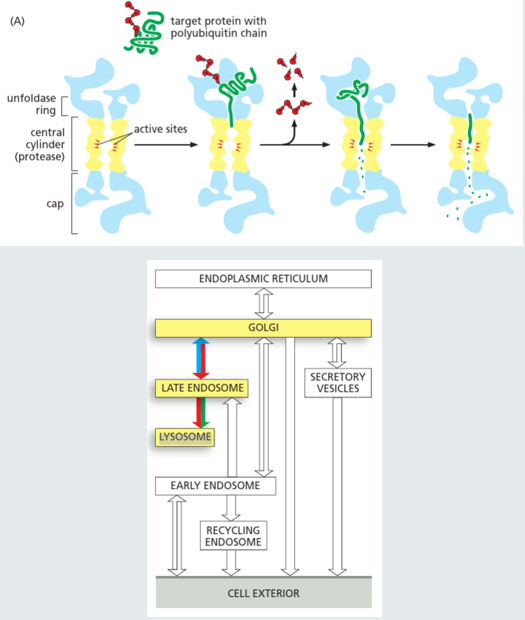
Proteins identified as "old" or damaged are recognized by the cell.
These proteins are tagged with polyubiquitin.
They are subsequently degraded within the proteasome, resulting in a mixture of the 20 standard amino acids.
Organelle and Exogenous Protein Degradation
Includes foreign (exogenous) proteins and aging or damaged sub-cellular organelles.
These are internalized into vesicles via endocytosis or autophagocytosis.
The vesicles fuse with lysosomes, where proteolytic enzymes degrade the proteins into amino acids.
Hormonal and Metabolic Regulation
Factors such as starvation and hormones (e.g., cortisol) significantly increase the rates of protein breakdown, particularly in skeletal muscle.
Amino Acid Catabolism and Nitrogen Removal
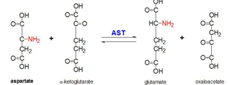
Transamination: The Redistribution of Nitrogen
Transamination involves the conversion of one amino acid to another by transferring the -amino group to -ketoglutarate (-KG).
General Reaction: .
Which keto acid is formed depends on the side chain of the original amino acid
This reaction is catalyzed by aminotransferases (also called transaminases), which are specific to individual amino acids.’
Specific Examples:
Aspartate aminotransferase: .
Alanine aminotransferase: .
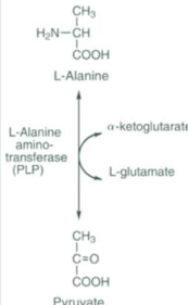
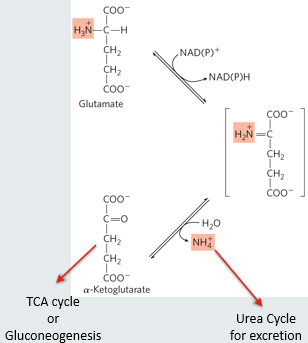
The resulting carbon skeletons (Keto Acids) are easily metabolized in the TCA cycle or used in gluconeogenesis.
Deamination: The Release of Ammonia
Deamination is the removal of the -amino group from glutamate.
Reaction: .
Catalyzed by glutamate dehydrogenase.
In humans, this reaction almost exclusively occurs with Glutamate and takes place in the liver mitochondrial matrix.
Ammonia is toxic! Brain is sensitive where ammonia toxicity causes cognitive impairment, ataxia, seizures
ammonia is converted to a non-toxic compound in the urea cycle
urea is transported via the blood to the kidney for excretion
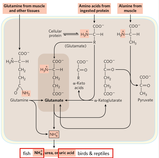
Transdeamination
This is the combined action of aminotransferases and glutamate dehydrogenase.
It allows -amino groups from various amino acids to be funneled into the Urea Cycle using glutamate as an intermediate.
Example (Alanine in the liver):
.
.
Ammonia Toxicity and Transport Mechanisms
The Danger of Free Ammonia
Free ammonia is generated elsewhere in the body and needs to be transported to the liver
processes in other tissues generate ammonia (e.g. nucleotide degradation)
Free ammonia is highly toxic, particularly to the brain, where it can cause cognitive impairment, ataxia, and seizures.
It cannot be safely transported in the blood as ammonium ().
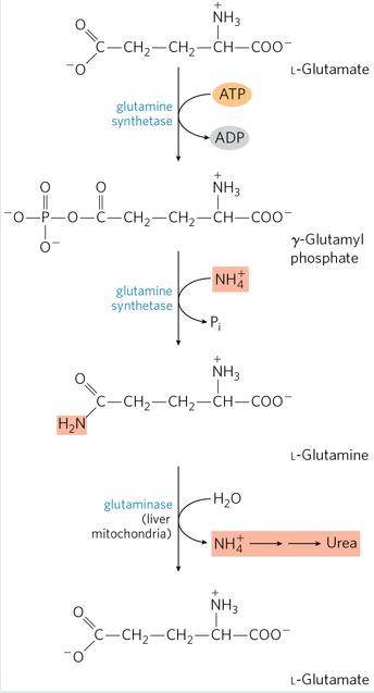
Glutamine: Transport from Extra-hepatic Tissues
Glutamate has 1N, glutamine has 2N (a helpful mnemonic: the name Glutamine contains an "n" for the extra nitrogen).
In extra-hepatic tissues, ammonia is added to glutamate to produce glutamine.
Glutamine is safely transported through the bloodstream to the liver.
In the liver, glutamine is converted back to glutamate, releasing ammonia for disposal in the Urea Cycle.
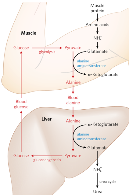
Alanine: Transport from Skeletal Muscle
During vigorous exercise, skeletal muscle utilizes protein and carbohydrates for energy.
Amino acid carbon skeletons enter the TCA cycle for ATP production, but ammonia must be safely removed.
Skeletal muscle tissue generates high levels of pyruvate (and lactate).
Transamination of pyruvate to alanine allows the ammonia to be safely transported to the liver.
The glucose-alanine cycle allows the liver to regenerate glucose from this alanine
Amino acid catabolism in the liver
glutamine transports ammonia to the liver where its converted to glutamate and ammonia
alanine from skeletal muscle is converted to glutamate + pyruvate
excess amino acids are converted to glutamate by transamination
glutamate is deaminated to generate a-ketoglutarate and ammonia
ammonia is converted to urea in humans in the urea cycle
urea is transported to the kidneys for excretion
The Urea Cycle
Ammonia is converted into Urea, a non-toxic compound, via the Urea Cycle.
Urea is transported through the blood to the kidneys for excretion.
Note: Different organisms excrete nitrogen differently (Fish excrete ammonia; Birds and Reptiles excrete uric acid; Land Vertebrates excrete urea).
Metabolic Fates of Carbon Skeletons
Major Products
The degradation of all 20 amino acids leads to 7 major carbon skeleton products: Pyruvate, Oxaloacetate, Fumarate, Succinyl-CoA, -Ketoglutarate, Acetoacetyl-CoA, and Acetyl-CoA.
Glucogenic Amino Acids
These produce skeletons (Pyruvate, Oxaloacetate, Fumarate, Succinyl-CoA, -KG) that enter TCA cycle to generate ATP, and enter gluconeogenesis to release glucose into the blood.
amino acids producing these carbon skeletons are termed glucogenic because they can produce glucose via gluconeogenesis
Ketogenic Amino Acids
These produce skeletons (Acetoacetyl-CoA, Acetyl-CoA) that can produce ketone bodies.
amino acids producing these carbon skeletons are termed ketogenic
Lysine and Leucine are the only two amino acids that are strictly ketogenic.
Amino Acid Biosynthesis and Classification
Biosynthetic Origins
Amino acids are synthesized from intermediates found in Glycolysis, the Citric Acid Cycle, and the Pentose Phosphate Pathway.
organisms vary in their ability to synthesis different amino acids
most plants and bacteria can synthesize all 20
mammals can only synthesize half of them
Essential vs. Non-essential Classification
Essential: Cannot be synthesized by mammals and must be obtained from dietary sources.
Non-essential: Can be synthesized by mammals and are not strictly required in the diet.
Conditionally Essential: Required under specific physiological conditions.
Categorization Table
Non-essential: Alanine, Asparagine, Aspartate, Glutamate, Serine.
Conditionally essential: Arginine, Cysteine, Glutamine, Glycine, Proline, Tyrosine.
Essential: Histidine, Isoleucine, Leucine, Lysine, Methionine, Phenylalanine, Threonine, Tryptophan, Valine.
Key Metabolic Players
Glutamate: Central to the process of amino acid degradation.
Glutamine: Primary transporter of ammonia to the liver.
Alanine: Transports ammonia from skeletal muscle to the liver.
Aspartate: Involved in several roles, including the Malate-Aspartate Shuttle.
readily generated and degraded in humans