Abdomen & Thorax
KNSS 220 Introduction to Athletic Therapy
Abdomen & Thorax
Abdomen Anatomy
Kidneys
Adrenal Glands
Anatomy of Human Urinary System
Organs Involved:
Ureters
Urinary bladder
Right ureter
Urethra
Left ureter
Anatomy of the Digestive System
Organs Involved:
Liver
Gallbladder
Pancreas
Stomach
Small intestine
Large intestine
Anatomy of the Lymphatic System
Spleen
Anatomy of the Reproductive System
Female Organs:
Ovaries
Uterus
Uterine tubes
Vagina
Male Organs:
Seminal vesicles
Prostate gland
Testes
Vas deferens
Epididymis
Urethra
Penis
Conditions and Injuries
1. Condition: Kidney Stones
Etiology:
The formation of hard deposits made of minerals and salts that develop in the kidneys, often due to dehydration, high sodium intake, or underlying metabolic disorders.
Signs and Symptoms
Severe pain in the back or side, often radiating to the lower abdomen and groin
Blood in urine (hematuria)
Frequent urination or urgency
Nausea and vomiting
Cloudy or foul-smelling urine
Management:
Encourage increased fluid intake to dilute urine.
Monitor for signs of urinary tract infection (UTI) and consider urinalysis.
Provide analgesics for discomfort as needed.
Consider prescribing antibiotics if a bacterial infection is confirmed.
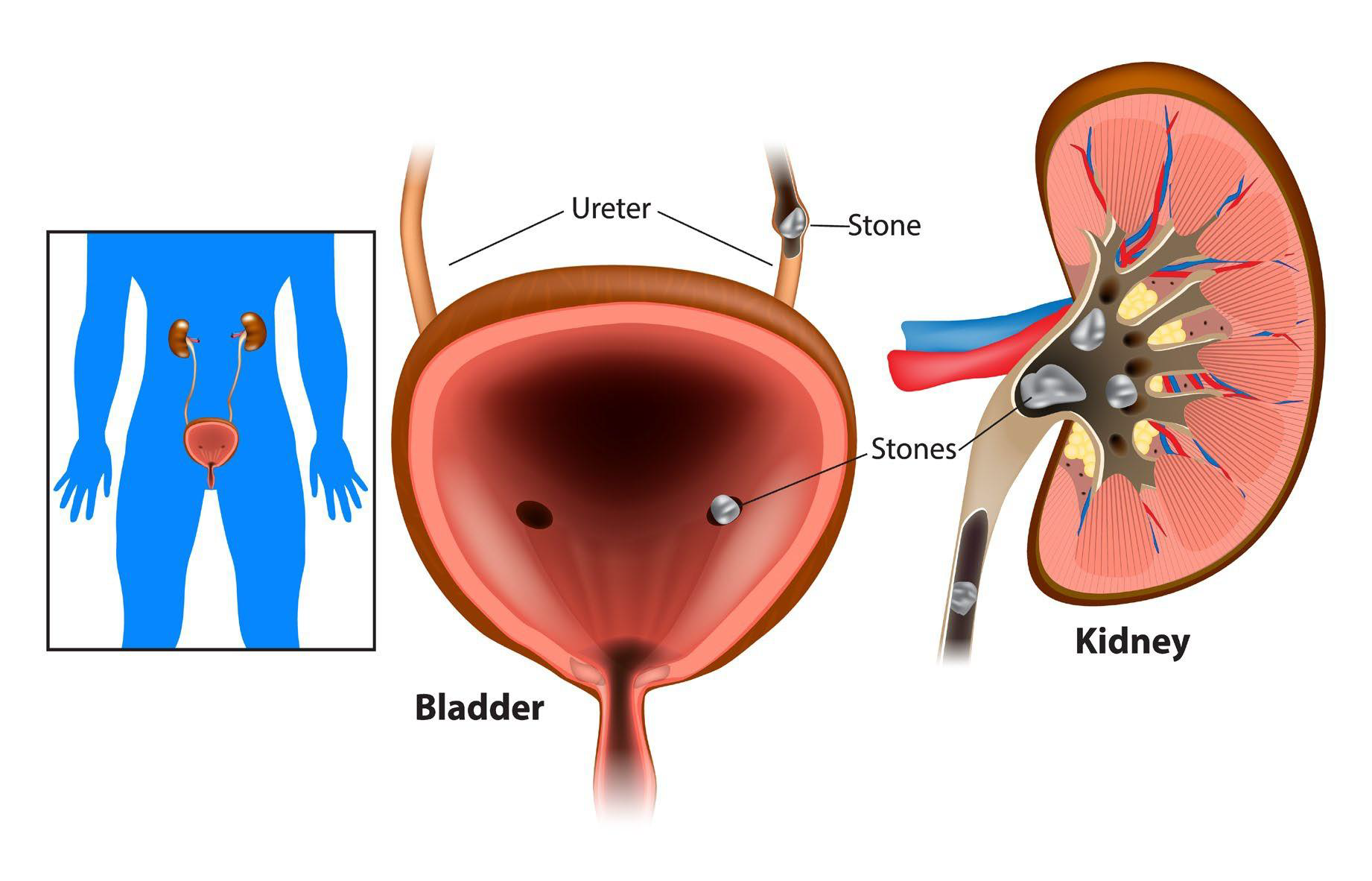
Involves the Bladder, Ureter, and Kidney
Stones:
Stone Location: Ureter
Types of Stones: Various stone types based on composition
2. Injuries: Kidney Contusion
Etiology:
The primary causes of kidney contusions include blunt trauma from falls, sports injuries, or vehicular accidents, leading to potential bleeding and tissue damage.
Signs and Symptoms
Hematuria: Presence of blood in the urine, often one of the most noticeable signs.
Flank pain: Patients may report sharp or dull pain in the side or back where the kidneys are located.
Abdominal pain: General discomfort in the abdominal area may occur due to associated injuries.
Nausea and vomiting: Some individuals may experience gastrointestinal symptoms as a reaction to pain or stress.
Management
Hydration: Ensuring adequate fluid intake is crucial, especially if vomiting is present, to prevent dehydration.
Diagnostic imaging: Utilizing ultrasound or CT scans can help identify any underlying conditions affecting the abdomen and thorax.
Surgical intervention: In cases of severe injury or complications, surgical options may be necessary to relieve symptoms and address the cause.
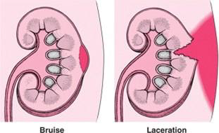
Understand bruises and lacerations related to kidney injuries
3. Condition: Urinary Tract Infection
Etiology:
Bacterial infection: The most common cause, often originating from bacteria in the bowel or skin.
Urinary stasis: Conditions that hinder normal flow of urine can encourage bacterial growth.
Catheterization: Indwelling catheters can introduce bacteria into the urinary system, leading to infection.
Signs and Symptoms:
Painful urination (dysuria): A common symptom experienced by patients.
Increased urgency and frequency: Patients may feel an overwhelming need to urinate.
Fever and chills: Indicating a systemic response to the infection.
Back pain: May occur if the infection ascends to the kidneys.
Cloudy or foul-smelling urine: A result of the bacterial presence.
Management
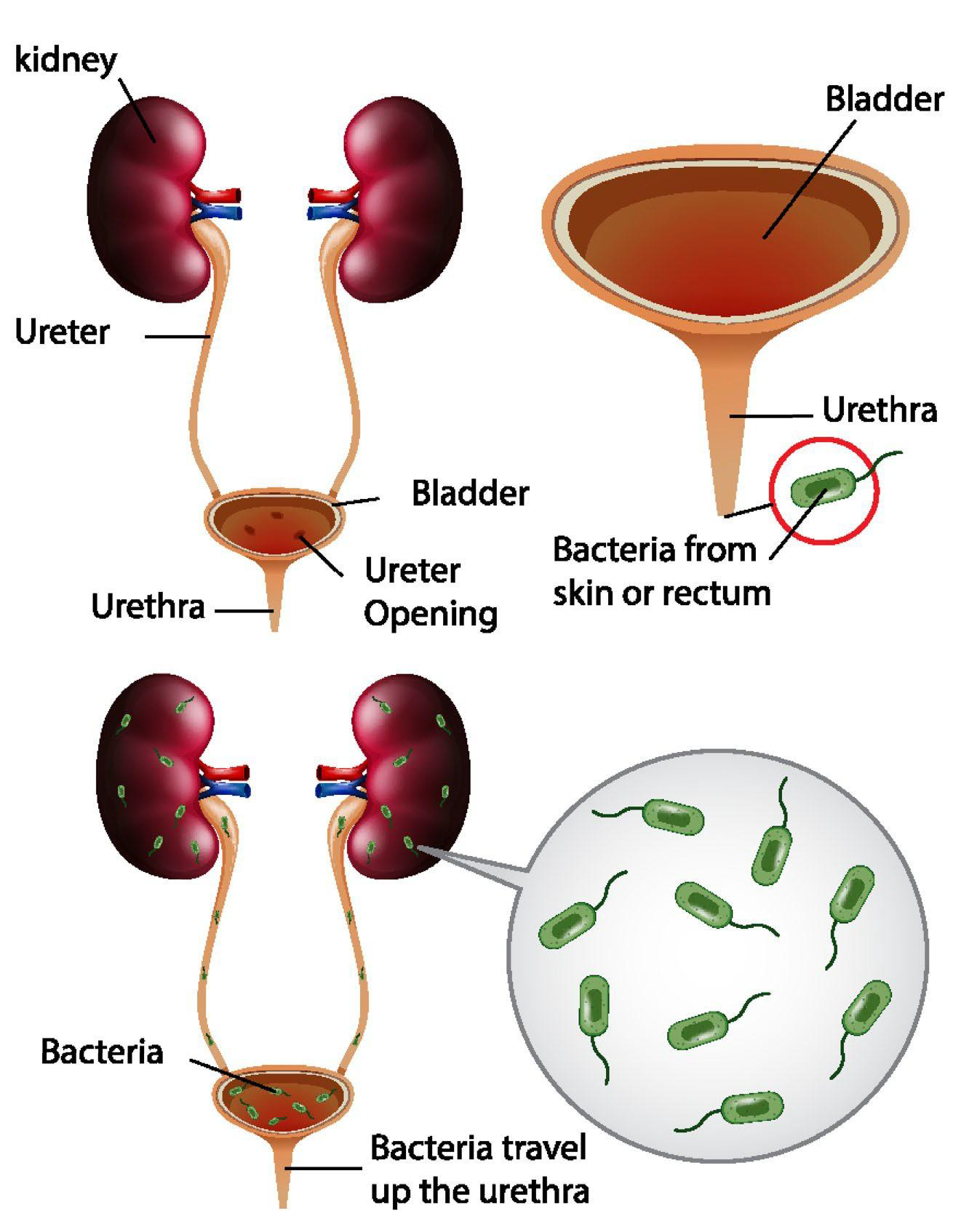
Pathway of bacteria from skin or rectum to the urethra
Involvement of kidney, ureter, bladder
Mechanism of Infection: Bacteria travel up the urethra to the bladder
4. Condition: Amenorrhea
Etiology:
Hormonal imbalances, such as those resulting from thyroid disorders or polycystic ovary syndrome (PCOS)
Stress and excessive exercise, which can disrupt normal menstrual cycles by affecting hormonal regulation.
Environmental factors, including nutritional deficiencies or excessive body weight changes, can also contribute to the onset of amenorrhea.
Signs and Symptoms
Irregular menstrual cycles or missed periods
Extreme weight loss or gain
Hot flashes or night sweats
Fatigue or weakness
Difficulty concentrating or mood swings
Hair loss or excessive hair growth (hirsutism)
Pelvic pain or discomfort
Changes in skin texture or appearance
Management:
Hormonal assessment to identify underlying endocrine disorders
Lifestyle modifications including diet and exercise
Medications such as hormonal therapies or anti-androgens
5. Condition: Indigestion (Dyspepsia)
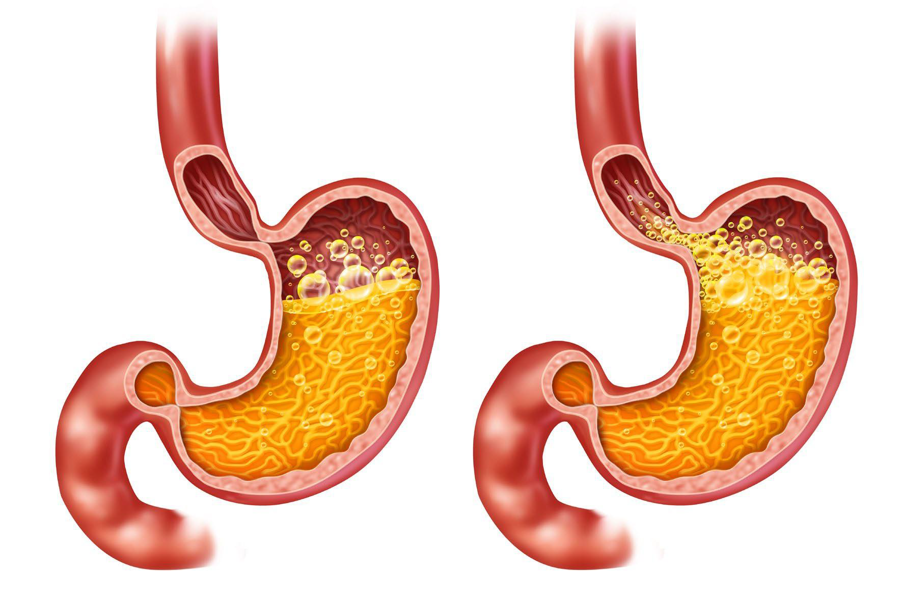
Etiology: Common causes of indigestion include overeating, consuming spicy or fatty foods, and stress, which can lead to gastric disturbances.
Signs and Symptoms: Indigestion manifests in various ways, including bloating, belching, nausea, and discomfort or pain in the upper abdomen. Other symptoms may include loss of appetite and a feeling of fullness after eating.
Management: Lifestyle modifications such as avoiding trigger foods, eating smaller meals, and managing stress through relaxation techniques are essential.
6. Condition: Appendicitis
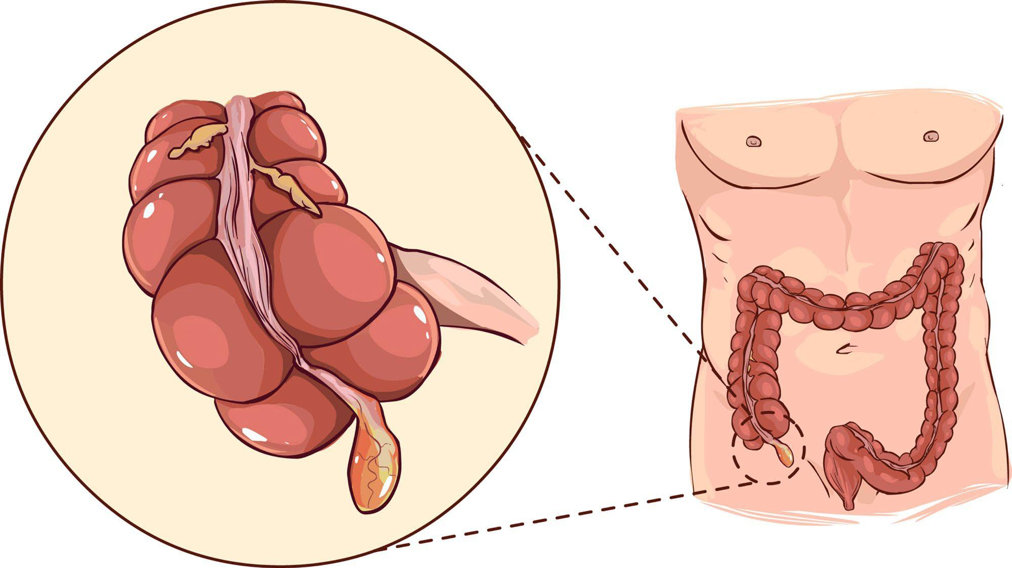
Etiology: Appendicitis typically arises from obstruction of the appendiceal lumen, which can result from fecaliths, lymphoid hyperplasia, or foreign bodies, leading to inflammation and infection of the appendix.
Signs and Symptoms: Common signs and symptoms of appendicitis include right lower quadrant abdominal pain, nausea, vomiting, loss of appetite, and fever. Additionally, patients may experience tenderness upon palpation of the abdomen and rebound tenderness, indicating irritation of the peritoneum.
Management:
Management of appendicitis typically involves surgical intervention, specifically an appendectomy.
Injuries Related to Abdomen
1. Scrotal Contusion
Etiology:
Scrotal contusion is usually caused by blunt trauma to the groin area, which can occur during contact sports, accidents, or falls.
Signs and Symptoms:
Localized pain and swelling in the scrotum
Bruising may appear
Discomfort during walking or sitting
Nausea or vomiting in severe cases
Management:
Assess the severity of the bruising and monitor for signs of internal injury.
Rest and Immobilization
Initiate IV fluid therapy to maintain hydration and electrolyte balance.
Consider pain management options to alleviate discomfort.
2. Abdominal Muscle Strain
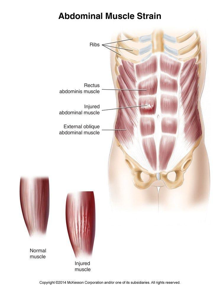
Normal Muscle Structures:
Abdominal muscle types involved include:
Rectus abdominis
External oblique abdominal muscle
Injured Muscles: Identifying which abdominal muscle is strained
Etiology:
The etiology of abdominal muscle strain can often be attributed to sudden twists, heavy lifting, or prolonged activities that require excessive force or repetitive motion, leading to muscle fatigue and damage.
Signs and Symptoms:
Pain or tenderness in the affected area, often worsening with movement or palpation
Swelling or bruising around the site of injury
Limited range of motion due to discomfort
Muscle spasms and stiffness in the abdominal region
Difficulty performing daily activities that require trunk movement.
Management:
Incorporating gentle stretching exercises to improve flexibility and reduce tension.
Utilizing heat therapy to alleviate muscle spasms and promote relaxation.
Consulting with a physical therapist for personalized rehabilitation programs that focus on enhancing trunk mobility. Implementing core strengthening activities to support abdominal and thoracic stability, thereby maintaining proper posture and reducing strain on surrounding muscles.
Abdomen Assessment
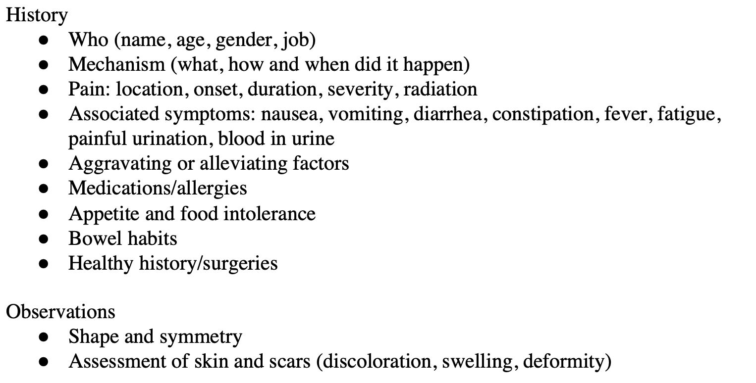
History Compilation
Who: Name, age, gender, job
Mechanism: What, how and when did it happen
Pain:
Location
Onset
Duration
Severity
Radiation
Associated symptoms:
Nausea
Vomiting
Diarrhea
Constipation
Fever
Fatigue
Painful urination
Blood in urine
Aggravating or Alleviating Factors
Medications/Allergies
Appetite & Food Intolerance
Bowel Habits
Health History/Surgeries
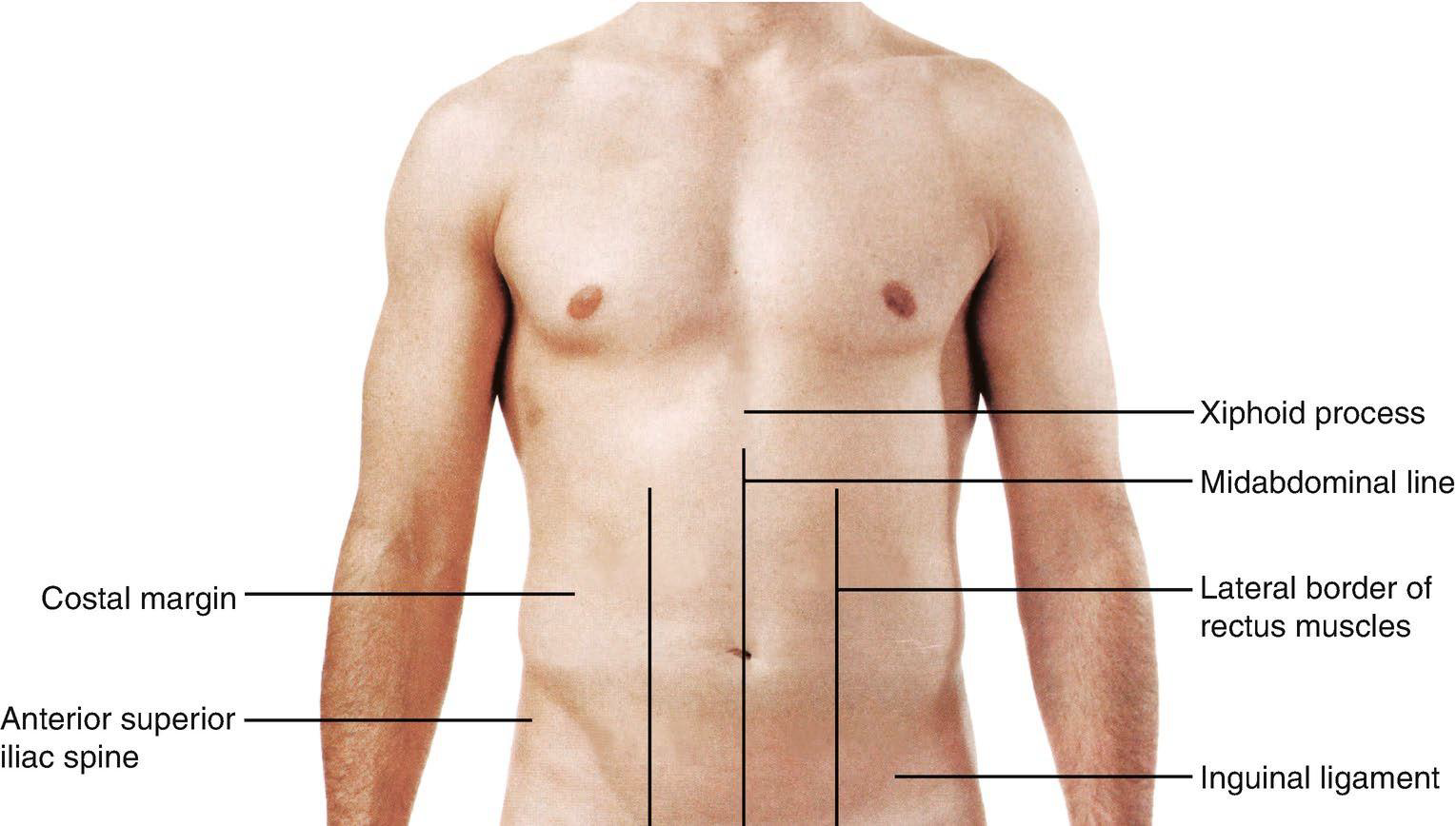
Observations During Assessment
Evaluate the following:
Costal margin
Xiphoid process
Midabdominal line
Lateral border of rectus muscles
Anterior superior iliac spine
Shape and symmetry
Skin Assessment:
Scars (discoloration, swelling, deformity)
Inguinal ligament
Abdomen Assessment Techniques
1. Auscultation

Listen to bowel sounds for:
Presence or absence
Frequency
Loudness
Quality
Duration
Use the 4 quadrants of the abdomen to auscultate specific organs
2. Percussion
Technique:
Place fingers on the abdomen and strike with the other hand
Sound Identifier:
Solid organ: dull sound
Hollow organ: tympanic sound
3. Palpation
Light Palpation: Assess tenderness across all 4 quadrants
Deeper Palpation: Identify masses in all 4 quadrants
Organs to Assess:
Liver
Spleen
Appendix
Kidneys
Pancreas
Stomach
Muscles to Assess:
Rectus abdominus
Obliques
Transverse abdominus
Four Quadrants of Palpation
Right Upper Quadrant:
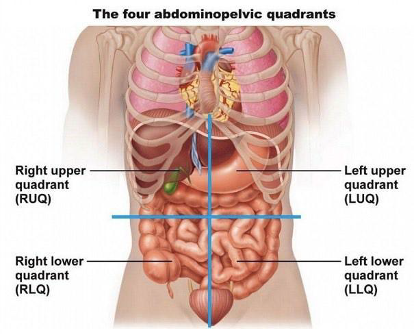
Liver
Gallbladder
Duodenum
Head of pancreas
Right adrenal gland
Portion of right kidney
Portions of ascending and transverse colon
Left Upper Quadrant:
Left lobe of liver
Spleen
Stomach
Body of pancreas
Left adrenal gland
Portion of left kidney
Portions of ascending and transverse colon
Right Lower Quadrant:
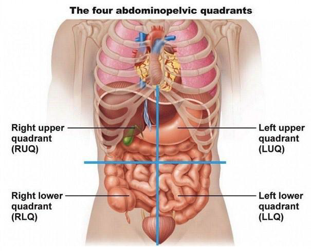
Lower pole of right kidney
Cecum and appendix
Portion of ascending colon
Bladder
Ovary
Uterus
Right spermatic cord
Right ureter
Left Lower Quadrant:
Lower pole of left kidney
Sigmoid colon
Portion of descending colon
Bladder
Ovary
Uterus
Left spermatic cord
Left ureter
Introduction to Athletic Therapy - Thorax
Thorax Anatomy
Bones Involved:
12 Thoracic Vertebrae
Rib Classifications:
Upper 7 ribs: True Ribs
Ribs 8-10: False Ribs
Ribs 11-12: Floating Ribs
Sternum Components
Parts:
Manubrium
Body
Xiphoid process
Muscles of the Thorax
Intercostal Muscles:
External intercostals (Function: inhalation)
Internal intercostals (Function: exhalation)
Diaphragm
Anatomy of the Lungs
Pathway: Pharynx → Larynx → Trachea → Bronchus → Bronchioles → Alveoli
Gas Exchange: Capillaries in the alveoli facilitate exchange of oxygen and nutrients in, and carbon dioxide and waste products out
Anatomy of the Heart
Chambers:
Left and Right Chambers
Blood Flow:
Deoxygenated Blood: Travels to the right atrium → right ventricle → lungs
Oxygenated Blood: Travels from lungs → left atrium → left ventricle → to the body
Blood Circulation
Pulmonary Arteries: Supply deoxygenated blood to lungs
Vena Cavae: Carry oxygen-poor, CO₂-rich blood to the right atrium
Pulmonary Veins: Bring oxygen-rich, CO₂-poor blood back to the left atrium
Systemic Circuit: Flow to and from all body tissues for gas exchange
Injuries Related to Thorax
1. Rib Contusion
Etiology:
This injury typically occurs due to direct trauma to the chest, such as a blow or impact to the rib cage, which can cause bruising of the rib muscles and surrounding tissues.
Signs and Symptoms:
Localized pain at the site of impact, which may worsen with movement or deep breaths
Swelling or bruising visible on the skin over the affected area
Tenderness when pressing on the affected ribs
Difficulty breathing, particularly taking deep breaths
Possible referred pain to the back or abdomen, depending on the location of the contusion.
Management
Rest and avoid activities that exacerbate the pain.
Application of ice packs to reduce swelling and alleviate discomfort.
Pain management with over-the-counter analgesics such as ibuprofen or acetaminophen.
In severe cases, consider evaluation for rib fractures or other complications.
2. Rib Fracture
Etiology:
Rib fractures are commonly caused by direct trauma, such as falls or sports-related injuries, but can also result from severe coughing or underlying conditions that weaken the bones.
Signs and Symptoms:
Localized pain at the site of injury, often worsening with movement or deep breaths
Swelling and bruising in the affected area
Difficulty breathing or shortness of breath (due to pain)
Crepitus (a crackling or popping sound) at the injury site
Potentially associated injuries to internal organs or vessels, depending on the location and severity of the fracture.
Management:
Administer oxygen as needed to mitigate hypoxia.
Stabilize the patient and prepare for potential transportation to a medical facility.
Order imaging studies to assess for internal injuries and confirm the extent of fractures.
Initiate fluid resuscitation if signs of hemorrhage are present.
3. Costochondral Separation
Etiology
Trauma to the chest wall, commonly from blunt force injuries such as falls or vehicle accidents.
Repeated stress or overuse in athletes, especially those involved in contact sports.
Broken rib directly causing a separation
Signs and Symptoms:
Localized pain at the site of injury, which may worsen with movement or palpation
Tenderness and swelling in the affected area
Possible difficulty breathing or deep breathing due to pain
Crepitus (a crackling sensation) may be felt upon examination
Bruising or discoloration of the skin over the site of injury.
Management:
Administer supplemental oxygen if necessary to improve oxygenation levels.
Monitor vital signs closely, focusing on respiratory rate and effort.
Provide pain management options to facilitate easier breathing, such as analgesics or positioning.
Consider further imaging studies, like a chest X-ray or CT scan, to assess for underlying injuries.
Sudden Death Syndrome in Athletes
1. Commotio Cordis
Etiology
Complex pathways involving cardiac rhythm analysis (QRS intervals, QT intervals)
2. Hypertrophic Cardiomyopathy
Etiology: Abnormal thickening of the ventricular wall
Important structural components include:
Right atrium
Arch of aorta
Left atrium
Left ventricle
Ventricular septum
3. Wolff-Parkinson-White Syndrome
Etiology:
This condition is caused by an abnormal electrical pathway in the heart, leading to episodes of rapid heartbeat due to reentrant tachycardia.
Description involves heart rhythm disruptions
4. Myocarditis
Etiology:
Myocarditis can result from various factors including viral infections, autoimmune diseases, and exposure to certain toxins or medications, which can lead to inflammation of the heart muscle and subsequent heart failure.
Inflammation of the heart muscle
Respiratory Tract Conditions
Conditions to understand include:
Asthma
Chronic Obstructive Pulmonary Disease (COPD)
Pneumonia
Upper respiratory infections like sinusitis, tonsillitis, pharyngitis, laryngitis, bronchitis
Thorax Assessment
Assessment Process:
Compiling history regarding:
Name, age, gender, job
Mechanisms of injury: what, how, and when
Pain characteristics: location, onset, duration, severity, radiation
Associated symptoms: difficulty breathing, coughing, shortness of breath, chest pain
Aggravating/Alleviating Factors
Medical History: Previous respiratory conditions (e.g., asthma, bronchitis, TB)
Observations:
Assess shape and symmetry
Skin condition (discoloration, swelling, deformity)
Thorax Assessment Techniques
1. Palpation
Comprehensive palpation of the entire chest wall:
Noting tenderness
Skin temperature
Moisture
Lumps or lesions
2. Special Tests
Rib Compression Test to identify potential rib injuries
3. Auscultation
Listen for respiratory and cardiovascular sounds:
Presence or absence, frequency, loudness, quality, duration
4. Percussion
Systematic tapping to compare sides and assess chest condition
Condition: Kidney Stones
Etiology:
Signs and Symptoms
Management:
Involves the Bladder, Ureter, and Kidney
Stones:
Stone Location:
Types of Stones:
2. Injuries: Kidney Contusion
Etiology
Signs and Symptoms
Management
3. Condition: Urinary Tract Infection
Etiology
Signs and Symptoms
Management
4. Condition: Amenorrhea
Etiology
Signs and Symptoms
Management
5. Condition: Indigestion (Dyspepsia)
Etiology
Signs and Symptoms
Management
6. Condition: Appendicitis
Etiology
Signs and Symptoms
Management
Injuries Related to Abdomen
1. Scrotal Contusion
Etiology:
direct blow to the scrotum, blunt force to the testicles
Signs and Symptoms
Management
2. Abdominal Muscle Strain
Normal Muscle Structures:
Abdominal muscle types involved include:
Rectus abdominis
External oblique abdominal muscle
Injured Muscles: Identifying which abdominal muscle is strained
Etiology:
The etiology of abdominal muscle strain can often be attributed to sudden twists, heavy lifting, or prolonged activities that require excessive force or repetitive motion, leading to muscle fatigue and damage.
Signs and Symptoms:
Pain or tenderness in the affected area, often worsening with movement or palpation
Swelling or bruising around the site of injury
Limited range of motion due to discomfort
Muscle spasms and stiffness in the abdominal region
Difficulty performing daily activities that require trunk movement.
Management:
Incorporating gentle stretching exercises to improve flexibility and reduce tension.
Utilizing heat therapy to alleviate muscle spasms and promote relaxation.
Consulting with a physical therapist for personalized rehabilitation programs that focus on enhancing trunk mobility. Implementing core strengthening activities to support abdominal and thoracic stability, thereby maintaining proper posture and reducing strain on surrounding muscles.