Lecture 12: Protein Sequencing, Analysis, and Interactions
Overview of Protein Analysis
Intracellular Complexity: There are approximately (10 billion) proteins inside a mammalian cell. Understanding their interactions is vital for mapping the molecular pathways of the cell.
Standard Analytical Techniques: Proteins are commonly purified and analyzed using:
Enzyme assays
Chromatography
SDS-PAGE
Western blotting
ELISA
Co-immunoprecipitation (Co-IP)
Specialized Techniques: Less frequent but valuable techniques include:
Protein sequencing
Pull-down assays
Calorimetry (ITC and DSC)
Yeast two-hybrid (Y2H) systems
Solid-Phase Peptide Synthesis (SPPS)
Purpose: Peptides can be chemically generated for use in interaction studies using solid-phase synthesis.
The Process:
Amino Acid 1: The first amino acid has its -amino group (left side) protected by a Fmoc group. Its carboxyl-terminal (right side) is attached to an insoluble polystyrene bead (resin) via a reactive group.
Deprotection: The Fmoc protecting group is removed. This results in a free -amino group
Activation and Coupling: The second amino acid (also Fmoc-protected) is activated at its carboxyl group using Dicyclohexylcarbodiimide (DCC). The free -amino group of the first amino acid attacks the activated carboxyl group of the second, forming a peptide bond.
Repetition: These reactions are repeated until the desired sequence is achieved.
Release: The completed peptide is deprotected one final time. Trifluoroacetic acid (TFA) is then used to cleave the ester linkage between the peptide and the resin, releasing the finished peptide.

Stepwise Yield Impact: The overall yield of a peptide synthesis drops significantly as the number of residues increases if the yield of each step is not very high.
Number of residues | Overall yield at per step | Overall yield at per step |
|---|---|---|
11 | ||
21 | ||
51 | ||
100 |
Protein Sequencing: Edman Degradation and Mass Spectrometry
N-terminal Sequencing (Edman Degradation):
History: In 1955, Frederick Sanger sequenced insulin for the first time using fluorodinitrobenzene.
Modern sequencing uses the method developed by Pehr Edman in the 1950s.
Reagent: Uses phenyl isothiocyanate.
Mechanism: Successive rounds of N-terminal modification, cleavage, and identification. phenyl isothiocyanate reacts with N-terminus and pulls it off and then you extract with an organic solvent and identify via electrophoresis or mass spectrometry
Applications: Can be used on bands selected from a gel or blot. However, samples must be extremely pure, as even skin keratin can contaminate the results.
Limitations: There is a limit of amino acids. It is generally not used for whole proteins because DNA sequencing is faster and less expensive. However, most proteins can be identified with sequences of less than 10 amino acids.

Mass Spectrometry (MS) Sequencing:
Methodology: Developed in the 1980s, this involves digesting the protein with specific proteases (e.g., trypsin, chymotrypsin, or pepsin). The resulting fragments are analyzed and the process is repeated with different enzymes to rebuild the protein sequence through overlap.
Ionization Techniques: MALDI-TOF (Matrix-Assisted Laser Desorption/Ionization Time-of-Flight), electrospray ionization, and tandem MS
Capabilities:
Determines the mass of peptides, which are compared to databases for sequence identification.
Identifies post-translational modifications (PTMs).
Maps protein interactions via crosslinking.
High sensitivity compared to Edman degradation.
Mechanism: taking pure protein, digesting, running on machine, getting separation peaks (mass to charge ratios)
Protease Digestion and Structural Analysis
Functional Analysis: Proteases are used to digest proteins into domains to analyze structural components, protein fingerprinting, conformational changes, and binding sites.
Protein Fingerprinting (Historical Case): In 1954, Vernon Ingram cleaved hemoglobin with trypsin (which cuts at Lys and Arg) to compare wild-type (WT) hemoglobin and sickle cell hemoglobin, identifying the first molecular basis for a genetic disease.
Domain Analysis Example (Vinculin): Protease digestion (e.g., using papain) is used to identify the domains of Vinculin
Mapping Drug Binding Sites (Anifrolumab): Anifrolumab is a monoclonal antibody binding to interferon- receptor 1 (IFNAR1). Proteolytic cleavage of INFAR1 with endoproteinase LysC shows anifrolumab’s binding sites
Protein-Protein Interactions
Similar to co-IP (antibody-protein interactions), but a tagged bait protein and an affinity column or beads are used instead of an antibody to detect protein interactions
Bait (known protein) and prey (targets)

Tandem Affinity Assay
A type of pull-down used to find novel protein interactions, uses two affinity steps in tandem to reduce false positives

Far Western Blot
A far-western blot is similar to a western blot, but a labeled protein probe (“bait”) is used instead of an antibody, and it is usually performed under native conditions (although sometimes SDS) to identify protein-protein interactions
The protein probe can be radiolabeled or tagged, or antibodies to it can be used

Mass Spectrometry
Mass spectrometry can determine protein interactions via crosslinking
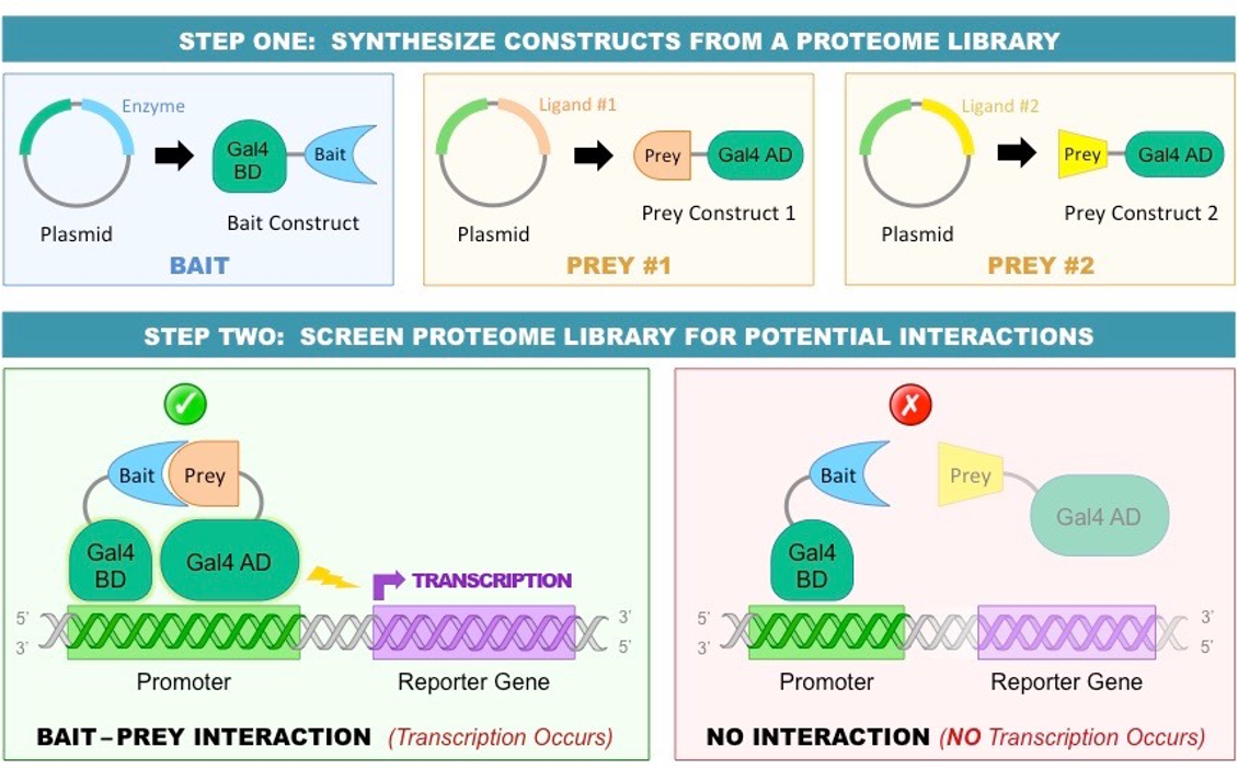
Yeast Two-Hybrid
Yeast two-hybrid allows for analysis of existing protein-protein interactions, and it can detect new interactions in yeast (also bacterial two-hybrid in bacteria)
If the bait and prey interact, then a reporter gene (lacZ) is transcriptionally activated color change
It can be used to screen a bait against a large cDNA prey library to study thousands of interactions at once

Yeast Gal4 is a transcription factor that expresses genes to breakdown galactose
DNA Binding domain: binds to the Gal1 promoter
Activating domain: binds to MED15 of the mediator complex, which activates RNA polymerase and transcription
A bait protein can be tagged with the DNA binding domain, and screened against a cDNA library of proteins tagged with the activation domain

The bait/prey proteins bring the two domains close together, activating RNA polymerase, causing color change
Advantage of this is that you can scan prey libraries using different bait/activating domain combinations
Bacterial/Yeast Two Hybrid can also be used to study protein Interfaces
Random mutations not at interface A/B interact Color change
Random mutations at interface Mutations prevent interaction No color change
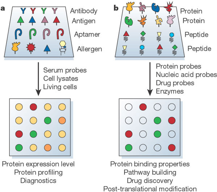
Protein Microarray (Gene Chip)
Protein microarrays can be used to study protein-protein interactions, transcription factor screening, drug, tumor analysis
Can be analytical or functional (proteins and peptides)
Proteins/peptides attached to chip and wash it through and see if it sticks
Method:
Tag protein and attach to chip (glass tray)
Add drug and wash across proteins
Add streptavidin that is fluorescent conjugate and shows where protein present
Calorimetry
Calorimetry measures heat change when two proteins interact
Isothermal Titration Calorimetry (ITC): measures the heat change when injecting a peptide or protein into a solution containing a binding partner
Differential Scanning Calorimetry (DSC): changes the temperature and measures resulting changes on a protein’s folding and structure; can be used to compare a protein vs a mutant
Can use calorimetry to determine enthalpy, entropy, free energy, dissociation constant, affinity, and molar ratios using ΔG = -RT ln Ka = ΔH – TΔS
Exothermic; ΔH < 0, releases heat
Endothermic; ΔH > 0, absorbs heat
Surface Plasmon Resonance (SPR)
Immobilize protein on membrane, run a binding partner such as a protein or peptide across it and measure association using reflected lightoli
Wash with buffer, measure rate of dissociation to determine dissociation constant (i.e. ‘tightness’ of binding)
Protein-DNA Interactions
Southwestern Blot:
Combines the principles of Southern and Western blotting (combines DNA and Protein interactions)
Proteins are separated by SDS-PAGE, SDS is removed, and the proteins are refolded (using urea).
The blot is probed with labeled, digested DNA to identify DNA-binding proteins like transcription factors or promoter-binding proteins.
Like a “western blot” which is Protein-Antibody interaction but the antibody is now DNA-binding protein
Electrophoretic Mobility Shift Assay (EMSA):
Also known as a "gel shift" assay.
Labeled DNA/RNA is mixed with protein; if they bind, the resulting complex is larger and moves more slowly through a gel than free DNA/RNA, creating a visible "shift."
DNA Footprinting:
DNA footprinting can identify the exact DNA sequence to which a protein binds
DNA and protein are mixed and treated with a restriction enzyme that can only digest unbound DNA
The protein protects its target DNA from digestion, which appears as a “footprint” on the gel where the restriction enzyme cannot cut
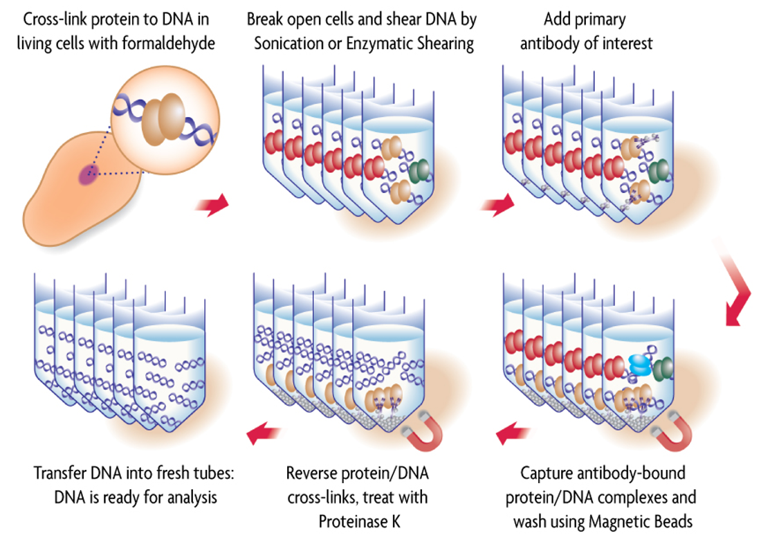
Chromatin Immunoprecipitation (ChIP):
Detects DNA-protein interactions in vivo.
Proteins are crosslinked to DNA in the cell, and antibodies (often on magnetic beads) are used to pull down the protein of interest along with its bound DNA fragments.
Reverse the crosslinking and then the DNA is then analyzed via PCR or sequencing.