Pelvis
Identify the following bones, bony landmarks, joints, and ligaments of the pelvis:
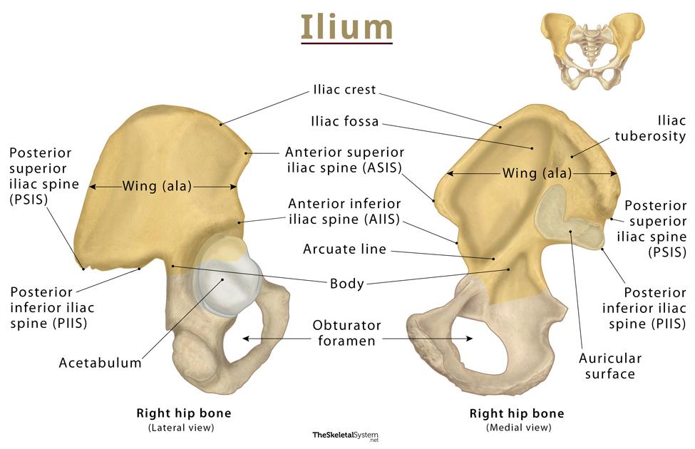
Ilium
-

Anterior Superior Iliac Spine (ASIS):
○ prominent projection at the anterior
end of the iliac crest.
■ Identifying the inguinal ligament,
which attaches here.
■ Locating the McBurney’s point,
used in diagnosing appendicitis.
■ Measuring leg length or aligning
surgical incisions.
● Anterior Inferior Iliac Spine (AIIS):
○ Located just below the ASIS
○ Site for muscle attachment (rectus
muscle - part of the
quadriceps) and the
ligament, one of the strongest
ligaments in the body.
—ASIS-
Palpable hip landmark
Attachment of the inguinal ligament
Used to find McBurney's point
Used to measure leg length
AIIS
Located just inferior to the ASIS
Origin of the rectus femoris
Attachment of the iliofemoral ligament
Important for hip stability and hip flexion
Iliac Crest:
○ the superior border that supplies
attachment for several
abdominal muscles and the
latissimus dorsi
● Posterior Superior Iliac Spine (PSIS):
○ posterior end of the iliac crest
○ Helpful in Identifying the
sacroiliac joint.
● Posterior Inferior Iliac Spine (PIIS):
○ Attachment site for the
sacrospinous ligament, which
helps stabilize the sacroiliac
joint.
Iliac Crest = Top ridge; attachment for abdominal muscles and latissimus dorsi; landmark for L4.
PSIS = Back upper bump; helps locate the sacroiliac joint.
PIIS = Back lower bump; attachment for a stabilizing pelvic ligament.
Ischium-The ischium is the lower back part of the hip bone.
It's the "sit bone" because it bears your weight when you're sitting.

Ischial tuberosity
○ Serves as the attachment site for several
important structures, including
hamstrings and sacrospinous ligaments
■ This is where the Hamstrings attach.
○ Supports body weight when sitting.
● Ischial Spine:
○ Sharp, bony projection located superior
to the ischial tuberosity.
○ Attachment of sacrospinous ligament
■ Helps convert the greater sciatic
notch into the greater sciatic
foramen
Ischial tuberosity
"Sit bone"
Supports body weight while sitting
Origin of the hamstrings
Common site of hamstring avulsion and ischial bursitis
Ischial spine
Sharp projection superior to the tuberosity
Attachment for the sacrospinous ligament
Helps form the greater sciatic foramen
Important landmark for assessing fetal station during labor
Pubic-

Pubic Crest:
○ Thickened anterior border of the pubic
bone.
○ Attachment site for the rectus abdominis
muscle and the inguinal ligament.
● Pubic Tubercle:
○ A small, palpable projection located
lateral to the pubic symphysis.
● Pectineal Line:
○ A sharp ridge on the superior ramus of
the pubis.
○ Attachment site for the pectineus muscle
and contributes to the pelvic brim.
Pubic Crest
Thickened anterior border of the pubis
Attachment for the rectus abdominis
Near the attachment of the inguinal ligament
Pubic Tubercle
Small palpable bump lateral to the pubic symphysis
Medial attachment of the inguinal ligament
Landmark for inguinal hernias
Pectineal Line
Ridge on the superior pubic ramus
Attachment for the pectineus muscle
Forms part of the pelvic brim, dividing the false and true pelvis
Sacrum / Coccyx


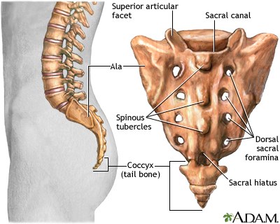
Sacrum:
○ Triangular bone formed by the fusion of five sacral
vertebrae.
○ Sacral Promontory:
■ Landmark in obstetrics for measuring pelvic
dimensions.
○ Sacral Canal: Houses the cauda equina
■ Used for epidural anesthesia.
○ Sacral Foramina: Allow passage of the sacral
nerves.
○ Median Sacral Crest: Provides attachment for
ligaments and muscles.
● Coccyx:
○ A small, triangular bone formed by the fusion of
three to five rudimentary vertebrae.
○ It provides attachment for the levator ani .
Sacrum
Five fused vertebrae (S1–S5)
Connects the spine to the pelvis and transmits body weight
Sacral Promontory
Landmark used in obstetrics to assess pelvic size
Sacral Canal
Contains the cauda equina
Site for caudal epidural anesthesia
Sacral Foramina
Openings for the sacral spinal nerves
Median Sacral Crest
Posterior ridge for muscle and ligament attachment
Coccyx
Fused tailbone (3–5 vertebrae)
Attachment for the levator ani, an important pelvic floor muscle
Can become painful after trauma (coccydynia)
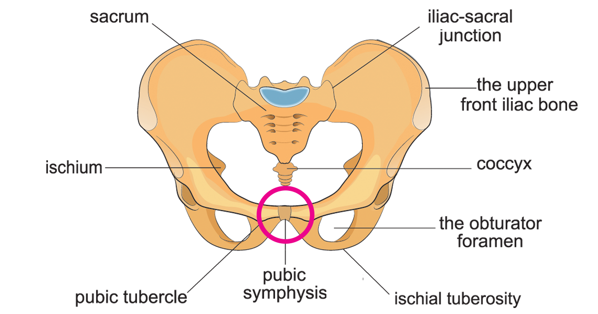
Pubic Symphysis:
The pubic symphysis is a cartilaginous joint that connects the left and right pubic bones.

A cartilaginous joint that connects
the two pubic bones
● Provides stability to the pelvic ring
● Allows slight movement during
activities like walking or childbirth.
Acetabulum: The acetabulum is a deep, cup-shaped socket on the outside (lateral side) of the pelvis.
Its job is to hold the head of the femur, forming the hip joint.

Deep, cup-shaped socket on the outer
pelvis that holds the head of the femur
(thighbone) to form the hip joint
● Formed by the fusion of all three pelvic
bones
● Crucial for weight-bearing and stability
Sacrospinous ligament:

Runs from the ischial spine to the lateral
sacrum.
● It converts the greater sciatic notch into
the greater sciatic foramen
○ This is the passage of the sciatic
nerve, pudendal nerve, and
piriformis muscle.
● It also opposes nutation (forward
rotation of the sacrum) during weight-
bearing and gait.
Feature | Description |
|---|---|
Attachment | Ischial spine → lateral sacrum |
Main function | Converts the greater sciatic notch into the greater sciatic foramen |
Structures passing through | Sciatic nerve, pudendal nerve, piriformis muscle (among others) |
Additional role | Stabilizes the pelvis and resists excessive sacral nutation during standing and walking |
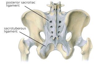
Sacrotuberous ligament:
The sacrotuberous ligament is a strong ligament that connects the:
Sacrum → Ischial tuberosity ("sit bone")

This ligament runs from the sacrum to
the ischial tuberosity.
● It converts the lesser sciatic notch into
the lesser sciatic foramen
○ which allows passage of the
internal pudendal nerve, artery,
and vein, as well as the tendon of
the obturator internus muscle.
○It resists nutation and prevents
excessive movement of the
sacrum.
Sacrospinous | Sacrotuberous |
|---|---|
Goes to ischial spine | Goes to ischial tuberosity (sit bone) |
Makes greater sciatic foramen | Makes lesser sciatic foramen |
Higher | Lower |
Identify the major foramen of the pelvic cavity and their contents and accurately categorize the neurovascular and muscular structures that traverse each opening
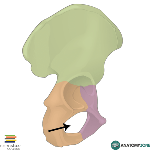
Obturator foramen:

Covered by the obturator
membrane
● Allows passage of the
obturator nerve, artery,
and vein.
Only three important structures travel through the obturator canal:
Obturator nerve → supplies the medial (inner thigh) muscles and sensation to part of the inner thigh.
Obturator artery → brings blood to the medial thigh.
Obturator vein → drains blood from the medial thigh.
Greater Sciatic notch: The greater sciatic foramen is a large opening in the back of the pelvis. It is the main exit from the pelvis to the buttock and lower limb.

Lesser sciatic notch
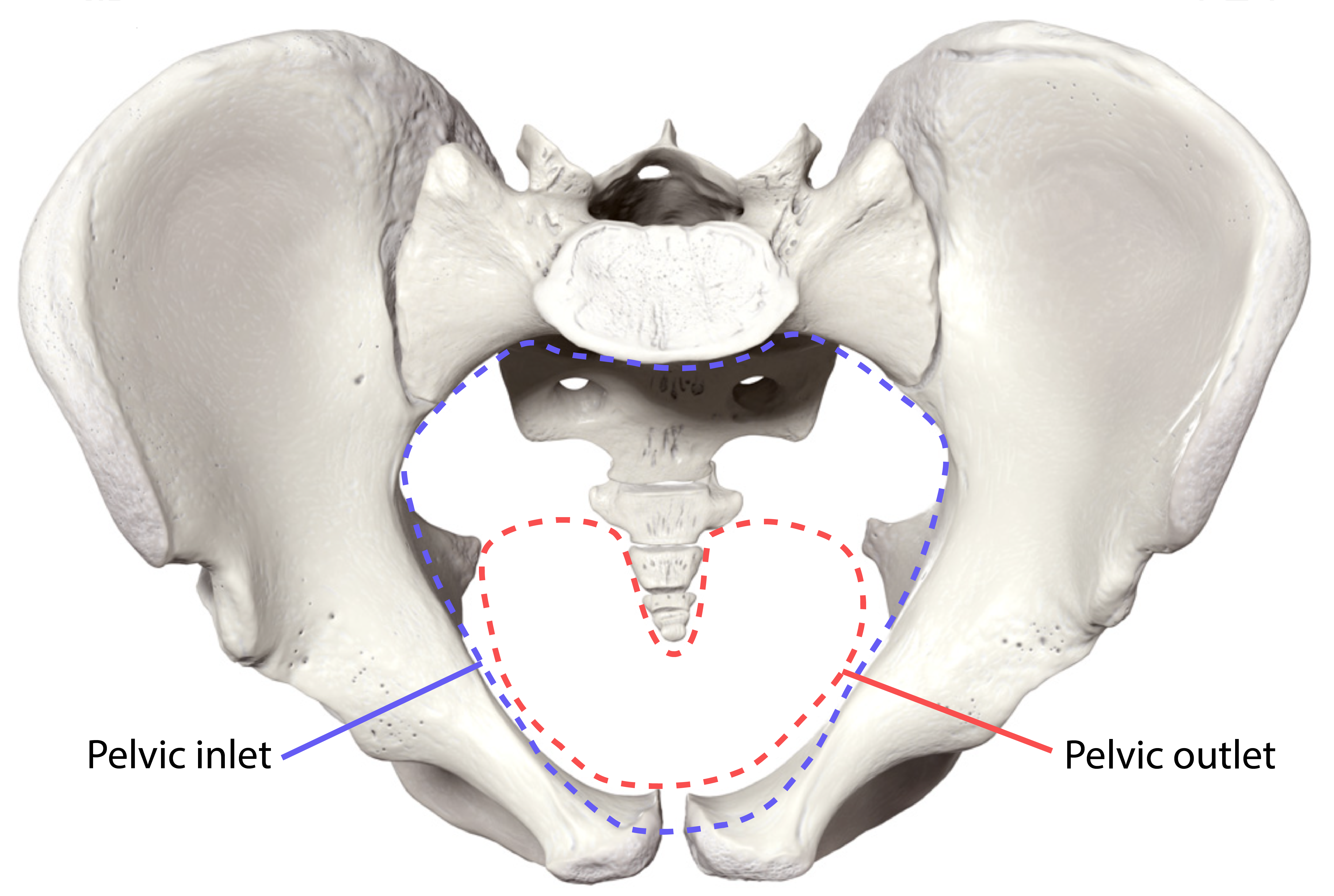
Pelvic inlet and outlet
Identify the contents of the pelvic cavity
Define the spaces of the urogenital triangle and describe the contents within the superficial and deep perineal pouch
Identify the following pelvic muscles, innervation and the corresponding actions
Muscles of the pelvic walls
Obturator internus muscles
Piriformis muscles
Muscles of the pelvic diaphragm
Levator Ani
Coccygeus muscles
Peroneal muscles
Bulbospongiosus
Ischiocavernosus
Identify the following pelvic vasculature
Anterior trunk of the internal iliac
Obliterated umbilical artery
Obturator artery
Uterine artery
Inferior vesical artery
Vaginal artery (not in males)
Middle rectal artery
Internal pudendal artery
Penile artery
Inferior gluteal artery
Posterior trunk of the internal iliac
Iliolumbar artery
Lateral sacral artery
Superior gluteal artery
Lymphatic nodes
Superficial inguinal nodes
Deep inguinal nodes
Identify the location and innervations of the pelvis
Sacral plexus
Superior and inferior gluteal nerve
Pudendal nerve
Sciatic nerve
Autonomic innervation
Sacral sympathetic trunk
Pelvic Splanchnic nerves
Hypogastric plexus
Identify the location, anatomical features, subdivisions, and innervations of the following structures:
Genitourinary System
Bladder
Urethra: differences between male and female anatomy, urethral sphincters
Gastrointestinal System
Rectum
Anus / Anal Canal: Pectinate line, Anal sphincter
Male Genitalia
Penis
Glans penis,
Body: Corpus Spongiosum, Corpora cavernosa
Root: Bulb of the penis, Crura of the penis
Scrotum
Testes: Seminiferous tubules
Epididymis
Dartos and cremaster muscle
Ductus (Vas) deferens
Ejaculatory ducts
Seminal vesicles
Prostate gland
Bulbourethral glands
Female Genetalia
Ovaries
Uterus
Uterine (Fallopian) Tubes
Cervix
Vagina
External genitalia:
Mons pubis
Labia majora and minora
Clitoris
Vestibule
Greater vestibular (Bartholin) gland
Ligaments: Broad, Round, Ovarian, Suspensory, Cardinal ligaments
Identify the following bones, bony landmarks, joints, and ligaments of the pelvis:
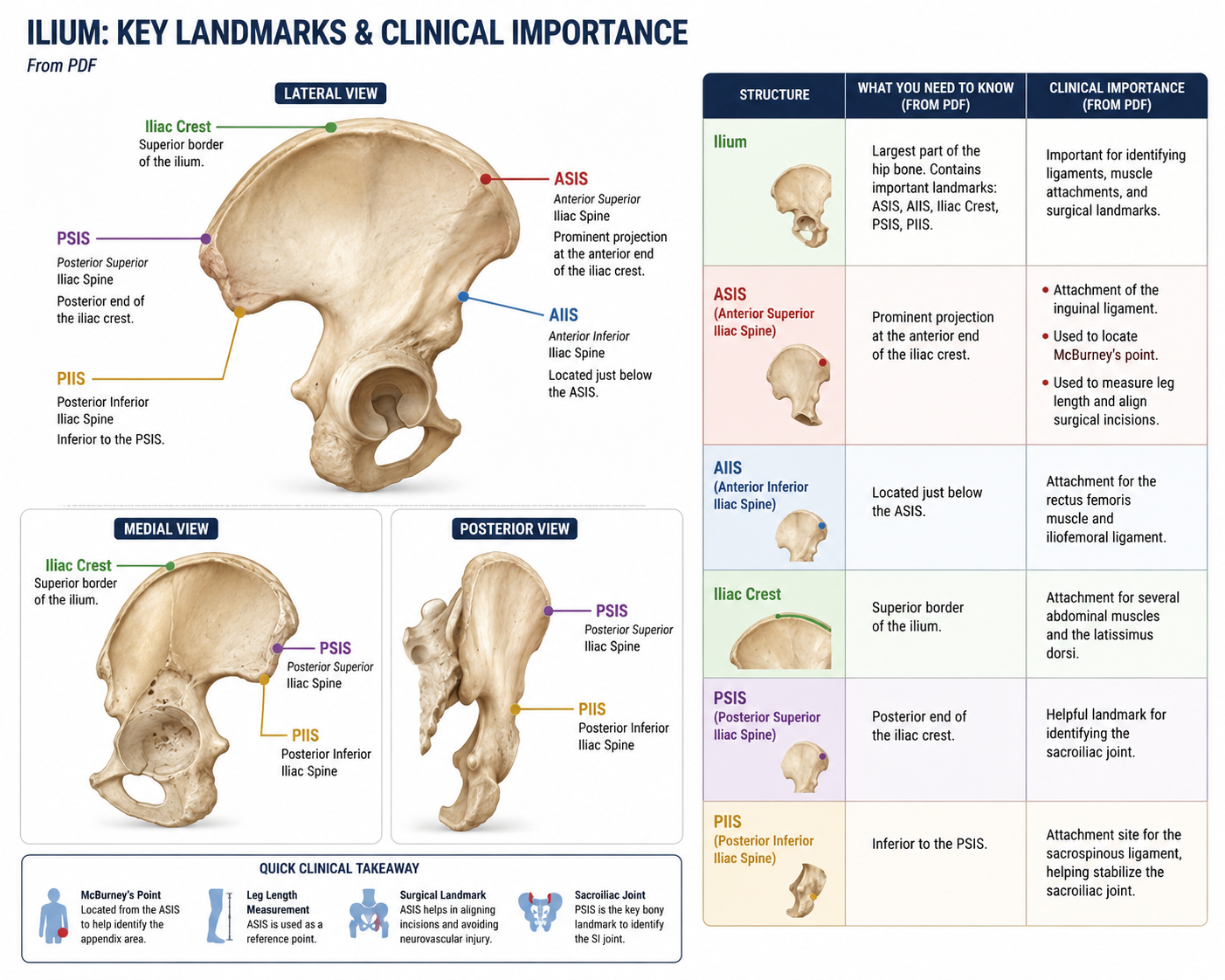
Ilium
Structure | What You Need to Know | Clinical Importance |
|---|---|---|
Ilium | Largest part of the hip bone. Contains important landmarks: ASIS, AIIS, Iliac Crest, PSIS, PIIS. | Important for identifying ligaments, muscle attachments, and surgical landmarks. |
ASIS (Anterior Superior Iliac Spine) | Prominent projection at the anterior end of the iliac crest. | • Attachment of the inguinal ligament. 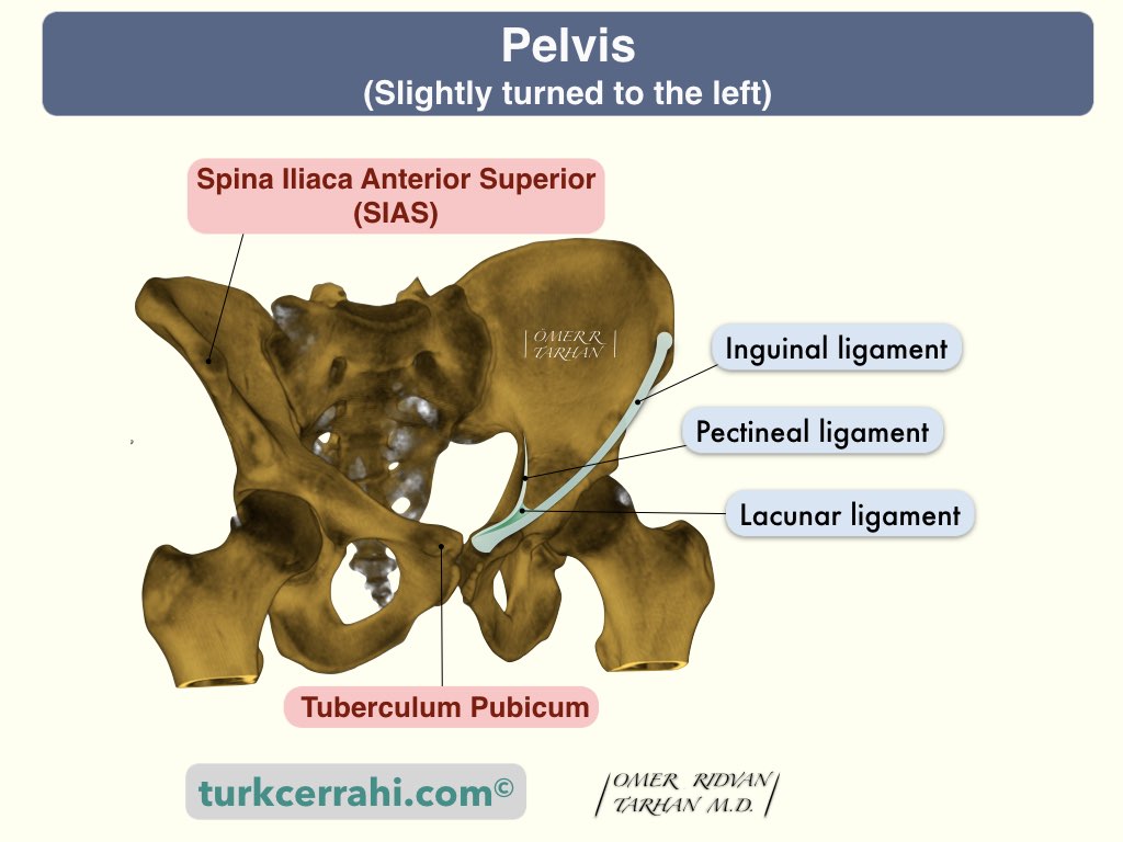 • Used to locate McBurney's point.  • Used to measure leg length and align surgical incisions. |
AIIS (Anterior Inferior Iliac Spine) | Located just below the ASIS. | Attachment for the rectus femoris muscle  and iliofemoral ligament.  |
Iliac Crest | Superior border of the ilium. | Attachment for several abdominal muscles and the latissimus dorsi.  |
PSIS (Posterior Superior Iliac Spine) | Posterior end of the iliac crest. | Helpful landmark for identifying the sacroiliac joint. :background_color(FFFFFF):format(jpeg)/images/library/13208/ZzTBXyAOEp5TcstuOZbGqg_Sacroiliac_Joint_01.png) |
PIIS (Posterior Inferior Iliac Spine) | Inferior to the PSIS. | Attachment site for the sacrospinous ligament, helping stabilize the sacroiliac joint. 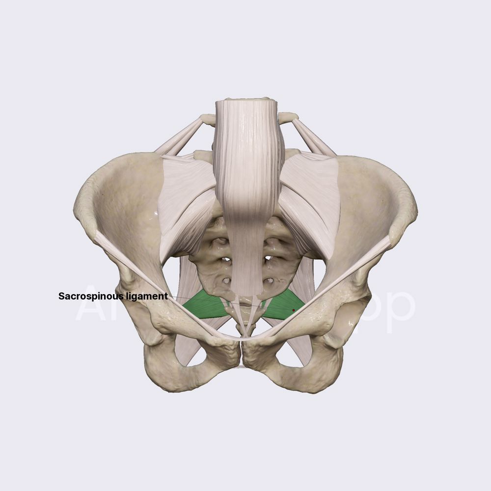 |
B. Ischium:
Structure | Description / Identification | Clinical Importance |
|---|---|---|
Ischium 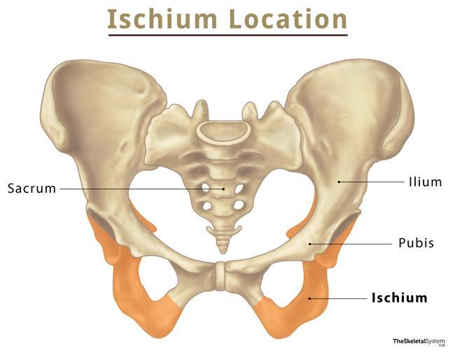 | Inferior and posterior portion of the hip bone. | Contains the ischial spine and ischial tuberosity. |
Ischial Tuberosity 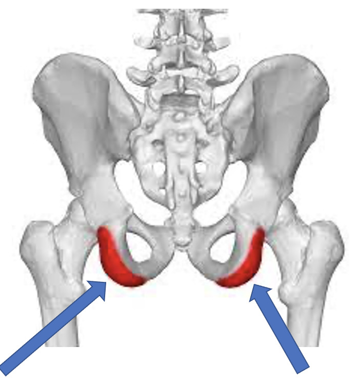 | Attachment site for the hamstrings and sacrospinous ligament. Supports body weight while sitting. | Common site for ischial bursitis and hamstring tendon injuries. |
Ischial Spine :background_color(FFFFFF):format(jpeg)/images/article/ischial-spine/mOxQpiZVpA5GtbVCNoCPQ_MUY7m3J7l62a95Nc8pmXCQ_Ischial_spine.png) | Sharp projection superior to the ischial tuberosity. Attachment of the sacrospinous ligament. Converts the greater sciatic notch into the greater sciatic foramen. | Landmark used in obstetrics to assess labor because it marks the narrowest part of the pelvic outlet. |

PUBIS
:watermark(/images/watermark_only_413.png,0,0,0):watermark(/logos/logo_url_sm.png,-10,-10,0):format(jpeg)/images/learnable/pubic-bone-6/QLmh9Ml2AJ9z3v3JE18w_480Pubic_bone.png)
Structure | Description / Identification |
|---|---|
Pubis | Anterior portion of the hip bone. |
Pubic Crest  | Thickened anterior border of the pubic bone. Attachment for the rectus abdominis and inguinal ligament. |
Pubic Tubercle | Small palpable projection lateral to the pubic symphysis. |
Pectineal Line | Sharp ridge on the superior pubic ramus. Attachment for the pectineus muscle and contributes to the pelvic brim. |
Sacrum / Coccyx
Structure | Description / Identification |
|---|---|
Sacrum | Triangular bone formed by fusion of five sacral vertebrae. |
Sacral Promontory | Landmark used for measuring pelvic dimensions in obstetrics. |
Sacral Canal | Houses the cauda equina; used for epidural anesthesia. |
Sacral Foramina | Allow passage of the sacral nerves. |
Median Sacral Crest | Attachment site for ligaments and muscles. |
Pubic Symphysis
Structure | Description / Identification | Clinical Importance |
|---|---|---|
Pubic Symphysis | Cartilaginous joint connecting the two pubic bones. Provides stability to the pelvic ring and allows slight movement during walking and childbirth. | Pregnancy hormones increase flexibility, which may lead to Symphysis Pubis Dysfunction (SPD) causing pain and instability. |
Acetabulum

Structure | Description / Identification |
|---|---|
Acetabulum | Deep, cup-shaped socket on the outer pelvis that holds the head of the femur to form the hip joint. Formed by the fusion of the ilium, ischium, and pubis. Crucial for weight-bearing and stability. |
Sacrotuberous Ligament
Feature | Description |
|---|---|
Attachments | Runs from the sacrum to the ischial tuberosity. |
Function | Converts the lesser sciatic notch into the lesser sciatic foramen. |
Structures Passing Through | • Internal pudendal nerve • Internal pudendal artery and vein • Tendon of the obturator internus muscle |
Additional Function | Resists nutation and prevents excessive movement of the sacrum. |
Sacrospinous Ligament
Feature | Description |
|---|---|
Attachments | Runs from the ischial spine to the lateral sacrum. |
Function | Converts the greater sciatic notch into the greater sciatic foramen. |
Structures Passing Through | • Sciatic nerve •Pudendal nerve • Piriformis muscle |
Additional Function | Opposes nutation (forward rotation of the sacrum) during weight-bearing and gait. |
High Yield
Objective | Know These Structures |
|---|---|
a. Ilium | ASIS, AIIS, Iliac Crest, PSIS, PIIS |
b. Ischium | Ischial Tuberosity, Ischial Spine |
c. Pubis | Pubic Crest, Pubic Tubercle, Pectineal Line |
d. Sacrum/Coccyx | Sacral Promontory, Sacral Canal, Sacral Foramina, Median Sacral Crest, Coccyx |
e. Pubic Symphysis | Cartilaginous joint connecting both pubic bones |
f. Acetabulum | Hip socket formed by all three pelvic bones |
g. Sacrotuberous Ligament | Sacrum → Ischial Tuberosity → Forms Lesser Sciatic Foramen |
h. Sacrospinous Ligament | Ischial Spine → Sacrum → Forms Greater Sciatic Foramen |
Identify the Major Foramen of the Pelvic Cavity and Their Contents
Obturator Foramen:
Feature | Information |
|---|---|
Location/Description | Large opening in the pelvic bone that is covered by the obturator membrane. |
Neurovascular Structures | • Obturator nerve • Obturator artery • Obturator vein |
Muscular Structures |
Greater Sciatic Notch (Greater Sciatic Foramen)

Category | Information |
|---|---|
Formation | Formed by the sacrotuberous ligament, sacrococcygeal ligaments, ilium, and sacrum/coccyx. |
Mnemonic (PDF) | "I Prefer Queens of Siam" |
Neurovascular Structures | • Inferior gluteal nerve • Pudendal nerve • Sciatic nerve • Posterior femoral cutaneous nerve |
Muscular Structures | • Piriformis muscle • Nerve to quadratus femoris • Nerve to obturator internus |
Additional Information | Shape and size of the greater sciatic notch vary between individuals and may be used in forensic anthropology to estimate sex. |
Lesser Sciatic Notch (Lesser Sciatic Foramen)

Category | Information |
|---|---|
Formation | Formed by the sacrotuberous ligament, sacrococcygeal ligaments, ilium, and sacrum/coccyx. |
Mnemonic (PDF) | "POO" |
Neurovascular Structures | • Internal pudendal nerve • Internal pudendal artery • Internal pudendal vein |
Muscular Structures | • Obturator internus tendon• Nerve to obturator internus |
Pelvic Inlet
Category | Information |
|---|---|
Also Called | Pelvic brim / Superior pelvic aperture |
Description | Superior opening of the pelvic cavity. |
Formed By | • Sacral promontory• Arcuate line of the ilium• Pectineal line of the pubis• Pubic symphysis• Pubic crest |
Neurovascular Structures Passing Through | Not listed in the PDF |
Muscular Structures Passing Through | Not listed in the PDF |
Pelvic Outlet
Category | Information |
|---|---|
Also Called | Inferior pelvic aperture |
Description | Inferior opening of the pelvic cavity. |
Formed By | • Coccyx• Ischial tuberosities• Pubic symphysis |
Neurovascular Structures Passing Through | Not listed in the PDF |
Muscular Structures Passing Through | Not listed in the PDF |
Contents of the Pelvic Cavity
Structure | Description / Function (from PDF) |
|---|---|
Urinary Bladder | Organ of the urinary system located within the pelvic cavity. |
Ureters | Tubes that enter the pelvic cavity and connect to the urinary bladder. |
Pelvic Genital Organs | Reproductive organs located within the pelvic cavity. |
Rectum | Terminal portion of the gastrointestinal tract located within the pelvic cavity. |
Blood Vessels | Pelvic arteries and veins supplying the pelvic organs. |
Lymphatics | Lymphatic vessels located throughout the pelvic cavity. |
Nerves | Pelvic nerves supplying the pelvic organs and surrounding structures. |
Define the spaces of the urogenital triangle and describe the contents within the superficial and deep perineal pouch
Urogenital Triangle:
Category | Information |
|---|---|
Location | Anterior portion of the perineum. |
Function | Contains the external genitalia and the openings of the urogenital system. |
Spaces | • Superficial Perineal Pouch (Space) • Deep Perineal Pouch (Space) |
Superficial Perineal Pouch (Space)
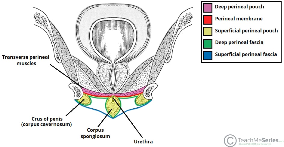
Category | Information |
|---|---|
Definition | A potential space located between the perineal membrane (superiorly) and the superficial fascia of the perineum (inferiorly). |
Location | Between the pubic symphysis and ischial tuberosities. |
Contents of the Superficial Perineal Pouch

Category | Structures |
|---|---|
External Genitalia | Root of the penis (male) / clitoris (female) |
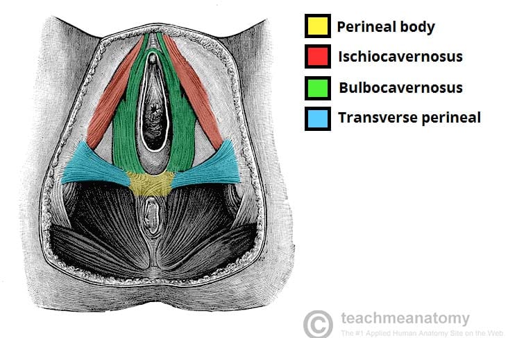
Perineal Muscles | • Bulbospongiosus muscle • Ischiocavernosus muscle • Superficial transverse perineal muscle |
Neurovascular Structures | • Internal pudendal artery • Internal pudendal vein • Pudendal nerve |
Deep Perineal Pouch (Space)
Category | Information |
|---|---|
Definition | A true space located between the perineal membrane (inferiorly) and the inferior fascia of the pelvic diaphragm (superiorly). |
Contents of the Deep Perineal Pouch
Category | Structures |
|---|---|
Urinary Structures | • Urethra |
Muscles | • External urethral sphincter• Urethrovaginal sphincter (females) |
Glands | • Cowper's (Bulbourethral) glands (males) |
Neurovascular Structures | • Internal pudendal artery• Internal pudendal vein• Pudendal nerve |
Perineal Membrane
Feature | Information |
|---|---|
Definition | Strong fibrous sheet of connective tissue that stretches across the urogenital triangle. |
Function | Separates the superficial and deep perineal pouches. |
Location | Lies between the levator ani muscle and the superficial perineal pouch. |
Attachments | Extends from the ischiopubic rami to the pubic symphysis. |
Perineal Body
Feature | Information |
|---|---|
Location | Midpoint at the junction of the urogenital triangle and the anal triangle. |
Function | Serves as the convergence point for several pelvic floor muscles. |
Importance | Supports the urethral and anal sphincters, contributing to urinary and fecal continence. |
Comparison of the Two Perineal Pouches
Feature | Superficial Perineal Pouch | Deep Perineal Pouch |
|---|---|---|
Type of Space | Potential space | True space |
Boundaries | Between the perineal membrane and superficial fascia | Between the perineal membrane and inferior fascia of the pelvic diaphragm |
Contains | Root of penis/clitoris | Urethra |
Bulbospongiosus muscle | External urethral sphincter | |
Ischiocavernosus muscle | Urethrovaginal sphincter (female) | |
Superficial transverse perineal muscle | Bulbourethral (Cowper's) glands (male) | |
Internal pudendal artery & vein | Internal pudendal artery & vein | |
Pudendal nerve | Pudendal nerve |
Identify the following pelvic muscles, innervation and the corresponding actions
Muscles of the pelvic walls
Obturator internus muscles
Piriformis muscles
i. Obturator Internus Muscle
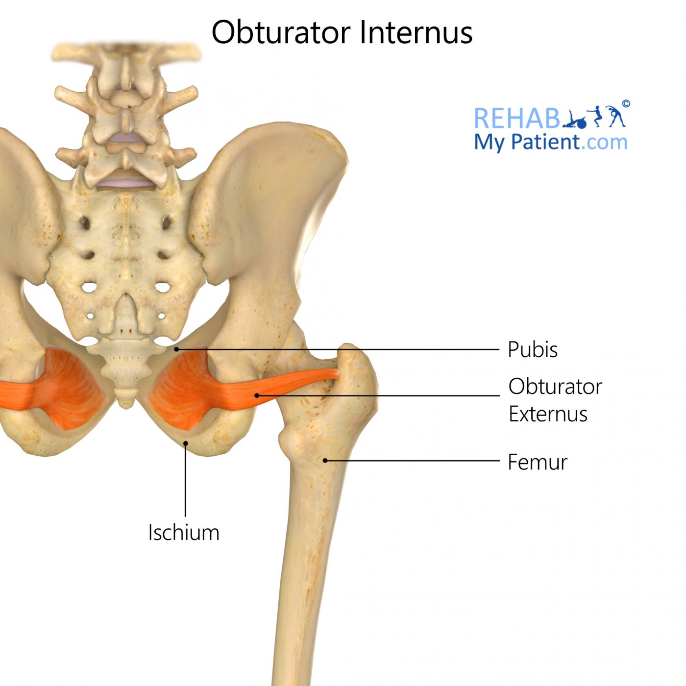
Category | Information |
|---|---|
Location | Muscle of the pelvic wall. |
Origin | Margins of the obturator foramen, obturator membrane, ischial ramus, and pubic ramus. |
Insertion | Greater trochanter of the femur. |
Innervation | Nerve to the obturator internus. |
Actions | • External rotation of the hip • Abduction of the hip when the hip is flexed • Stabilization of the hip |
Clinical Correlation | The pudendal nerve passes near the obturator internus as it exits the pelvis through the lesser sciatic foramen. Compression or irritation can lead to pudendal neuralgia, causing chronic perineal pain. |
ii. Piriformis Muscle
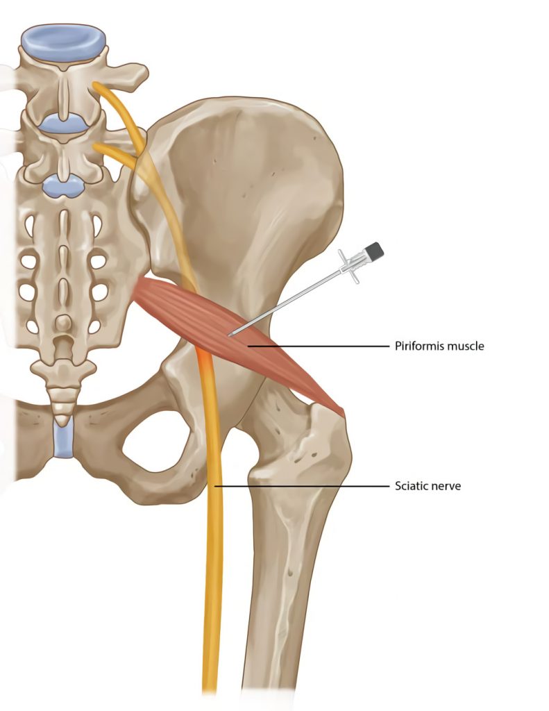
Category | Information |
|---|---|
Location | Muscle of the pelvic wall. |
Origin | Anterior surface of the sacrum and the gluteal surface of the ilium near the iliac spine. |
Insertion | Greater trochanter of the femur. |
Innervation | Nerve to piriformis. |
Actions | • External rotation of the hip • Abduction of the hip when the hip is flexed • Stabilizes the head of the femur in the acetabulum |
Clinical Correlation | Piriformis syndrome: Compression or irritation of the sciatic nerve by the piriformis muscle can produce sciatica-like pain in the buttock and leg. |
Comparison Table
Feature | Obturator Internus | Piriformis |
|---|---|---|
Origin | Obturator foramen, obturator membrane, ischial ramus, pubic ramus | Anterior sacrum and gluteal surface of ilium |
Insertion | Greater trochanter of femur | Greater trochanter of femur |
Innervation | Nerve to obturator internus | Nerve to piriformis |
Primary Actions | External rotation, hip abduction (when flexed), stabilization | External rotation, hip abduction (when flexed), stabilization of femoral head |
Clinical Correlation | Pudendal neuralgia | Piriformis syndrome (sciatic nerve compression) |
Muscles of the Pelvic Diaphragm
Feature | Information |
|---|---|
Forms | The pelvic floor (funnel-shaped pelvic diaphragm). |
Components | Levator ani and coccygeus muscles, along with their fascia. |
Extent | Stretches from the pubis (anteriorly) to the coccyx (posteriorly) and from one lateral pelvic wall to the other. |
Clinical Correlation | Repetitive stress (such as labor and delivery) can stretch and damage the levator ani muscles, resulting in pelvic floor insufficiency, uterine prolapse, and urinary incontinence. |
I. Levator Ani

Component | Origin | Insertion |
|---|---|---|
Pubococcygeus | Back of the pubic bone and anterior part of the obturator fascia | Coccyx, sacrum, and the tendinous center of the perineum |
Iliococcygeus | Fascia of the obturator internus muscle | Coccyx and the anococcygeal raphe |
Puborectalis | Lower part of the pubic symphysis and the superior fascia of the urogenital diaphragm | Forms a sling around the rectum, creating the anorectal angle, which is critical for maintaining fecal continence. |
Summary:
Category | Information |
|---|---|
Innervation | Not specified in the PDF. |
Actions | • Supports the pelvic floor through its three muscle components.• Puborectalis forms the anorectal angle, which is essential for fecal continence. |
Clinical Correlation | Weakness of the levator ani can lead to Pelvic Organ Prolapse (POP), commonly seen in postmenopausal women or after multiple vaginal births. |
II. Coccygeus (Ischiococcygeus) Muscle
:background_color(FFFFFF):format(jpeg)/images/library/13107/GJEqBx0Mc2i5kkAGg19g_Coccygeus_01.png)
Category | Information |
|---|---|
Origin | Ischial spine and bony projection of the ischium |
Insertion | Inferior end of the sacrum and the coccyx |
Innervation | Anterior rami of spinal nerves S4 and S5 |
Actions | • Supports the posterior part of the pelvic floor • Stabilizes the pelvic floor, helping maintain the position of the pelvic organs • Flexion of the coccyx (important during defecation and childbirth) |
Clinical Correlation | Pelvic floor muscle dysfunction can pull on the coccyx and cause coccyx pain, which may improve with pelvic floor physical therapy. |
Comparison Table:
Feature | Levator Ani | Coccygeus |
|---|---|---|
Location | Pelvic diaphragm | Pelvic diaphragm |
Components | Pubococcygeus, Iliococcygeus, Puborectalis | Single muscle |
Origin | Pubic bone, obturator fascia, obturator internus fascia, pubic symphysis, superior fascia of the urogenital diaphragm (varies by component) | Ischial spine and ischium |
Insertion | Coccyx, sacrum, tendinous center of perineum, anococcygeal raphe, rectal sling (varies by component) | Inferior sacrum and coccyx |
Innervation | Not specified in the PDF | Anterior rami of S4–S5 |
Actions | Supports pelvic floor; Puborectalis forms the anorectal angle for fecal continence | Supports and stabilizes the posterior pelvic floor; flexes the coccyx |
Exam Summary
Muscle | Remember This |
|---|---|
Levator Ani | Three parts: Pubococcygeus, Iliococcygeus, Puborectalis. The Puborectalis forms the anorectal angle, which is essential for fecal continence. Weakness can cause pelvic organ prolapse. |
Coccygeus | Origin: Ischial spine → Insertion: Sacrum & coccyx → Innervation: S4–S5 → Action: Supports and stabilizes the pelvic floor and flexes the coccyx during defecation and childbirth. |
Perineal Muscles
i. Bulbospongiosus:

Category | Bulbospongiosus |
|---|---|
Muscle Group | Superficial perineal muscle |
Location | Located in the superficial perineal pouch of the urogenital triangle. |
Origin | Perineal body. |
Insertion | Male: Wraps around the bulb of the penis and inserts into the perineal membrane and the corpus spongiosum of the penis.Female: Surrounds the bulbs of the vestibule and inserts into the perineal membrane and the corpus cavernosum of the clitoris. |
Innervation | Perineal branch of the pudendal nerve. |
Actions | • Assists in ejaculation by rhythmically contracting to expel semen from the urethra (male). • Constricts the vaginal orifice and assists in clitoral erection by compressing the bulbs of the vestibule (female). • Provides pelvic floor support. • Contributes to urinary continence by compressing the urethra. • Contributes to fecal continence by supporting the external anal sphincter. |
II. Ischiocavernosus
Category | Ischiocavernosus |
|---|---|
Muscle Group | Superficial perineal muscle |
Location | Located in the superficial perineal pouch of the urogenital triangle. |
Origin | Ischial tuberosity and ischiopubic ramus. |
Insertion | Crura of the penis or clitoris. |
Innervation | Perineal branch of the pudendal nerve. |
Actions | • Maintains erection.• Helps with ejaculation.• Assists in emptying the last bits of urine.• Stabilizes the erectile structures. |
High-Yield Comparison
Feature | Bulbospongiosus | Ischiocavernosus |
|---|---|---|
Muscle Type | Superficial perineal muscle | Superficial perineal muscle |
Location | Superficial perineal pouch | Superficial perineal pouch |
Origin | Perineal body | Ischial tuberosity and ischiopubic ramus |
Insertion | Corpus spongiosum (male) / Corpus cavernosum of clitoris (female) | Crura of penis or clitoris |
Innervation | Perineal branch of the pudendal nerve | Perineal branch of the pudendal nerve |
Main Actions | Ejaculation, constricts vaginal orifice, assists clitoral erection, supports pelvic floor, aids urinary and fecal continence | Maintains erection, assists ejaculation, empties the last urine, stabilizes erectile structures |
Identify the following pelvic vasculature
Anterior trunk of the internal iliac
Obliterated umbilical artery
Obturator artery
Uterine artery
Inferior vesical artery
Vaginal artery (not in males)
Middle rectal artery
Internal pudendal artery
Penile artery
Inferior gluteal artery
The anterior division of the internal iliac artery primarily gives rise to visceral branches that supply the pelvic organs, perineum, external genitalia, and portions of the gluteal region.
Artery | Location / Course | Structures Supplied (Function) | Important Features / Clinical Notes |
|---|---|---|---|
Obliterated Umbilical Artery | Branch of the anterior division of the internal iliac artery. | Gives rise to the superior vesical artery, which supplies the urinary bladder. | Distal portion becomes the medial umbilical ligament after birth. |
Obturator Artery | Branch of the anterior division; travels toward the obturator canal. | Supplies the obturator internus muscle and the medial thigh. | Passes through the obturator foramen with the obturator nerve and vein. |
Uterine Artery(Female only) | Branch of the anterior division. | Supplies the uterus and vagina. | Crosses over the ureter, an important relationship to remember during surgery to prevent ureteral injury ("water under the bridge"). |
Inferior Vesical Artery(Male) | Branch of the anterior division. | Supplies the urinary bladderand prostate. | Present in males according to the PDF. |
Vaginal Artery(Female only) | Branch of the anterior division. | Supplies the vagina and urinary bladder. | Not present in males. |
Middle Rectal Artery | Branch of the anterior division. | Supplies the rectum. | The PDF notes it is often associated with internal hemorrhoids. |
Internal Pudendal Artery | Branch of the anterior division that travels into the perineum. | Supplies the perineum, external genitalia, and anal canal. | Gives rise to the inferior rectal artery, perineal artery, and dorsal artery of the penis/clitoris. |
Penile Artery | Branch of the internal pudendal artery. | Supplies the penis. | Listed in the PDF as a branch of the internal pudendal artery. |
Inferior Gluteal Artery | Branch of the anterior division. | Included as a branch of the anterior division of the internal iliac artery. | Listed in the pelvic vasculature section of the PDF. |

Branches of the Internal Pudendal Artery
Branch | Supplies |
|---|---|
Inferior Rectal Artery | Anal canal |
Perineal Artery | Perineum |
Dorsal Artery of the Penis / Clitoris | Penis or clitoris |
Penile Artery | Penis |
Artery | Remember This |
|---|---|
Obliterated Umbilical Artery | Becomes the medial umbilical ligament and gives rise to the superior vesical artery. |
Obturator Artery | Supplies the obturator internus muscle and medial thigh. |
Uterine Artery | Crosses over the ureter; supplies the uterus and vagina. |
Inferior Vesical Artery | Supplies the bladder and prostate. |
Vaginal Artery | Supplies the vagina and bladder (female only). |
Middle Rectal Artery | Supplies the rectum; associated with internal hemorrhoids in the PDF. |
Internal Pudendal Artery | Main blood supply to the perineum, external genitalia, and anal canal. |
Penile Artery | Branch of the internal pudendal artery supplying the penis. |
Inferior Gluteal Artery | Branch of the anterior division of the internal iliac artery. |
Posterior Trunk (Posterior Division) of the Internal Iliac Artery
The posterior division of the internal iliac artery primarily gives rise to parietal branches, which supply the pelvic walls, gluteal region, sacrum, and supporting musculoskeletal structures rather than the pelvic organs.
Artery | Location / Course | Structures Supplied (Function) | Important Features |
|---|---|---|---|
Iliolumbar Artery | Branch of the posterior division of the internal iliac artery. | Listed as a posterior division branch supplying the pelvic supporting structures. | One of the three major branches of the posterior trunk. |
Lateral Sacral Artery | Branch of the posterior division of the internal iliac artery. | Listed as a posterior division branch supplying the pelvic supporting structures. | Travels along the sacrum. |
Superior Gluteal Artery | Branch of the posterior division of the internal iliac artery. | Supplies the gluteal muscles. | Largest branch of the posterior division shown in the pelvic vasculature diagrams. |
High-Yield Summary:
Posterior Division Branch | Remember This |
|---|---|
Iliolumbar Artery | Posterior division branch that supplies pelvic supporting structures. |
Lateral Sacral Artery | Posterior division branch that courses along the sacrum and supplies pelvic supporting structures. |
Superior Gluteal Artery | Posterior division branch that supplies the gluteal region. |
Easy Way to Remember
Posterior Branch | Think of... |
|---|---|
Iliolumbar | Ilium + lumbar region |
Lateral Sacral | Sacrum |
Superior Gluteal | Gluteal muscles (buttock) |
Memory Trick: "I Love Glutes"
I = Iliolumbar artery
L = Lateral sacral artery
G = Superior gluteal artery
This mnemonic matches the three branches listed in your pelvic anatomy

Lymphatic nodes
Superficial inguinal nodes
Deep inguinal nodes
The pelvic lymphatic system contains superficial (palpable) and deep (non-palpable) lymph nodes that drain different regions of the pelvis, lower limb, and external genitalia. Knowing what each node drains helps identify the possible source of infection or cancer spread.
Superficial Inguinal Nodes
Category | Study Guide |
|---|---|
Location | Located in the superficial inguinal region (groin). They are superficial and palpable, located near the femoral pulse. |
Palpable? | Yes – can normally be palpated during a physical examination. |
Drainage | • Anterior abdominal wall• Inguinal region• External genitalia |
Clinical Correlation | A "lump in the groin" may indicate disease involving structures drained by these nodes. The PDF specifically notes evaluating for STIs or anal cancer, not just leg pathology. |
Deep Inguinal Nodes
Category | Study Guide |
|---|---|
Location | Located in the deep inguinal region. They are part of the deep (non-palpable) pelvic lymphatic system. |
Palpable? | No – deep and non-palpable. |
Drainage | • Receive lymph from the superficial inguinal nodes.• Drain the deep structures of the leg. |
Relationship | Act as the next drainage station after the superficial inguinal nodes before lymph continues centrally. |
Other Deep Pelvic Lymph Nodes
Lymph Node Group | Drains |
|---|---|
External iliac nodes | Pelvic viscera |
Internal iliac nodes | Pelvic viscera |
Pararectal nodes | Colorectal area |
Sacral nodes | Colorectal area |
Lumbar lymph nodes | Primary sites for testicular cancer and ovarian cancer metastasis (not the inguinal nodes). |
Feature | Superficial Inguinal Nodes | Deep Inguinal Nodes |
|---|---|---|
Location | Superficial groin | Deep groin |
Palpable | ✅ Yes | ❌ No |
Drain | Anterior abdominal wall, inguinal region, external genitalia | Superficial inguinal nodes and deep structures of the leg |
Clinical Pearl | Enlarged with infections or disease affecting structures they drain (e.g., STI or anal cancer per the PDF) | Receive drainage from superficial nodes and deep leg structures |

Identify the location and innervations of the pelvis
Sacral plexus
Superior and inferior gluteal nerve
Pudendal nerve
Sciatic nerve
Autonomic innervation
Sacral sympathetic trunk
Pelvic Splanchnic nerves
Hypogastric plexus
Sacral Plexus:
The sacral plexus is formed by the lumbosacral trunk (L4–L5) and the anterior rami of S1–S4. It lies on the anterior surface of the piriformis muscle on the posterior pelvic wall and supplies the pelvis, gluteal region, perineum, and lower limb.
Category | Study Guide |
|---|---|
Location | Lies on the anterior surface of the piriformis muscle in the posterior pelvis. |
Formation | Lumbosacral trunk (L4–L5) + S1–S4 ventral rami. |
Function | Provides motor and sensory innervation to the pelvis, gluteal region, perineum, and lower limb. |
Superior and Inferior Gluteal Nerves
Nerve | Location / Course | Innervates |
|---|---|---|
Superior Gluteal Nerve | Branch of the sacral plexus that exits the pelvis to the gluteal region. | Gluteus medius, gluteus minimus, and tensor fasciae latae. |
Inferior Gluteal Nerve | Branch of the sacral plexus that exits the pelvis to the gluteal region. | Gluteus maximus. |
Superior gluteal nerve = gluteus medius & minimus + TFL
Inferior gluteal nerve = gluteus maximus
Pudendal Nerve:
Category | Study Guide |
|---|---|
Location / Course | Branch of the sacral plexus that travels to the perineum. |
Innervates | • Perineum • External genitalia • External anal sphincter • Perineal muscles (including bulbospongiosus and ischiocavernosus). |
Clinical Importance | The perineal branch supplies the superficial perineal muscles. |
Sciatic Nerve:
Category | Study Guide |
|---|---|
Location / Course | Largest branch of the sacral plexus that exits the pelvis to enter the lower limb. |
Innervates | Lower limb (listed as a major branch of the sacral plexus in the PDF). |
Autonomic Innervation of the Pelvis
The pelvic autonomic nervous system consists of sympathetic and parasympathetic fibers that regulate involuntary functions of the pelvic organs. The PDF identifies three major autonomic structures:
Sacral sympathetic trunk
Pelvic splanchnic nerves
Hypogastric plexus
Sacral Sympathetic Trunk:
Category | Study Guide |
|---|---|
Location | Located within the pelvis as part of the autonomic nervous system. |
Type | Sympathetic. |
Function | Contributes sympathetic fibers to the pelvic organs through the pelvic autonomic plexuses. |
Pelvic Splanchnic Nerves:
Category | Study Guide |
|---|---|
Location | Pelvic autonomic nerves arising within the pelvis. |
Type | Parasympathetic. |
Function | Provide parasympathetic innervation to the pelvic organs. |
Hypogastric Plexus:
Category | Study Guide |
|---|---|
Location | Pelvic autonomic nerve plexus within the pelvis. |
Function | Network that distributes autonomic fibers (sympathetic and parasympathetic) to the pelvic organs. |
High-Yield Summary Table
Structure | Location | Innervates / Function |
|---|---|---|
Sacral Plexus | Anterior surface of piriformis | Pelvis, gluteal region, perineum, lower limb |
Superior Gluteal Nerve | Branch of sacral plexus | Gluteus medius, gluteus minimus, tensor fasciae latae |
Inferior Gluteal Nerve | Branch of sacral plexus | Gluteus maximus |
Pudendal Nerve | Branch of sacral plexus to perineum | Perineum, external genitalia, external anal sphincter, perineal muscles |
Sciatic Nerve | Branch of sacral plexus | Lower limb |
Sacral Sympathetic Trunk | Pelvis | Sympathetic fibers to pelvic organs |
Pelvic Splanchnic Nerves | Pelvis | Parasympathetic fibers to pelvic organs |
Hypogastric Plexus | Pelvis | Distributes autonomic fibers to pelvic organs |
Superior gluteal → Medius, Minimus, TFL
Inferior gluteal → Maximus
Pudendal → Perineum
Sciatic → Lower limb
Sacral sympathetic trunk = Sympathetic
Pelvic splanchnic nerves = Parasympathetic
Hypogastric plexus = Autonomic distribution center
