Chapter 12: Central Nervous System
1. Big Picture: What Is the CNS?
The central nervous system (CNS) is the brain and spinal cord. It receives information, processes it, and helps coordinate responses.
Four major brain regions
Region | Simple description / key job |
Cerebral hemispheres (cerebrum) | Largest part; conscious awareness, thought, memory, language, sensation, and voluntary movement. |
Diencephalon | Thalamus, hypothalamus, and epithalamus; relays information and regulates internal balance; connects the cerebrum (thinking and voluntary movement part) to the rest of the nervous system |
Brain stem | Midbrain, pons, and medulla oblongata; connects brain and spinal cord and controls vital automatic functions (heartbeat, breathing, etc.). |
Cerebellum | Fine-tunes movement, coordination, posture, and balance. |

Gray matter vs. white matter
Gray matter: mostly neuron cell bodies and short, nonmyelinated processes; where much information processing occurs.
White matter: mostly axons (many myelinated); carries signals between regions.
The dense coating of fatty myelin is what gives white matter its color
Basic pattern found in CNS: central cavity surrounded by gray matter, with white matter external to gray matter
2. Major Brain Regions: Cerebrum
Cerebrum
The cerebrum is the largest, superior part of the brain
Divided into the cerebral hemispheres (left and right) - connected by corpus collosum
Each hemisphere has three layers:
Cerebral cortex - the “executive suite” of brain; site of the conscious mind: awareness, sensory perception, voluntary motor initiation, communication, memory storage, understanding
Thin (2–4 mm) superficial layer of gray matter
Composed of neuron cell bodies, dendrites, glial cells, and blood vessels, but no axons
Internal white matter
Basal ganglia deep within white matter

Surface markings
Term | Meaning |
Gyrus (gyri) | A raised ridge or fold. |
Sulcus (sulci) | A shallow groove between gyri. |
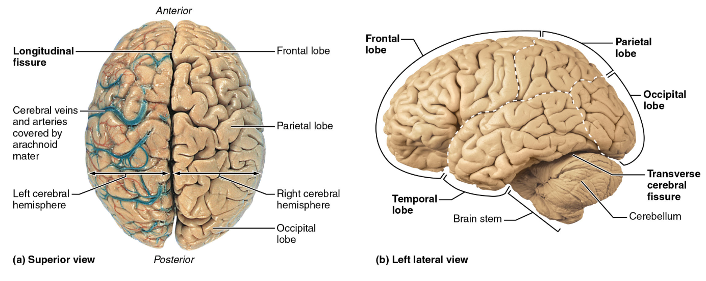
1) Central sulcus | Separates frontal and parietal lobes; primary motor cortex is just in front of it and primary somatosensory cortex just behind. |
2) Lateral sulcus | Marks the upper boundary of the temporal lobe. Separates the temporal lobe from the frontal and parietal lobes. |
3) Parieto-occipital sulcus | Separates parietal and occipital lobes. |
Fissure | A deep groove. |
1) Longitudinal fissure | Separates the left and right cerebral hemispheres. |
2) Transverse cerebral fissure | Separates cerebrum from cerebellum. |

Five lobes
Each cerebral hemisphere is divided by several sulci into five sections or lobes
Each lobe has a different function
First four are named after overlying skull bones
Insular lobe is buried under portions of temporal, parietal, and frontal lobes
Lobe | Main functions |
Frontal | Executive function, like planning and organizing); higher order thinking, behavior, and problem-solving; motor control (i.e. motor cortex) |
Parietal | Somatic sensory processing, including touch and information from muscles/joints; sensory integration (i.e. sensory cortex); spatial reasoning/awareness |
Temporal | Hearing, smell, memory, and language comprehension |
Occipital | Vision and visual interpretation |
Insula | Taste, vestibular awareness, and visceral sensory awareness |

Cerebral Cortex
Four features of the cerebral cortex:
Contains three types of functional areas:
motor areas - voluntary movement (frontal lobe)
sensory areas - control awareness of sensation (occipital, parietal, and temporal lobes)
association areas - combine information from multiple sensory areas, give it meaning, connect it to memory and experience, and help choose a response (throughout the cortex)
Each hemisphere controls or receives information from the opposite (contralateral) side of the body
Left hemisphere of the brain controls right side and vice versa
Lateralization (specialization) of cortical function can occur in only one hemisphere
Left hemisphere: Language, math, and logic. About 90% of people have left-sided language dominance
Right hemisphere: Visual-spatial skills, intuition, emotion, and artistic/musical skills.
Conscious behavior involves entire cortex in one way or another


3. Major Brain Regions: Diencephalon
The diencephalon contains three paired gray-matter structures that surround the third ventricle: thalamus, hypothalamus, and epithalamus.
Thalamus — the relay station
Sorts, edits, and relays much incoming information to the cerebral cortex.
Relays sensory information and signals involved in movement, emotion/visceral function, memory, learning, and cortical arousal.
Think: the thalamus routes information to the right cortical destination.
Hypothalamus — homeostasis and body regulation
Located below the thalamus; links the nervous system to the endocrine (hormone) system via the pituitary gland
Controls autonomic nervous system functions such as blood pressure, heart rate, digestive movement, and pupil size.
Helps coordinate physical responses to emotions and is part of the limbic system
Regulates various bodily functions: body temperature, hunger and satiety, thirst and water balance, sleep-wake cycles, biological rhythms, and endocrine function.
Disruption may contribute to wasting, obesity, sleep problems, dehydration, or emotional imbalance.
Epithalamus
Contains the pineal gland, which secretes melatonin and helps regulate the sleep-wake cycle

4. Major Brain Regions:Brain Stem
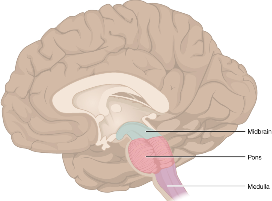
The brain stem consists of the midbrain, pons, and medulla oblongata.
Primary features:
Stalk-like base of the brain that connects the cerebral hemispheres and cerebellum to the spinal cord
Similar in structure to spinal cord but contains nuclei embedded in white matter
Acts as the body's main autopilot center and information highway, keeping essential survival systems running without conscious effort
Vital Life Support: Automatically regulates essential survival functions, including breathing, heart rate, and blood pressure.
Information Highway: Serves as the main pathway for all sensory signals traveling up to the brain and all motor commands traveling down to the body.
Consciousness & Alertness: Houses the reticular formation, a nerve network that controls sleep-wake cycles, arousal, and overall consciousness.
Protective Reflexes: Coordinates automatic survival reflexes such as swallowing, coughing, sneezing, vomiting, and gagging.
Cranial Nerve Hub: Serves as the attachment point for 10 of the 12 pairs of cranial nerves, which control eye movement, facial expressions, hearing, balance, and taste.
Midbrain
Between the diencephalon and pons
Acts as a relay center for visual and auditory information and helps control movement.
Pons
Located in the middle of the brain stem (its name means "bridge")
Connects different parts of the brain and regulates breathing and sleep
Medulla oblongata
Lowest part of brain stem; blends into spinal cord at the foramen magnum
Manages vital, automatic survival functions:
Cardiac center adjusts heart rate and force of contraction
Vasomotor center adjusts blood vessel diameter and helps regulate blood pressure.
Respiratory centers help generate breathing rhythm and control rate/depth of breathing with pontine center
Other reflex centers coordinate vomiting, hiccupping, swallowing, coughing, and sneezing.
5. Major Brain Regions:Cerebellum
Located behind (dorsal to) the pons and medulla; about 11% of brain mass
Coordinates precise, smooth skeletal-muscle movements and is important for balance
Receives intended movement information from cerebral cortex, plus body-position information from proprioceptors and visual/equilibrium pathways.
Compares intended movement with information about actual position and momentum, then sends corrections to motor cortex and brain-stem nuclei.
Memory cue: the cerebrum plans a movement; the cerebellum fine-tunes it so it is smooth and coordinated

7. Protection of the Brain: Meninges, CSF, and Blood-Brain Barrier
Meninges
The meninges are three connective-tissue coverings around the brain and spinal cord. They protect the CNS, protect blood vessels, contain CSF, and form partitions in the skull.
Layer (outside → inside) | Key details |
Dura mater | Tough outer layer. In the skull it has periosteal and meningeal layers; the meningeal layer continues around the spinal cord. Where the layers separate, dural venous sinuses collect venous blood and drain toward the jugular veins. |
Arachnoid mater | Middle, web-like layer. The subarachnoid space beneath it contains CSF and major brain blood vessels. Arachnoid granulations return CSF to venous blood. |
Pia mater | Very delicate inner layer that hugs the brain surface and follows its folds; contains small blood vessels that nourish brain tissue. |

Ventricles and Cerebrospinal Fluid (CSF)
Ventricles are connected, fluid-filled chambers inside the brain. They are lined by ependymal cells and contain cerebrospinal fluid (CSF).
CSF: why it matters
Buoyancy: CSF makes the brain effectively much lighter, reducing pressure from its own weight.
Protection: cushions the CNS against blows and other trauma.
Chemical Stability: carries nutrients and chemical signals and helps remove wastes.
Production and circulation
Produced by the choroid plexus - a cluster of capillaries covered by pia mater and ependymal cells in the ventricles.
Ependymal cells help regulate CSF composition, remove wastes, and their cilia help move the fluid.
CSF is constantly produced, circulated, and drained to maintain optimal pressure in the brain's ventricular system and regulate the environment for neuronal function.
Blood-Brain Barrier (BBB)
The blood-brain barrier (BBB) is a highly selective protective barrier that separates the blood from the brain tissue. Its job is to control what substances can enter the brain, protecting it from harmful substances while allowing essential nutrients to pass through.
The BBB is primarily formed by:
Endothelial cells: Line the brain's capillaries.
Tight junctions: Connect endothelial cells tightly, preventing substances from freely passing between them.
Astrocytes: Surround capillaries and help maintain BBB function.
Pericytes: Help regulate and support the barrier.
What does it do?
Protects the brain from toxins, pathogens, and harmful chemicals.
Maintains a stable environment for neurons to function.
Controls which nutrients and substances enter brain tissue.
Helps regulate the movement of waste products out of the brain.
Why is it important?
Without the BBB, harmful substances in the bloodstream could enter brain tissue and interfere with normal neuronal function.
However, it also creates a challenge in medicine because many medications cannot cross the BBB, making brain diseases harder to treat.