Lecture 5: Cell Growth
Cell Growth
Cell growth: cell division
1st step of cell division = DNA replication
DNA Structure:
Five prime (5') end: Last nucleotide contains a phosphate group.
Three prime (3') end: Last nucleotide contains a hydroxyl (OH) group.
DNA Replication:
DNA Replication: copying of DNA
happens only when cell is preparing for cell division
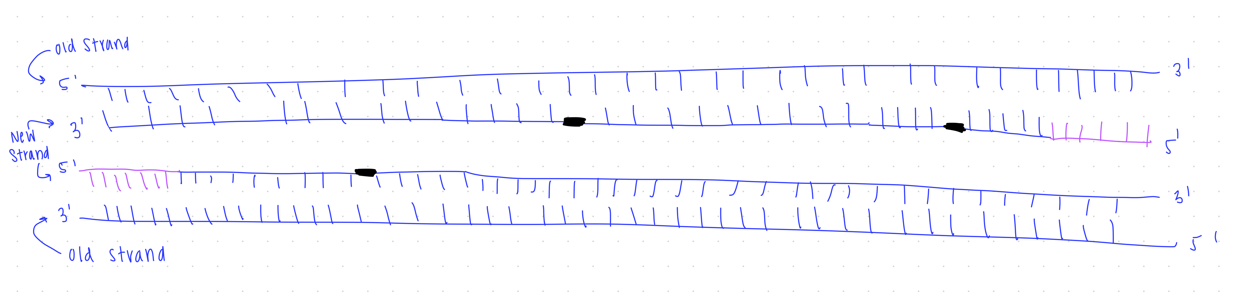
Step 1: Unwinding the DNA
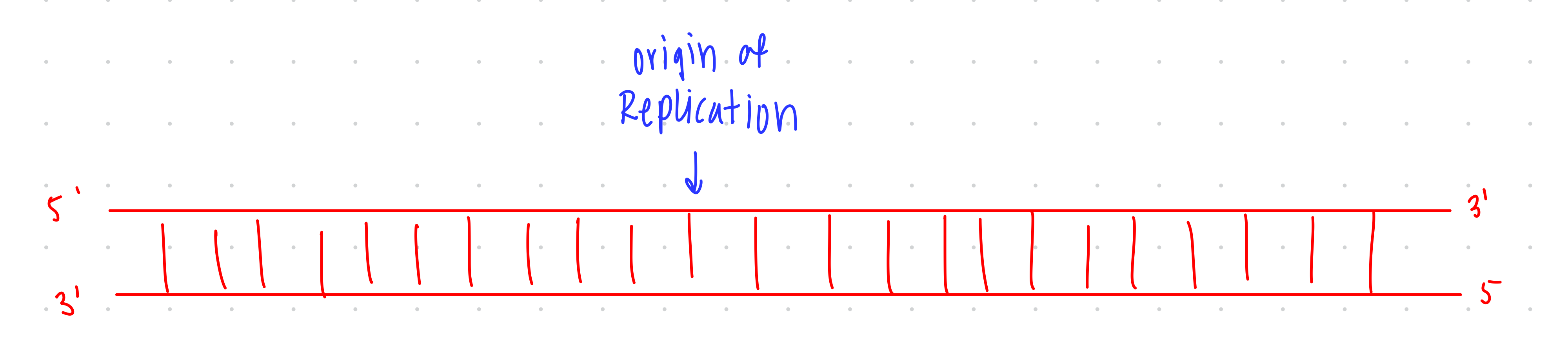
helicase opens the DNA at the origin of replication
Helicase: en enzyme that opens DNA by separating the two strands, forming a replication bubble.
Topoismerase: enzyme that cuts and relaxes DNA so it doesn’t get overwound, alleviate twisting tension during replication.
Step 2: Priming for DNA Synthesis
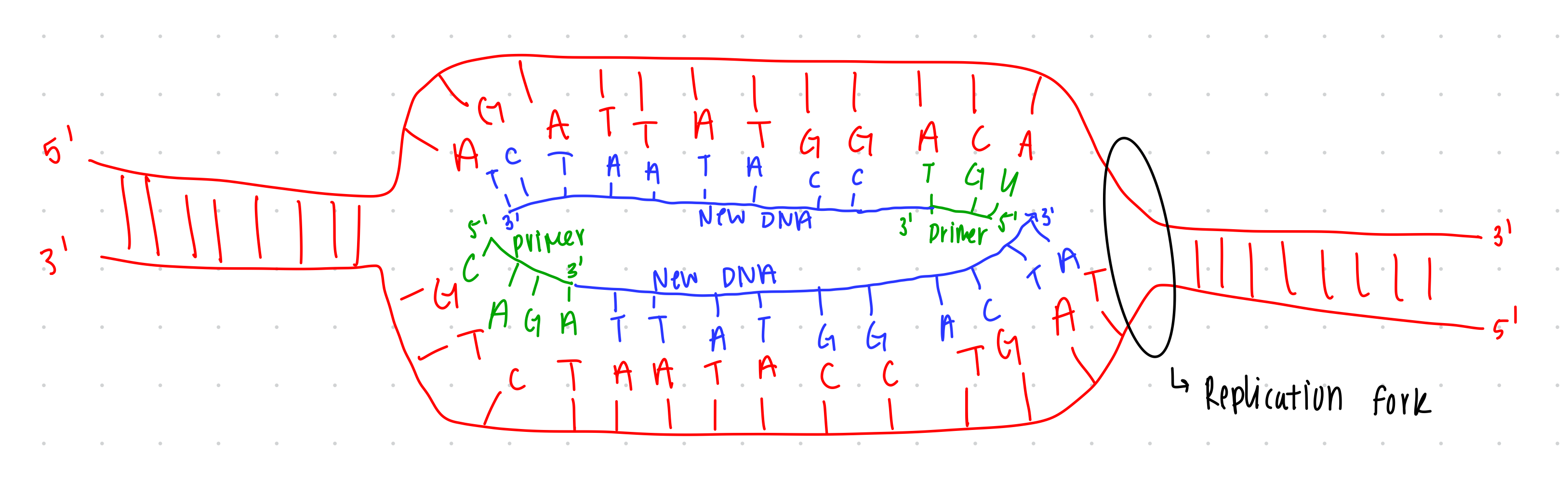
RNA primase enzyme builds RNA primer, which provides 3’ end for DNA polymerase enzyme to build new DNA off of.
RNA primase: an enzyme that Lays down a short RNA primer complementary to DNA.
Base pairing rules:
A pairs with U (instead of T in DNA) and C pairs with G.
Both RNA and DNA run in antiparallel directions (5' to 3').
Step 3: DNA Synthesis
Helicase opens at replication fork, while DNA polymerase adds nucleotides to 3’ end of RNA primer, building new DNA.
DNA polymerase: enzyme that adds nucleotides to build DNA, after RNA primer strand
Step 4: Leading Strand
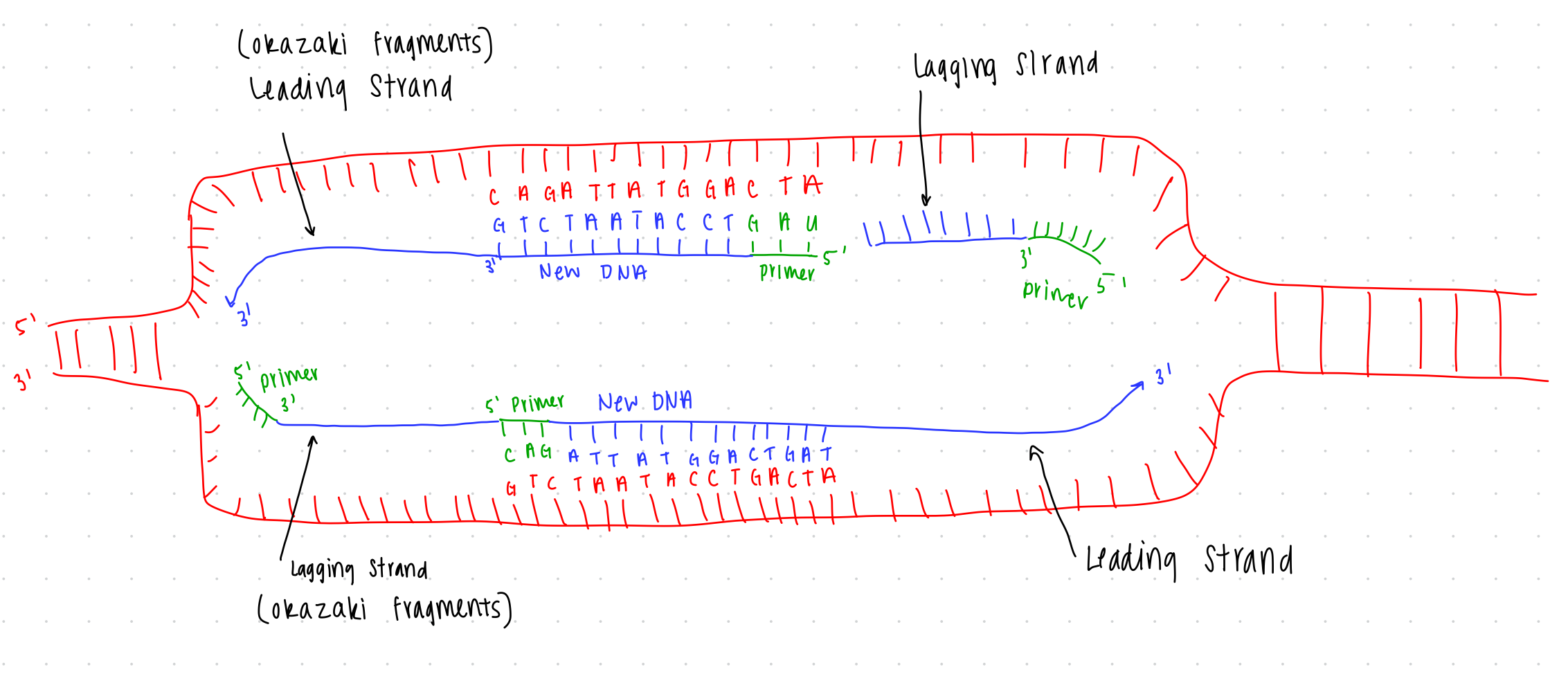
At each replication fork, the leading strands is built continuously as fork opens
Leading strand is synthesized towards replication fork
Leading strand: the strand that is built continuously, as the replication fork opens
Step 5: Lagging Strand
The lagging strand is built in pieces.
as replication fork opens, RNA primase adds more primers, creating additional 3’ ends
The necessity of RNA primer: DNA polymerase can only extend from an existing 3' end.
DNA polymerase back fills DNA, forming lagging strands
Lagging strand (Okazaki fragments): the strand that is built in pieces, needing additional primers as the replication form opens
Step 6: DNA formation
steps 4-6 repeat until the end of the molecule is reached
Step 7: Removal of RNA primers
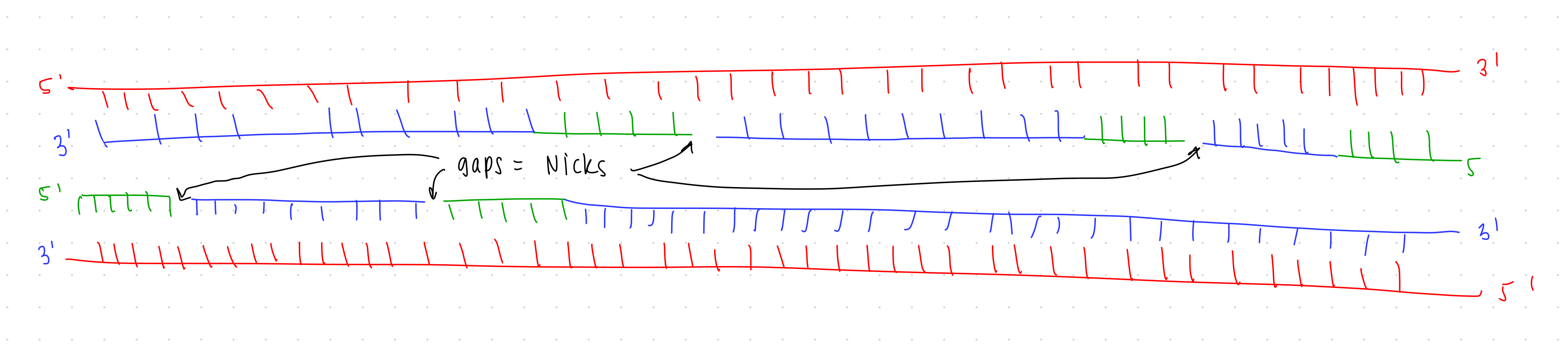
RNA primers removed and replaced with DNA.
Primer replacement with DNA is essential as RNA cannot remain in the final DNA product.
Step 8: DNA Ligase
Ligase seals the nicks.
DNA Ligase: Seals nicks where RNA was replaced by DNA, creating a complete DNA strand.
Step 9: Ending Strands of DNA
Organism with linear chromosomes either cut off the end OR rebuild the end with telomerase enzyme
Telomere: extra DNA on the ends of chromosomes, does not encode proteins
telomerase: enzyme that replaces list telomerase after DNA replication
Step 10: Final Result
Final result: Two daughter DNA molecules each with half original (parent) and half newly synthesized DNA
Semi-conservative Replication: Each new DNA molecule contains one old strand and one new strand.
Differences in Eukaryotic vs. Prokaryotic DNA Replication
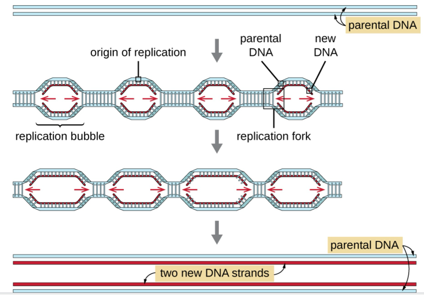
Eukaryotic replication:
Typically occurs at multiple origins of replication.
Replication bubbles spread bidirectionally until they meet.
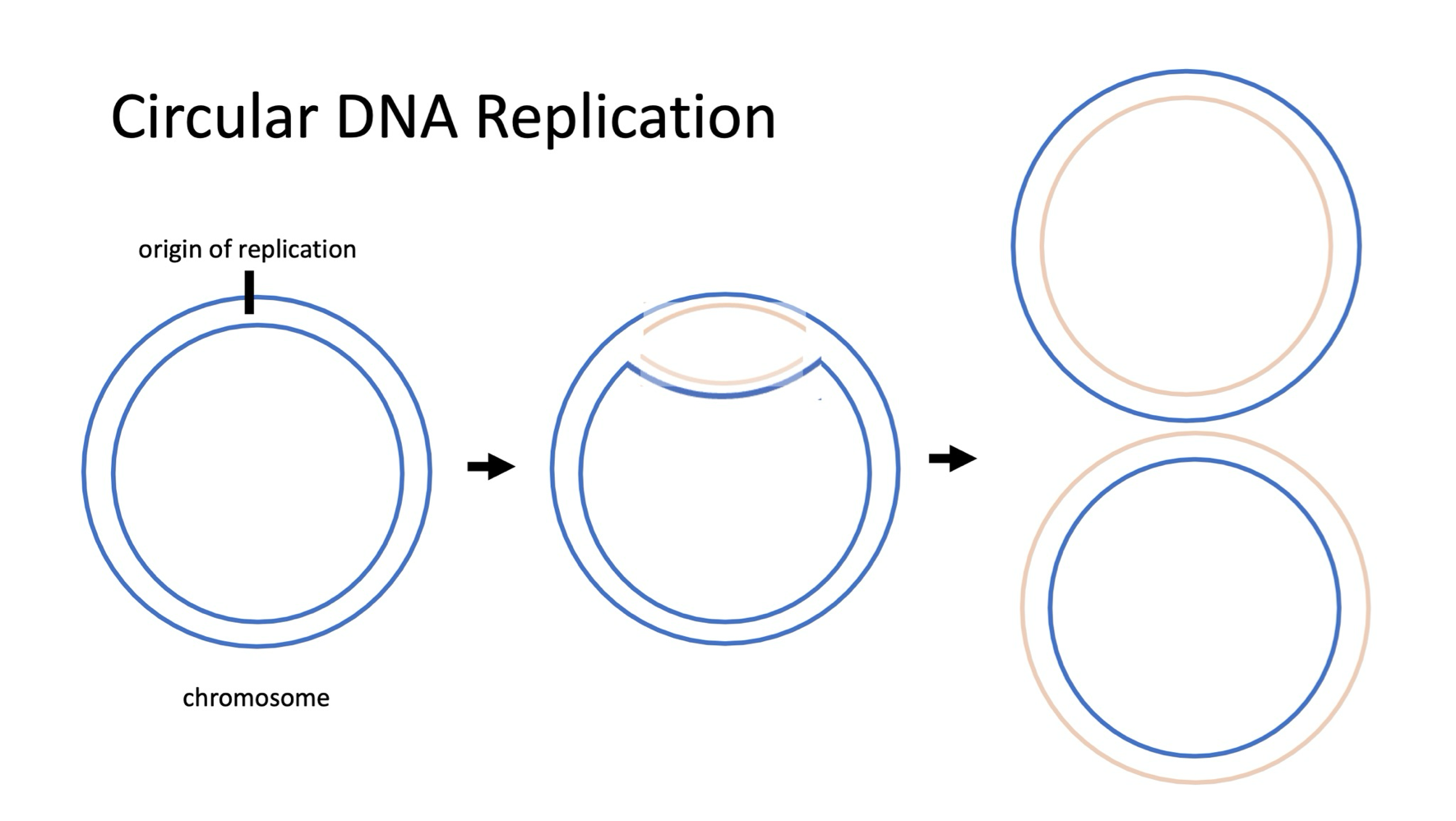
Prokaryotic replication:
Characterized by a single origin of replication.
Overview of Cell Division in Prokaryotic and Eukaryotic Cells
Prokaryotic Cell Division
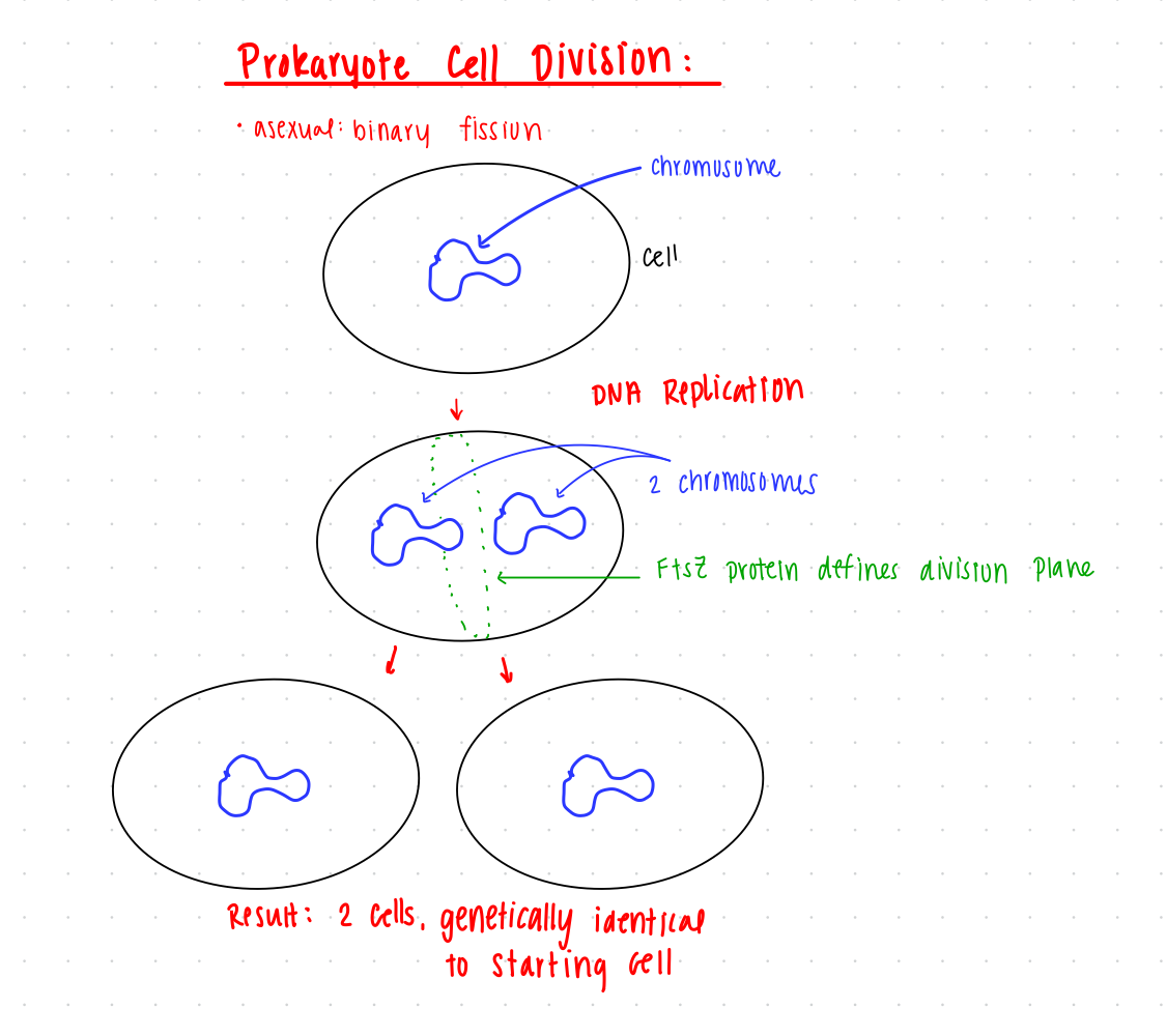
Prokaryotes cell division: asexually with binary fission.
asexual: makes a duplicating copy of the cell and the DNA
make clones of themselves
Steps involved in binary fission:
DNA Replication: The chromosome of the prokaryotic cell is copied.
Cell Division: Following DNA replication, the cell divides.
FtsZ ring forms at the division site between the two cells, defining division plane
The process completes when the septum has formed, resulting in two new cells, that are genetically identical
Eukaryotic Cell Division
Eukaryotes cell division: asexually (mitosis) or sexually (meiosis), depending on the species.
sexual: two different parent cells contributing half of their DNA to the creation of a new cell
new cell is genetically unique from parent cell
Steps to Mitosis (asexual reproduction):
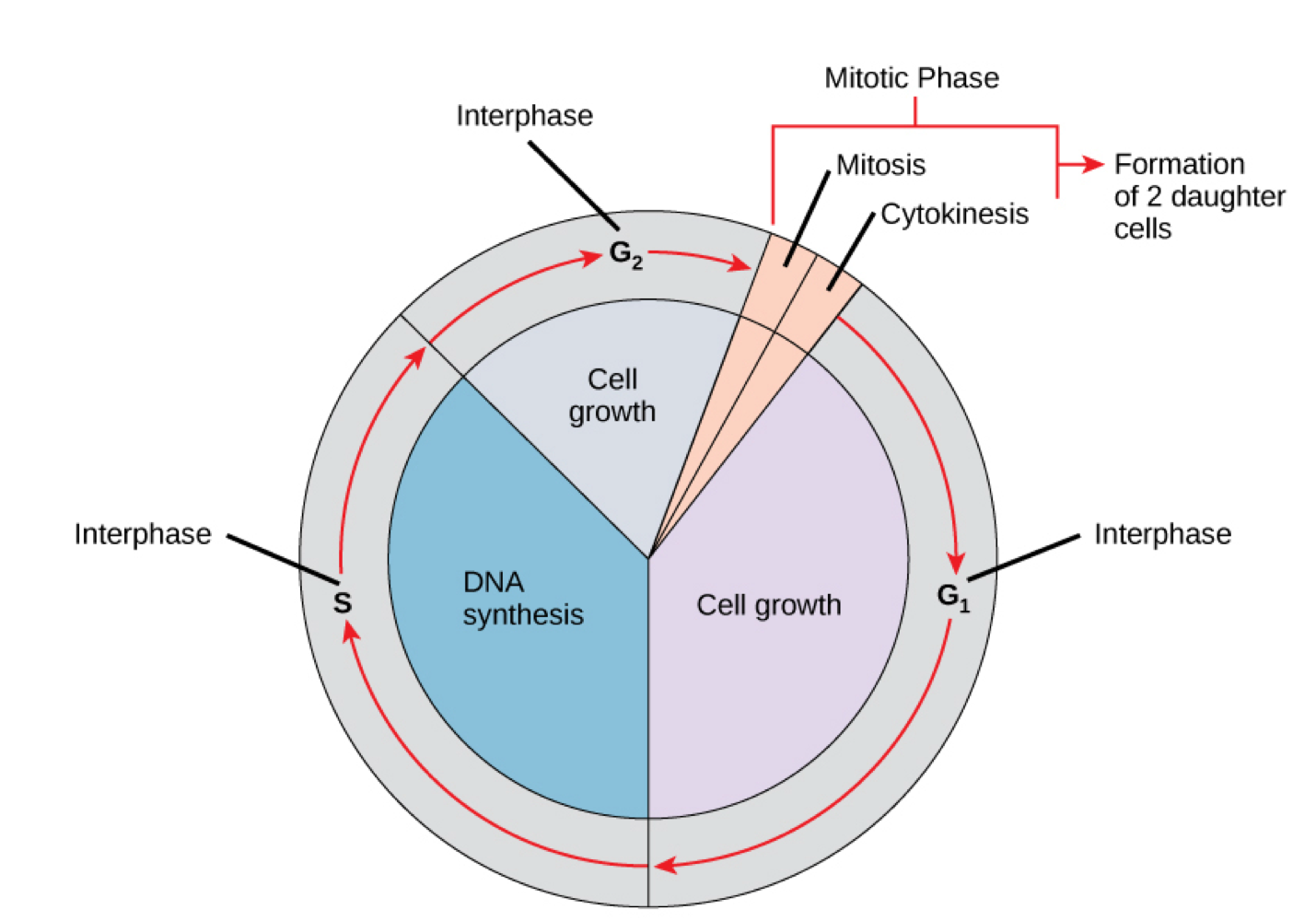
Interphase: getting ready to divide
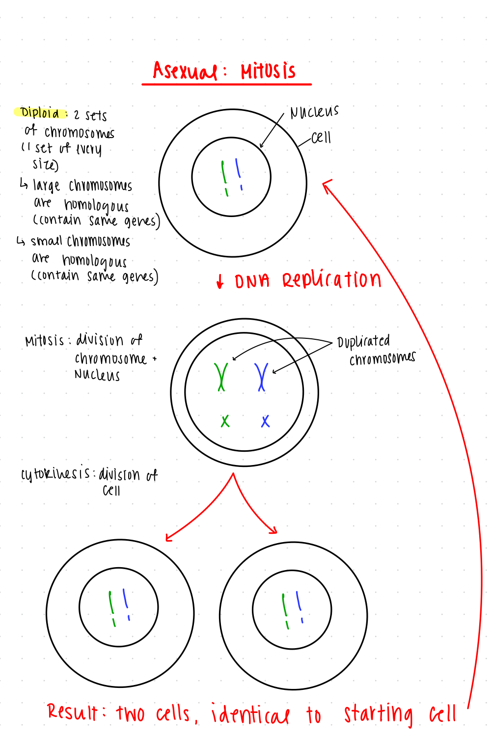
Mitosis: division of chromosomes and nucleus
begins with a diploid cell
diploid: 2 sets of chromosomes (1 set of every size)
large chromosomes are homologous
small chromosomes are homologous
homologous: contain the same genes
Replication of DNA in nucleus
chromosomes are duplicated
Cytokinesis: Cell division
separates into two cells that are identical to starting cell
Steps to Meiosis (sexual reproduction):
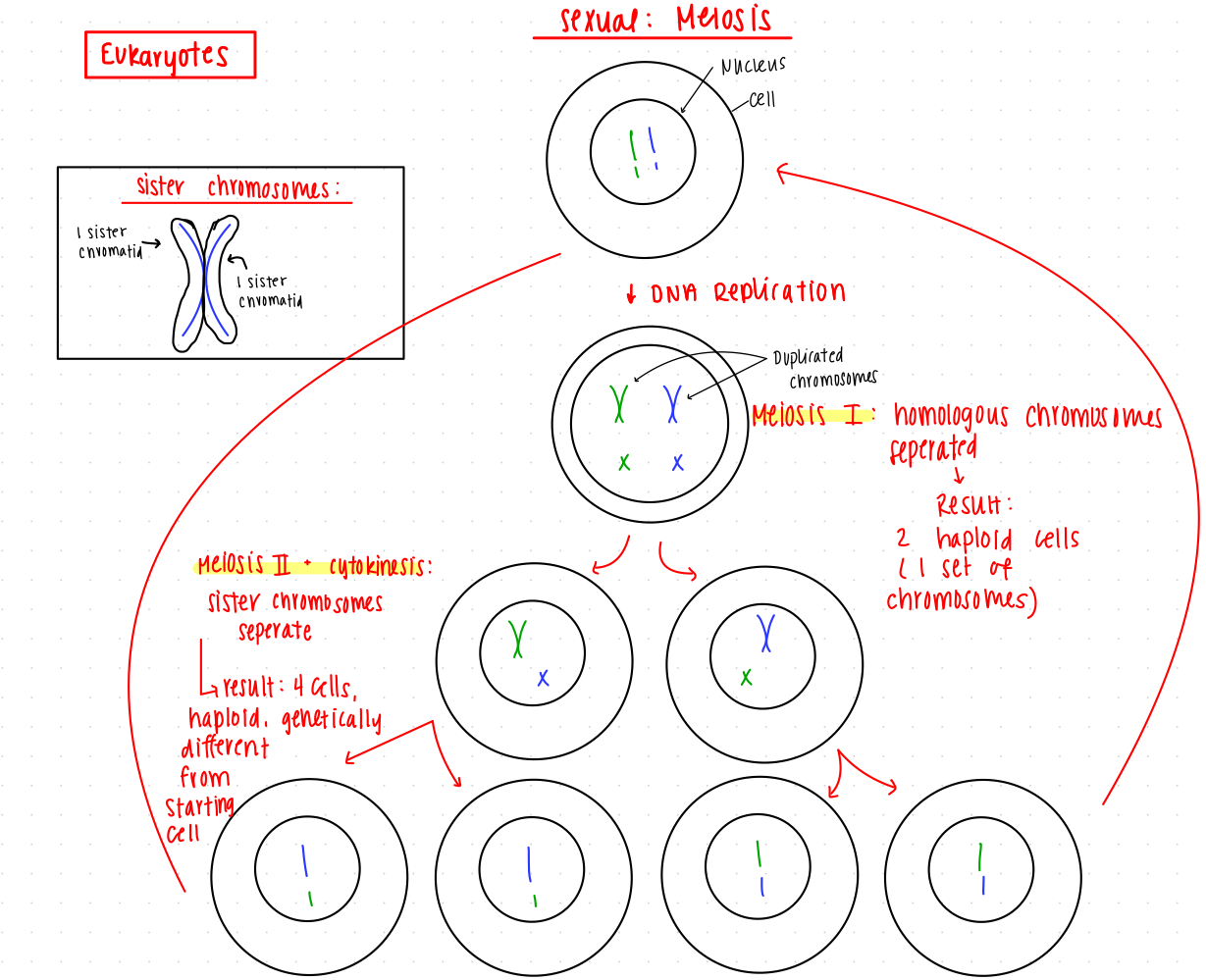
Starts with a diploid cell from mitosis
DNA replication
forms sister chromatids
sister chromatids: two identical copies of a single chromosome
Meiosis I: separation of homologous chromosomes instead of sister chromatids, resulting in 2 haploid cells
Meiosis II and cytokinesis: sister chromatids separate, resulting in 4 haploid cells that are genetically different from starting cell
The haploid cells can develop into:
Gametes: Involved in fertilization with another individual, leading to genetic recombination to form a diploid individual.
Spores: Can result from a zygote formed after sexual reproduction, allowing growth back into haploid mycelia or entering asexual reproduction.
Planktonic Cells
Planktonic: Free floating or motile cells in aquatic environments
Example: Diatom
Biofilms
Biofilms: adherent microorganisms embedded in an extracellular matrix.
The extracellular matrix is generally comprised of carbohydrate molecules and various other substances that create a gooey substance surrounding the biofilm.
Examples:
Biofilm on the inside of a catheter: The surface of catheters is ideal for biofilm adhesion, leading to infections.
Green scum around a rain barrel: This is typically another form of biofilm growth.
Dental plaque: A common biofilm that accumulates on teeth.
Development of Biofilms:
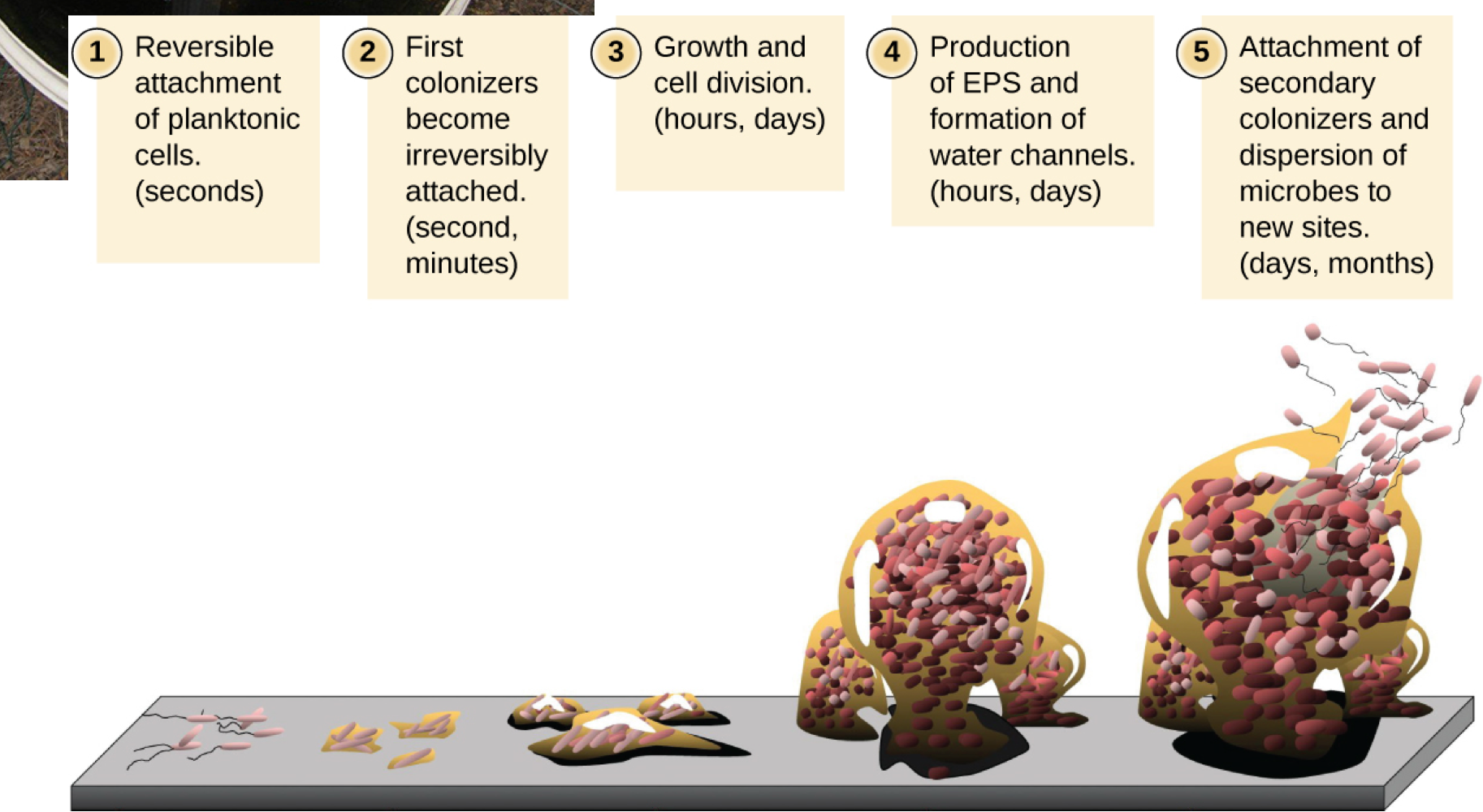
Planktonic cells land on a surface
reversible attachment
occurs in seconds
First colonizers express proteins that promote adhesion
becoming irreversibly attached.
occurs in seconds or minutes
Growth and cell division
occurs in hours or days
Production of EPS and formation of water channels
Cells begin to secrete extracellular matrix material and proliferate, forming multicellular structures
occurs in hours or days
Attachement of secondary colonizers and dispersion of microbes to new sites
Mature biofilm cells revert to a planktonic state allowing them to disperse and colonize new surfaces.
occurs in days or months
Biofilm and Human Health
Biofilms pose significant health risks
involved in about 80% of bacterial infections.
Common locations include:
On the skin and wounds.
Surface of respiratory and digestive epithelia.
Comparison with Planktonic Infections: Planktonic infections occur when bacteria are free-floating in the bloodstream, making them different from biofilm-related infections.
Challenges in Treatment:
Adherence: Biofilms' natural adherence complicates their removal from surfaces and devices.
Drug Resistance
Drug Pumps: Biofilm cells have drug pumps, actively pumping drugs out of cells
Reduced Metabolic Activity: Biofilm cells are less metabolic active
prevents drugs from working
many drugs target cell growth/metabolism work work well
Extracellular Matrix Barrier: Certain drugs struggle to penetrate the matrix, limiting their efficacy.
Laboratory Growth Conditions for Microbes
Closed vs. Open Systems:
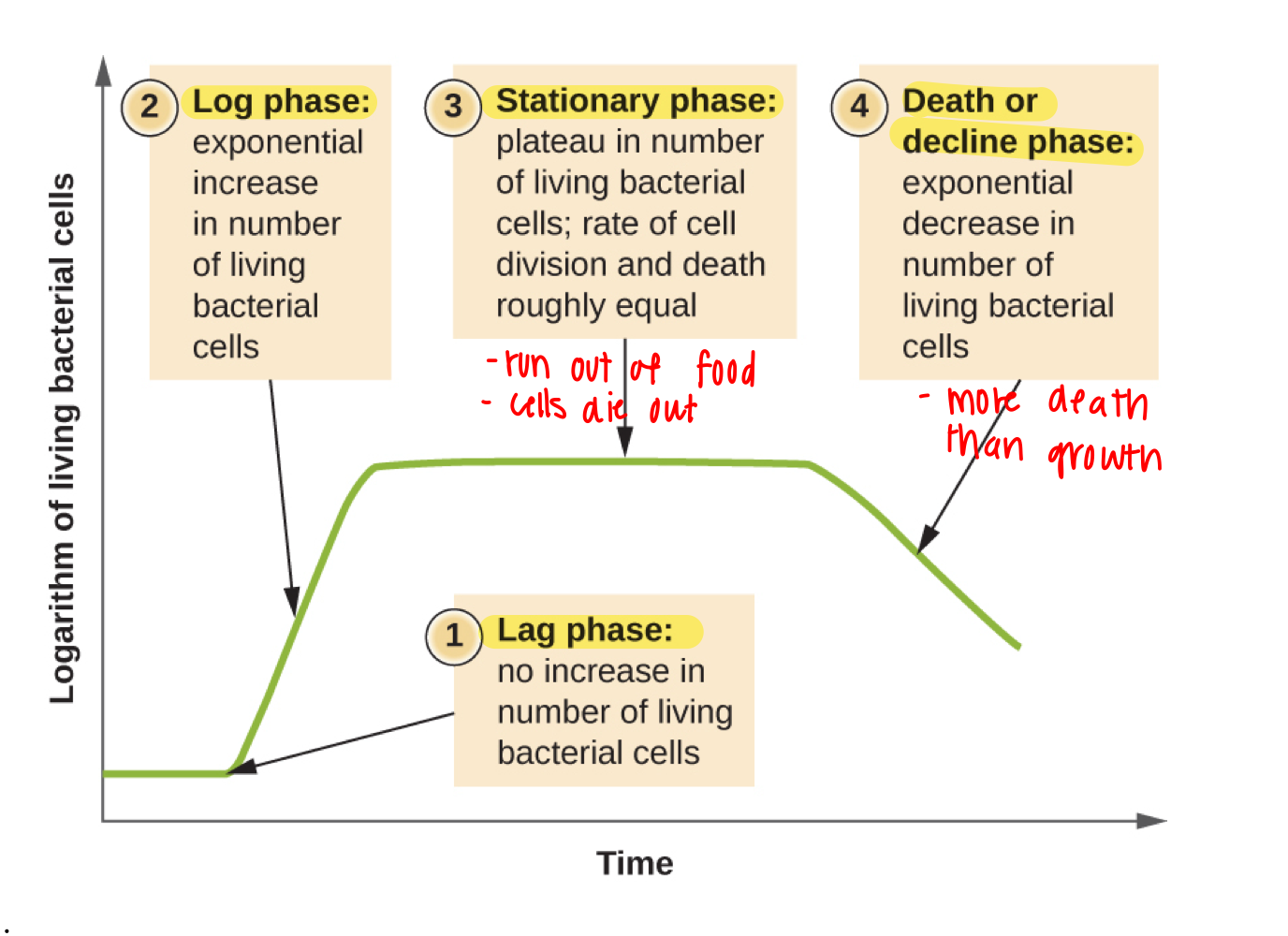
Closed System: Limited nutrients lead to initial rapid growth followed by predictably ceasing with nutrient depletion, leading through phases:
Lag Phase: Cells prepare for growth without division.
no increase in number of living bacterial cells
Log Phase: Cells divide rapidly as nutrients are abundant.
exponential increase in number of living bacterial cells
Stationary Phase: Balance between cell death and growth; as nutrients wane, waste accumulates.
plateau in number of living bacterial cells
rate of cell division and cell death are roughly equal
Death Phase: Higher death rate as resources become too low and wastes are toxic.
exponential decrease in number of living bacterial cells
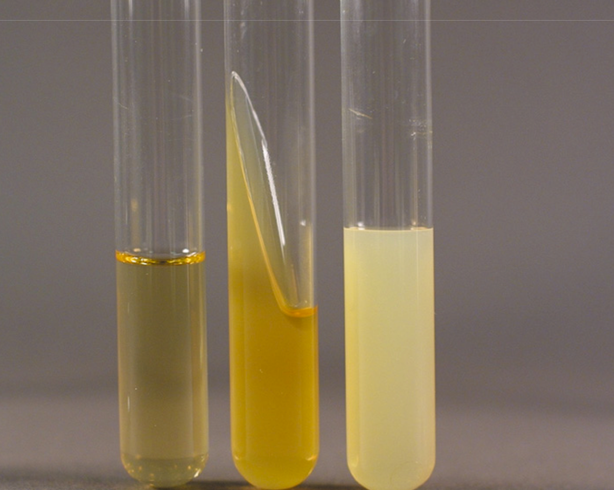
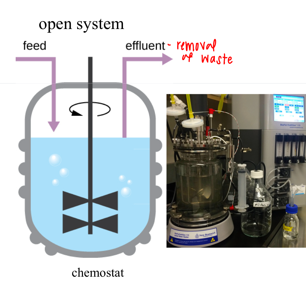
Open System (Chemostat): Fresh nutrients are consistently supplied and wastes removed, allowing indefinite growth potential.
Generation Time
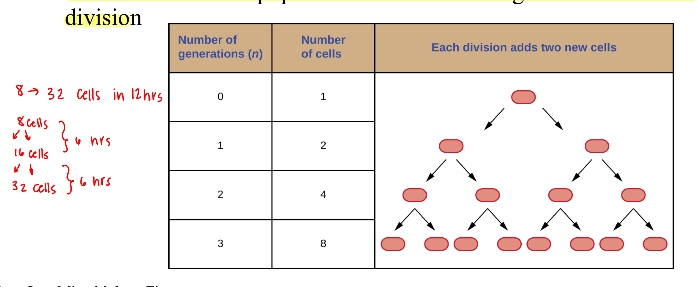
Generation Time: The duration required for a population of cells to double through one round of cell division
Example Calculation: Transitioning from 8 to 32 cells in 12 hours involves two ``rounds of division, indicating a generation time of 6 hours.
Breakdown: This is derived from needing 6 hours for the first division (8 to 16) and another 6 hours for the second (16 to 32).
Microbial Growth Conditions and Preferences
Oxygen Requirements:
Obligate Aerobes: Require oxygen.
Obligate Anaerobes: killed by oxygen.
Facultative Anaerobes: grow best with oxygen but can grow without it
Aerotolerant Anaerobes: Do not require oxygen but are not hurt by it
Microaerophiles: Require a precisely low amount of oxygen levels.
pH Preferences:
Acidophiles: grow best in acidic environments (low pH)
Neutrophiles: grow best in neutral pH.
Alkalophiles: grow best in alkaline/basic environments (high pH)
Temperature Preferences:
Psychrophile: grow best in cold
Psychrotroph: tolerate cold
Mesophile: grow best in moderate temperature
Thermophile: grow best in hot temperatures
Salinity Preferences:
Halophiles: grows best in high salt concentrations
halotolerant: tolerates high salt environments but do not require it to grow
Non-halophiles: grows best in low salt conditions.