HOMEOSTASIS
Overview
Page 1
Learning Objectives
Explore the basic structure of the integumentary system.
Discuss the function of the skin.
Explore the skin's role in immune function.
Examine the impact of environmental factors on skin health.
Examine various skin lesions.
Discuss the principles of skin assessment and care.
Introduction
The integumentary system functions as the body’s shield, acting as a barrier that is the first defense against harm. Together, the skin, hair, nails, and their associated glands help to protect against infection, regulate temperature, prevent fluid loss, and facilitate sensory perception. The skin also plays a role in vitamin D production, and the skin’s appearance, texture, temperature, and integrity can reflect changes in overall health. Understanding how this system works is essential for recognizing early manifestations of illness, injury, or systemic conditions.
In nursing, skin assessments are part of nearly every physical exam and can reveal critical information about circulation, oxygenation, hydration, nutrition, and more. Changes in the skin may be the first visible manifestations of pressure injuries, infection, or chronic disease. Nurses must know how to assess the skin accurately across all skin tones, document findings clearly, and intervene quickly to prevent complications. A strong foundation in the structure and function of the integumentary system supports safe, effective, and equitable care.
This module will provide nursing students with a foundational understanding of the integumentary system's structure and function, preparing them for more advanced topics in integumentary pathophysiology and care.
Overview Scenario
Consider the following scenario as you read the module content.
Scenario: Clarence is a 68-year-old client who has type 2 diabetes mellitus. He presents at the clinic for an annual checkup and asks the nurse to assess a wound on his foot that he noticed 2 weeks ago. The nurse notes that there is a deep, open sore on the top of Clarence’s toe. Clarence admits that he often wears tight shoes that rub against his skin. He is worried because the wound seems to be getting worse, not better.
Reflective Questions: What type of skin lesion is Clarence likely experiencing? What factors increase Clarence’s risk of wound development.
Introduction to the Structure of the Skin
Page 1
The skin is the heaviest organ in the body, making up about 15% of total body weight. This may seem surprising, but there is more to the skin than the part that can be visualized. Beneath the surface, many different structures comprise the integumentary system and play important roles in its functioning.
The integumentary system is one of the most important systems in the human body, and as such it has a complicated structure that enables its function. The skin has three main layers, contains appendages, and has several cells that support protection, sensation, and balance. The following sections will demonstrate the importance of each layer, how hair and nails form and grow, how glands contribute to system function, and how different cells keep the skin healthy.
Layers of the Skin
The skin is made up of three layers that work together to protect and support the body: the epidermis, the dermis, and the subcutaneous tissue (sometimes called the hypodermis). Each layer has its own structure and function, and all three are essential for maintaining health.
The layers of the skin are illustrated in the following image.
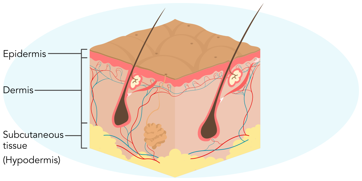
Layers of the SkinThe skin is composed of three layers: the epidermis, dermis, and the subcutaneous tissue (hypodermis).
The epidermis is the visible, outermost layer of skin. It acts as a barrier against germs, chemicals, and water loss. The epidermis is made mostly of cells called keratinocytes, which produce keratin. The epidermis also contains melanocytes that produce melanin. A single melanocyte distributes melanin to 30 to 40 keratinocytes. The outermost layer is made of corneocytes, which create a tough protective barrier.
keratin
melanin
corneocyte
3D Experience
Explore the visualization of epidermal skin layers
Read the text in the left panel to gain important information.
Move through the different views by using the arrows.
Click a label to closely inspect an area. Click the label again to zoom out.
Scroll up and down or use touch gestures to zoom in and out.
Click and drag or use touch gestures to rotate the visual.
Click the icon with three dots to access additional information about navigating the 3D visual.
Epidermal Skin Layers
Refer to the 3D visualization of epidermal skin layers and apply what you have learned to the following activity.
Match each epidermal cell type with the description of its function.
Drag the options on the left to the corresponding category on the right (or click the option on the left and then the corresponding category on the right).
Produce keratin and lipids; absorb UVB light for vitamin D activation; constantly replicate
Primary immune cells in the epidermis; engulf pathogens and migrate to lymph nodes
Synthesize melanin; determine skin color; stimulated by UVB light
Sensory cells for light touch; concentrated in fingertips, palms, soles, and mucosa
Beneath the epidermis is the dermis, a thicker layer that provides strength and flexibility. It consists of two sublayers, the superficial papillary dermis and the deep reticular layer. The dermis contains connective tissue—primarily collagen and elastin—which helps give skin its structure and flexibility. It also contains blood vessels, lymphatic vessels, and nerve endings, as well as hair follicles, sweat glands, and sebaceous glands.
papillary dermis
deep reticular layer
collagen
elastin
sebaceous gland
The deepest layer is the subcutaneous tissue, also known as the hypodermis. This layer is made mostly of loose areolar tissue and adipose tissue. The subcutaneous tissue cushions the body, provides insulation to maintain body temperature, and stores energy. The subcutaneous tissue also anchors the skin to the muscles and bones beneath it.
areolar tissue
adipose tissue
Together, these three layers form a strong, flexible covering that protects the body and supports many important functions.
Apply what you have learned about the layers of the skin to the following activity.
Match each layer of the skin with its corresponding characteristic or function.
Drag the options on the left to the corresponding category on the right (or click the option on the left and then the corresponding category on the right).
Contains keratinocytes and melanocytes, providing a barrier against germs and water loss
Contains connective tissue, blood vessels, and nerve endings, providing strength and flexibility
Composed mainly of fat and loose areolar tissue, providing insulation and energy storage
Cells of the Skin
Healthy skin depends on a team of cells that work together every day. These include cells that specifically contribute to skin integrity and function, such as keratinocytes and melanocytes. However, other cells also reside in skin tissues, including cells associated with the immune and sensory systems.
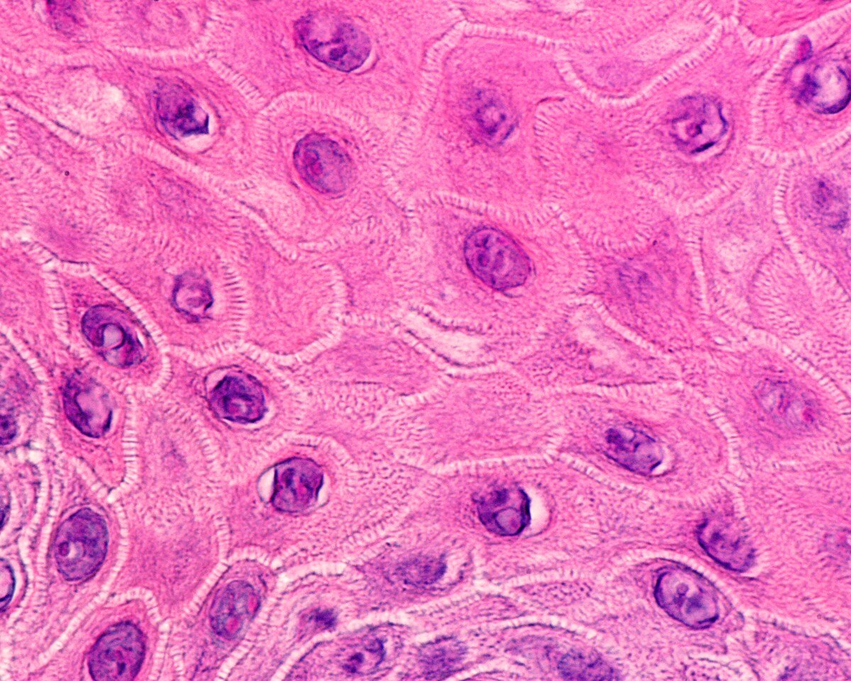
As previously mentioned, the most common type of skin cells are keratinocytes, which are depicted in the accompanying image. Without the keratin produced by keratinocytes, skin would lack strength and be prone to injury. Keratin also provides waterproofing, which enables the body’s water content to stay inside, where it belongs. Keratinocytes are formed in the deepest part of the epidermis, then move upward as they mature. When they reach the skin’s surface, they are flat and tough. They are then ready to shed and make room for new cells. This steady cycle supports healing, protects against germs, and keeps the surface smooth.
© Jose Luis Calvo/Shutterstock
KeratinocytesKeratinocytes flatten and pack themselves tightly together, giving structure and protection to the skin.
Mixed among the keratinocytes are melanocytes, which are dendritic cells. Melanocytes exist in the stratum basale, and their primary purpose is to produce melanin granules that pass through the dendritic extensions to the surrounding keratinocytes, forming a unit of melanin.
Making the Connection: The Importance of Melanin
When melanin is passed from the melanocyte to the keratinocyte, it forms a protective shield over the keratinocyte’s nucleus. This protects the cell’s DNA from the harmful effects of ultraviolet (UV) rays. There are two primary types of melanin: eumelanin and pheomelanin. In populations closer to the equator, where UV radiation is higher, individuals produce more eumelanin, a pigment that absorbs more UV radiation, reducing DNA damage. Conversely, populations in higher latitudes (further from the equator), have developed higher levels of pheomelanin, which can generate free radicals with UV exposure, increasing the risk of skin cancer.
free radical
Because UV radiation is the driver of vitamin D synthesis, clients who have higher melanin content and those who are in low-UV environments are at risk for lower vitamin D levels, requiring routine screening and supplementation if vitamin D levels are decreased. Excessive UV exposure can be detrimental, causing cellular damage increasing one's risk of skin cancer.
Langerhans cells are also found in the epidermis. These immune cells work by detecting harmful invaders like bacteria, viruses, or allergens. When they recognize a potential threat, they capture it, break it down, and carry pieces of it to nearby lymph nodes. There, they help activate other immune cells to start a defense response.
Additionally, the skin is host to a type of specialized cell, called a Merkel cell, that supports the sense of touch. Merkel cells sit close to nerve endings in the epidermis, especially in areas like the hands, feet, hair follicles, and oral and genital mucosa, and help with transmitting sensations of gentle pressure and fine texture.
Use what you have learned about skin cells to answer the following question.
A nurse is caring for a client who sustained a superficial skin injury. The client asks how the body’s defense system works. Which of the following cells should the nurse identify as being primarily responsible for initiating an immune response by capturing and presenting antigens to other immune cells?
a
Keratinocytes
b
Melanocytes
c
Langerhans cells
d
Merkel cells
Skin Appendages
Page 1
Skin Appendages
A skin appendage is any structure that grows from the skin, including hair, nails, and various glands. Appendages grow from the epidermis and anchor within the dermis, and together they add strength, protection, and control to the body’s outer covering. Although they appear different on the surface, each type of appendage shares a common building block of keratin, and they depend on nearby blood vessels and nerves in the dermis for nutritional and structural support.
Hair begins in a tiny pocket in the dermis called a hair follicle. The accompanying image illustrates the hair follicle and its location in the skin layers.
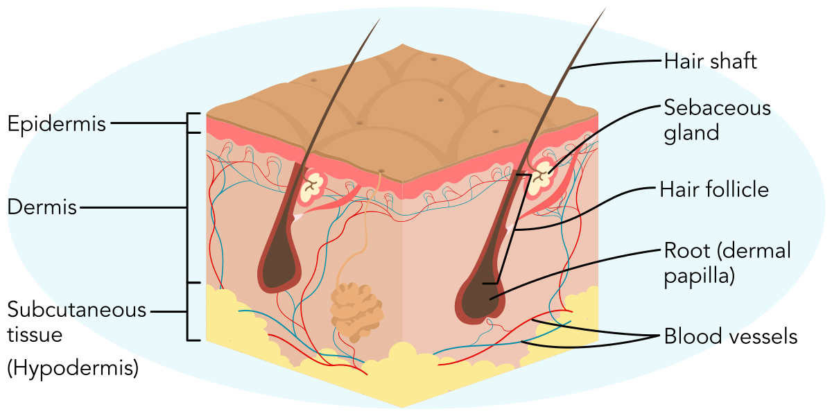
Hair FollicleEach hair follicle extends into the dermis, where it is connected to blood vessels that support it.
The part of the hair that is visible is called the hair shaft (or strand), while the hair root sits beneath the surface of the skin inside the follicle. At the base of the follicle, keratinocytes divide and harden to form the hair shaft, which grows upward to become visible. Hair color comes from melanin made by cells in the follicle, and growth follows a natural cycle with active growth, a short resting period, and shedding. The cycle length is variable and dependent upon multiple factors. Age, sex, and genetics may influence this cycle, as can the location of the body part where the hair is growing.
Located near the follicle are the sebaceous glands and a small muscle called the arrector pili. This muscle attaches to the follicle and can control both the sebaceous glands and the hair itself in response to environmental triggers. For example, the arrector pili can pull the hair upright, which creates the sensation of “goosebumps” when an individual feels cold or afraid.
Meanwhile, nails protect the tips of the fingers and toes and help with fine motor tasks. Like hair, nails are made of keratin. New nail growth starts in the nail matrix, which is a wedge-shaped structure that sits beneath the cuticle. The nail matrix generates the visible part of the nail, known as the nail plate. The nail plate lies on the nail bed. The keratin in the nail bed helps it adhere to the nail plate. As cells in the matrix add new layers, the plate slides forward in a smooth, steady movement. The hard plate shields delicate tissue and supports grip.
cuticle
The parts of the nail and nail bed are depicted in the following image.
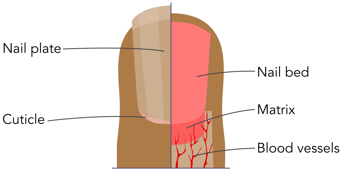
Parts of the Fingernail and Nail BedBeneath the keratin-based nail plate and bed are blood vessels that provide nutrients to support continuous nail growth.
Knowledge of the intricate structures of the nails provides foundational understanding of their function as well as the disease processes that impact them.
Apply your knowledge about the nail to the following question.
A nurse is teaching a client about the functions of skin appendages. Which of the following structures is primarily responsible for producing the visible part of the nail?
a
Nail bed
b
Nail matrix
c
Cuticle
d
Arrector pili
Throughout the skin, there are various glands that help the body stay balanced and protected. They release substances that support temperature control, moisture, and other natural functions that keep the skin healthy. There are three main types of exocrine glands within human skin: sebaceous glands, sweat glands (sometimes called sudoriferous glands), and mammary glands.
exocrine
Sebaceous glands tend to be found clustered in areas like the face, scalp, and upper trunk, and they often connect to hair follicles. They secrete an oily mixture called sebum that coats the hair and surrounding skin. Sebum is meant to keep the surface of the skin soft and moist and add a light barrier against water and some microbes. However, in puberty, sebum production increases in response to rising hormone levels. Increases in sebum can contribute to the development of acne.
There are two main types of sweat glands: eccrin and apocrin glands. Eccrine glands are spread across most of the body, including the palms, soles, and forehead. They release a watery sweat that evaporates and cools the body, which helps maintain a stable temperature during heat or exercise. Apocrine glands, on the other hand, are found mainly in the underarms and groin. These glands are larger and become active at puberty. They have a thicker secretion that has little odor at first. Rather, the body odor that is associated with sweat comes from skin bacteria that break the sweat down.
Mammary glands are in the breast tissue. They are a specialized kind of apocrine sweat gland that has been modified to produce milk. As such, the primary function of mammary glands is lactation in females. Although males have mammary glands from birth, testosterone levels during puberty inhibit the mammary glands, leading them to become a vestigial organ in males.
vestigial organ
Making the Connection: Lactational Mastitis
Mastitis is inflammation of tissue in the mammary glands that can lead to infection. Mastitis can happen to anyone, but it usually occurs in clients who are breastfeeding due to a buildup of milk within the breast. Manifestations include pain, tenderness, redness, and swelling. Treatment usually begins with supportive care like breast massage, lymph drainage, and pain control. Antibiotics may be provided if these methods are not sufficient.
Together, hair, nails, and glands extend the work of the skin. They protect sensitive areas, support touch and grip, help control temperature and moisture, and respond to hormonal and environmental changes. Their shared origin in the epidermis and their close connection to the dermis explain why skin health strongly influences how these appendages appear and function.
Apply what you have learned about skin glands to the following activity.
Match each type of gland with its corresponding function or characteristic.
Drag the options on the left to the corresponding category on the right (or click the option on the left and then the corresponding category on the right).
Located primarily in the scalp and face, these glands secrete sebum to prevent skin dryness.
Found on the forehead and palms, these glands release sweat to cool the body during exercise.
Present in the underarms, these glands produce a secretion that can lead to body odor.
Situated in breast tissue, these glands are responsible for milk production during lactation.
Function of the Skin
Page 1
The skin sheds about 30,000 cells every minute. Even with this constant turnover, it continues to regulate temperature, guard against infection, and help the body sense the world. How does it manage all of that without missing a step?
The integumentary system does more than just cover the body. It also protects against germs, helps keep the right amount of fluid inside, and works with blood vessels and sweat to hold temperature steady. It renews itself from the inside out through a steady cycle of cell growth and shedding. It makes pigments that provide some protection against damage from sunlight and repairs damage after injury. It also connects individuals to the world through touch, pressure, pain, and temperature by sending constant signals that help humans interact with their environment and stay safe.
Life Cycle of Epidermal Cells
Cells in the body are constantly replaced as part of the cell cycle. This is especially true of epidermal cells, which develop through a process known as keratinization. During this process, keratinocytes begin in the stratum basale, where new cells form. The cells then rise toward the surface, layer by layer, over the course of several weeks. As they move up through the other layers, they make keratin and release lipids that fill the tiny spaces between cells. By the time they reach the top, they have flattened and hardened into corneocytes. This gives the outer layer of skin a structure similar to bricks and mortar, with the corneocytes as the bricks and the lipids as the mortar that seals the wall.
lipid
This constant turnover builds a smooth, flexible barrier that helps keep water in and germs out. Enzymes at the surface loosen the bonds between old cells so they can shed in thin, even flakes, making room for the fresh cells coming up from below. When the keratinocyte process speeds up, which happens with conditions such as psoriasis, the surface can appear thick and scaly because cells arrive before they fully mature. With aging, the process slows down and the lipid seal thins, which may cause the skin to crack, feel dry or irritated, and become less able to protect against environmental factors.
psoriasis
Use what you have learned about skin keratinization to answer the following question.
A nurse is educating a newly licensed nurse about the process of keratinization in the skin. Which of the following statements best describes the role of lipids in the keratinization process?
a
Lipids provide structural support to keratinocytes, allowing them to rise through the epidermal layers.
b
Lipids act as enzymes that facilitate the shedding of old skin cells at the surface.
c
Lipids accelerate the maturation of keratinocytes, leading to faster cell turnover.
d
Lipids fill spaces between cells, creating a seal that helps retain moisture and block pathogens.
Role of Melanin
Melanin plays an important role in protecting the body by shielding the nucleus of skin cells from UV radiation. After melanocytes create the melanin, they then package it into tiny bundles and pass it on to nearby keratinocytes. People across the world have a similar number of melanocytes. What determines differences in skin color mainly comes from how much melanin these cells make, how large the bundles are, and how they are arranged.
Melanin also helps shield the skin from UV radiation. When exposed to sunlight, melanocytes begin to produce extra melanin to protect the body from UV damage, resulting in a deeper skin tones for some people. Increased melanin absorbs and scatters some of the energy from sunlight and helps neutralize harmful byproducts like free radicals that form after exposure.
Making the Connection: UV Protection
It should be noted that there is no such thing as a “healthy tan.” Too much UV radiation damages the cells and increases the risk of skin cancer even if there is no sunburn. Everyone, including clients who have deeper skin tones, needs to practice sun safety. Educate clients of all skin tones on the importance of applying sunscreen with SPF 30 or higher about 20 minutes before sun exposure and advise clients to wear protective clothing and spend time in the shade when the sun is strongest.
Changes in melanin can shape common skin findings. If melanocytes make less pigment or the body loses these cells in certain areas, pale patches can appear in a condition called vitiligo. Increased pigmentation of the skin can occur due to irritation or injury, which is referred to as postinflammatory hyperpigmentation (PIH). Hormonal changes can also increase pigmentation, causing darker patches to form in a condition known as melasma. Understanding how melanin is made and how it moves through the epidermis explains both expected skin color and many of these common conditions.
Thermoregulation
Podcast Experience

In this episode of Pint-Sized Patho, the host explains how the skin, blood vessels, and sweat glands coordinate thermoregulation through processes like vasodilation, vasoconstriction, sweating, and piloerection to maintain a stable internal body temperature.
How Skin Regulates Body Temperature
00:00
01:24
To download a podcast episode, click the three dots at the end of the player. Choose "download" and click "okay." Select where to save the file.
Podcast Transcript
After you listen to the episode, consider the following reflective questions.
What is one way understanding the skin’s role in temperature regulation might help you better assess or care for a client in a clinical setting?
The skin helps maintain a safe internal temperature by adjusting blood flow and triggering sweat production in response to changes in the environment. This process is called thermoregulation, and it depends on a close partnership among the skin, blood vessels, sweat glands, and nervous system.
When the body becomes too warm, blood vessels in the dermis widen in a process known as vasodilation. This brings more blood to the surface, where heat can escape into the air. At the same time, sweat glands release sweat onto the skin, where it evaporates, cooling the surface and lowering body temperature.
When the body needs to conserve heat, the opposite happens. Blood vessels narrow through vasoconstriction, which reduces blood flow near the surface and limits heat loss. The arrector pili muscles also play a role in thermoregulation. When the ambient temperature is warm, the arrector pili is relaxed. However, in colder conditions, the muscle contracts, causing the hair to stand straight, as depicted in the accompanying image.
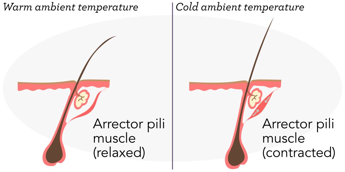
Arrector Pili MuscleThe arrector pili muscle attaches to the hair follicle and assists with the thermoregulation process.
When the arrector pili at the base of hair follicles contracts, the hair stands up. This causes the hair to trap a thin layer of air close to the skin, which helps retain body heat when an individual is cold.
Apply what you have learned about thermoregulation to the following question.
Which of the following physiological responses in the process of thermoregulation occur when the body becomes too cold?
Select all that apply.
a
Blood vessels increase blood flow to bring more heat to the skin's surface.
b
The arrector pili muscles contract, causing hair to trap body heat close to the skin.
c
Vasoconstriction occurs in the blood vessels to limit heat loss.
d
Melanin production is increased to help absorb more heat from the sun.
e
Keratinocytes pack more closely together to prevent heat from escaping the body.
Infection Prevention
One of the most important jobs of skin is to act as a barrier against infection. Its outer layer forms a physical shield that blocks bacteria, viruses, and fungi from entering the body, while natural oils and sweat add chemical protection by creating an environment that is less friendly to harmful microbes. The skin also works with the immune system, using specialized cells to detect and respond to threats. This topic will be explored later in this module with a detailed discussion of how these defenses work together to keep the body safe.
Fluid Loss Prevention
Along with blocking germs, the skin also protects the body’s water supply. The surface cells flatten and pack tightly, and natural oils spread between them to seal tiny spaces. A light film of oil and sweat slows evaporation. The surface is also slightly acidic, which supports the enzymes that keep this seal working well. Together, these features limit everyday water loss while keeping the skin flexible.
When this barrier is damaged, as illustrated in the following image, water escapes faster and the skin can dry, sting, or crack. Severe burns are the clearest example. Because the destroyed surface cannot hold fluid, this puts the person at risk for dehydration. But milder problems, such as very dry or inflamed skin, can also weaken the seal and increase water loss. Gentle cleansing, steady moisturization, and protection from further irritation help restore the surface and reduce fluid loss so healing can move forward.
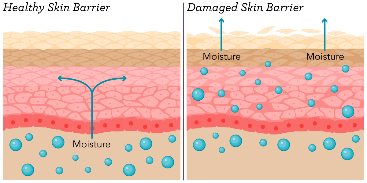
Skin BarrierA damaged skin barrier allows moisture to escape, raising the risk of dehydration.
Wound Healing and Tissue Repair
When the skin is injured, it begins repairing itself almost immediately. This process happens in four stages and involves both the epidermis and the deeper layers. It begins with hemostasis, in which the body responds by stopping bleeding. Blood vessels constrict, and clotting factors form a plug that seals the wound. Next is the inflammatory stage, wherein the body sends immune cells (including neutrophils and macrophages) to clear away debris and reduce the risk of infection. This early cleanup sets the stage for new growth.
After the inflammatory stage, the proliferation phase begins. Keratinocytes, a type of epithelial cell, in the epidermis start dividing and moving across the wound to close the gap, while fibroblasts in the dermis produce collagen that rebuilds the framework beneath the surface. New blood vessels grow into the area to bring oxygen and nutrients, which support healing. Finally comes the remodeling stage, when the tissue strengthens and remodels over time.
fibroblast
The following diagram illustrates the phases of the wound healing process in the skin.
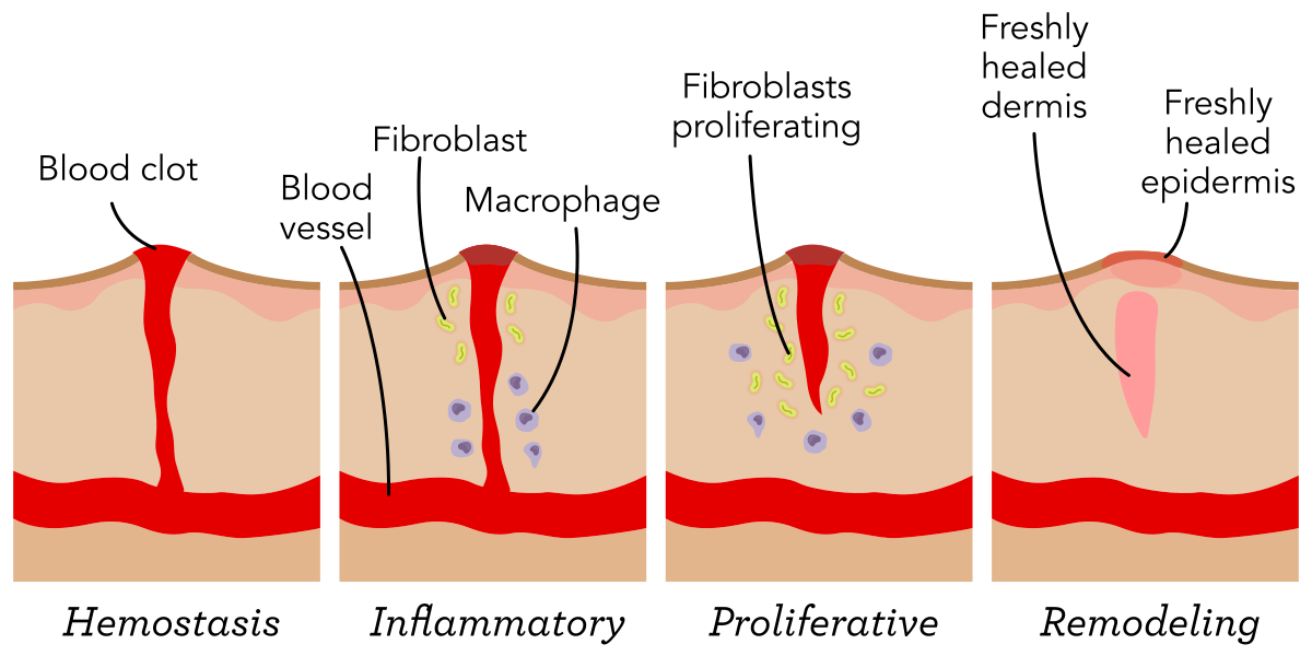
Wound HealingThere are four phases in the wound healing process: hemostasis, the inflammatory stage, the proliferative stage, and remodeling.
Wound healing is usually a smooth process, but it can slow down with infection, poor circulation, poor nutrition, or certain health conditions, such as diabetes mellitus. Understanding how wounds heal helps to recognize expected progress and identify problems early, which is essential for quick intervention and better client outcomes.
Animation Experience
This animation covers the wound healing process using an example of a cut to the skin and outlining the various phases of healing.
The Phases of Wound Healing
Animation Transcript
Refer to the animation to apply what you have learned about the phases of wound healing to the following activity.
Sensory Functions
In addition to functioning as a barrier, the skin also functions as a sensory organ that connects the body to the outside world. Scattered throughout the dermis and deeper layers are specialized receptors that detect touch, pressure, pain, and temperature. These receptors work with nerve endings to send signals to the brain, allowing the individual to respond quickly to changes in the environment.
Sensory Receptors
Sensory receptors are discussed in multiple modules. This module focuses on their role in the integumentary system. For more information about their role in the sensory system, please refer to the module Structure and Function of the Sensory System.
Light touch is sensed by mechanoreceptors near the surface, especially in areas like the fingertips and lips, where sensitivity is highest, while deeper in the skin, other receptors respond to steady pressure or vibration, helping to perceive texture and movement. Thermoreceptors detect warmth and cold, while nociceptors alert the body to potential harm, such as extreme heat, sharp objects, or injury. Together, these sensory systems allow the skin to protect the body not only by forming a barrier, but also by warning of danger and guiding safe interaction with the world.
Role of the Skin in Immune Function
Page 1
Podcast Experience
In this episode of Pint-Sized Patho, the host describes how the integumentary system protects the body through layered defenses, including a physical and chemical barrier, pathogen-detecting Langerhans cells, and immune responses in the dermis that eliminate threats and promote healing.
How Your Skin Fights Pathogens
00:00
01:40
To download a podcast episode, click the three dots at the end of the player. Choose "download" and click "okay." Select where to save the file.
Podcast Transcript
After you listen to the episode, consider the following reflective questions.
Redness, warmth, and swelling can happen quickly after a skin injury. Based on what you learned about mast cells and histamine, how would you explain these manifestations to a client?
Every day, the skin encounters countless microorganisms, allergens, and irritants. To keep the body safe, it works closely with the immune system. Specialized cells within the skin detect threats, trigger responses, and help prevent infection. When injury or infection occurs, the skin responds quickly through inflammation and other defense mechanisms. It also participates in allergic reactions and hypersensitivity, which are immune responses that can sometimes cause discomfort. Understanding these processes helps explain why the skin is essential for overall health and why its immune functions are so important in protecting the body.
Inflammation and Immune Function
Inflammation and hypersensitivity reactions are discussed in multiple modules. This module focuses on how they manifest in the integumentary system. For more information about inflammation, please refer to the module Inflammation. For more information about hypersensitivity reactions, please refer to the module Immunity.
First Line of Defense
As mentioned throughout this module, the skin is the first line of defense against harmful organisms and substances. Additionally, the surface of the skin is slightly acidic, which discourages the growth of many microorganisms. Natural oils and sweat also help by washing away dirt and carrying antimicrobial substances that kill or slow the growth of germs.
In addition to these chemical defenses, the skin’s structure provides strong protection. The epidermis forms a physical barrier that blocks most bacteria, viruses, and fungi from entering the body. The tightly packed cells of the epidermis and the presence of keratin make it difficult for pathogens to penetrate. Even hair and nails help by adding extra layers of protection in sensitive areas. Together, these features create an effective shield that works constantly to keep the internal environment safe.
Immune Cells of the Skin
The skin is an active part of the immune system. The immune cells that are present in the skin are represented in the following image.
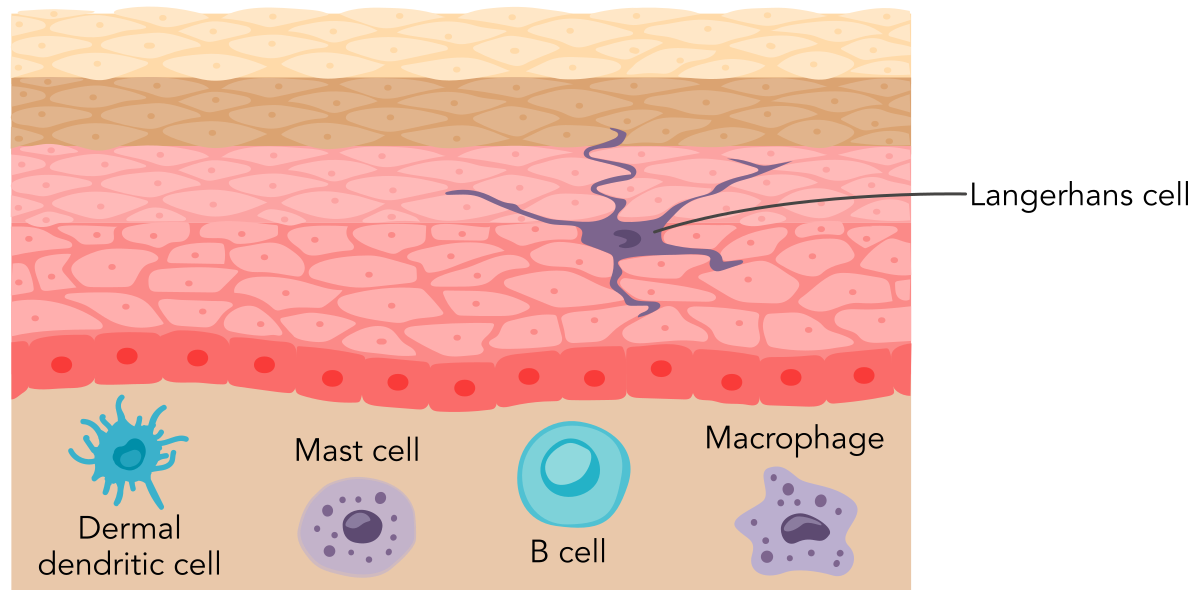
Immune CellsThere are several immune cells that are present in the skin. Each one plays a different role in protecting the health of the skin and body.
Within the epidermis, there are specialized immune cells called Langerhans cells. These cells act like sentinels, constantly monitoring for harmful substances such as bacteria, viruses, and allergens. When they detect a threat, they capture and process the foreign material, then travel to nearby lymph nodes to alert other immune cells. This process helps the body mount a defense before an infection can spread.
Other immune cells are also present in the skin. Macrophages and dendritic cells in the dermis work alongside Langerhans cells to destroy pathogens and remove damaged tissue. Mast cells, which are also found in the dermis, play a role in allergic reactions and inflammation by releasing chemicals like histamine. Together, these immune cells create a strong defense network that works continuously to protect the body from infection.
histamine
Use what you have learned about the immune function of the skin to answer the following question.
A nurse is discussing the immune functions of the skin with a client. Which of the following best describes the role of Langerhans cells in the epidermis when they detect a harmful substance?
a
Capture and process foreign material, then travel to lymph nodes to alert other immune cells
b
Release histamine to initiate an inflammatory response
c
Increase blood flow to the affected area to bring more immune cells
d
Destroy pathogens directly through phagocytosis
Inflammatory Response
When the skin is injured or invaded by harmful organisms, it reacts quickly through inflammation. This response is the body’s way of preventing infection and starting the healing process. The first step is an increase in blood flow to the affected area, which brings white blood cells (WBCs) and nutrients needed for repair. This extra blood flow causes redness and warmth. Blood vessels become more permeable, which allows immune cells and proteins to move into the tissue, leading to swelling and sometimes pain.
WBCs, such as neutrophils and macrophages, play an important role during this stage by invading organisms and clearing away damaged tissue. Cytokines help coordinate these actions by attracting more immune cells to the site. Although inflammation can cause discomfort, it is an essential defense mechanism that protects the body and promotes healing.
cytokine
Hypersensitivity Reactions
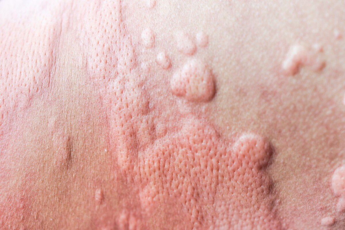
The skin is often the first place where allergic reactions appear. When the immune system reacts too strongly to something harmless like pollen or some foods, medications, or chemicals, the skin can develop redness, itching, and swelling. This happens because immune cells in the skin, including mast cells, release chemicals like histamine that increase blood flow and make blood vessels more permeable. This leads to the familiar manifestations of an allergic reaction, such as urticaria, which is depicted in the following image. Manifestations may come and go quickly, disappearing after only a few minutes.
urticaria
© OoddySmile Studio/Shutterstock
UrticariaUrticaria is also known as hives. It presents as itchy, raised bumps on the skin that indicate a hypersensitivity reaction.
Hypersensitivity reactions can vary in severity. Some are mild, such as a rash from contact with a plant or cosmetic product, while others can be more serious, involving widespread urticaria, edema, or anaphylaxis. In all cases, the skin plays an active role by signaling that the body perceives a threat. Understanding these responses is important because they demonstrate how closely the skin and immune system work together, even when the reaction is exaggerated.
Making the Connection: Hypersensitivity and Real-Life Skin Assessment
Type I hypersensitivity reactions are central to nursing because the skin is often the first organ to reveal an allergic response. When IgE antibodies on mast cells encounter an allergen, histamine and other mediators are released, producing rapid‑onset manifestations such as urticaria, pruritus, flushing, and angioedema. These visible changes provide early clues that the immune system has been activated. In everyday practice, nurses frequently encounter these reactions. For example, a client may develop acute urticaria within minutes of receiving an antibiotic, presenting with raised, erythematous wheals and intense itching. Current clinical reviews emphasize that acute urticaria is commonly triggered by medications and represents a classic IgE‑mediated hypersensitivity response, reinforcing the importance of rapid recognition and intervention. By grounding immunology concepts in real‑world skin assessment, nurses strengthen their ability to identify hypersensitivity reactions early, intervene appropriately, and educate clients about triggers and prevention.
pruritus
Hypersensitivity reactions begin when antibodies, already bound to mast cells during prior sensitization, encounter an allergen and trigger rapid degranulation. This release of histamine, leukotrienes, and other mediators produces the immediate vascular changes that can be visualized on the skin, including vasodilation, increased capillary permeability, and sensory nerve stimulation. These mechanisms explain why manifestations such as erythema, edema, and pruritus can appear within minutes of exposure. For nurses, recognizing that these visible skin findings reflect an underlying immune cascade helps connect what is observed at the bedside with the biologic processes driving the reaction, reinforcing the importance of early assessment and intervention.
erythema
Apply what you have learned about allergic reactions to the following question.
A nurse is educating a client about allergic reactions that manifest on the skin. Which of the following best explains the role of mast cells in the development of skin manifestations during an allergic reaction?
a
Mast cells produce antibodies that neutralize allergens on the skin.
b
Mast cells release histamine, increasing blood flow and vessel permeability, leading to redness and swelling.
c
Mast cells directly attack allergens, preventing them from entering the bloodstream.
d
Mast cells secrete enzymes that break down allergens on the skin surface.
Impact of Environmental Factors on the Skin
Page 1
Impact of Environmental Factors on the Skin
Podcast Experience
In this episode of Pint-Sized Patho, the host explains how the skin preserves the body’s internal fluid balance through keratinized cells, lipid barriers, and an acidic surface environment that work together to prevent water loss and protect against dehydration.
How Skin Prevents Dehydration
00:00
01:31
To download a podcast episode, click the three dots at the end of the player. Choose "download" and click "okay." Select where to save the file.
Podcast Transcript
After you listen to the episode, consider the following reflective questions.
How does this information change the way you think about everyday nursing actions like bathing, skincare, or wound care?
The skin is constantly exposed to the outside world, which means it faces many environmental challenges every day. Sunlight, pollutants, and irritants can all affect how the skin appears and functions. Over time, these factors can lead to changes in skin health, including damage that may increase the risk of disease. Nutrition and hydration play an important role in keeping the skin strong and able to repair itself. In addition, the natural aging process changes the way the skin appears and works, making it more likely to be affected by environmental stress. Understanding these influences helps explain why caring for the skin is essential for overall health.
UV Exposure
Sunlight is important for health. It is the primary source of vitamin D in the body, which is crucial for bone health. However, too much exposure can damage the skin. UV rays from the sun penetrate the epidermis and reach deeper layers of the stratum basale and stratum spinosum. Short-term overexposure can cause a tan or sunburn, both of which are an inflammatory response to cell injury. With sunburn, the skin becomes red, warm, and painful because blood vessels widen and immune cells move in to repair the damage. Severe sunburn can lead to blistering and peeling as the body removes damaged cells. In clients who have darker skin tones, redness from sunburn may be difficult to notice. Instead, skin color may appear as usual or slightly darker.
Long-term exposure to UV radiation is even more harmful. Repeated damage to skin cells can change their DNA, which increases the risk of skin cancer. The most common types are basal cell carcinoma, squamous cell carcinoma, and melanoma. Of these, melanoma is considered the most concerning because it can extend beyond the original area and affect additional parts of the body.
Making the Connection: Recognizing Melanoma
Skin cancer is the most common type of cancer in the U.S. Melanoma is a dangerous form of skin cancer that can spread quickly if not detected early. It affects clients of all skin tones. The characteristic findings of early melanoma are recognized with the following well-known ABCDE mnemonic:
A: Asymmetry: the lesion is not the same shape on the right side as compared to the left, or on the top as compared to the bottom of the lesion
B: Border: irregular, ragged, notched, or blurred edges
C: Color: nonuniform or varied
D: Diameter: larger than 6 millimeters
E: Evolving: changes in size, shape, or color
In clients who have darker skin, it often appears in areas like the palms, soles, or under the nails, which can lead to delayed diagnosis and treatment. Risk factors include multiple sunburns, a history of blistering sunburns as a child, tanning bed use, having many moles, and a family history of skin cancer. When caught early, melanoma has a 97% 5-year survival rate, which makes early detection and prevention crucial.
UV exposure also speeds up skin aging by breaking down collagen and elastin, leading to wrinkles and loss of elasticity. These effects demonstrate why protecting the skin from excessive sun exposure is essential for maintaining health.
Apply your knowledge about UV radiation’s effect on the skin to the following question.
A nurse is educating a client about the effects of UV radiation on the skin. Which of the following statements would the nurse include as part of the teaching?
a
“UV radiation causes immediate redness and warmth, indicating long-term skin damage.”
b
“UV radiation increases vitamin D production, which can lead to skin cancer.”
c
“UV radiation causes the skin to become darker, which is a sign of cancer.”
d
“UV radiation penetrates the epidermis and causes DNA changes in skin cells.”
Pollutants and Irritants
The skin is continually in contact with a wide range of substances in the environment that can cause harm. Pollutants in the air, such as smoke, chemicals, and particulates, can settle on the skin and lead to irritation or damage like clogged pores, inflammation, delayed wound healing, and collagen breakdown. Some pollutants also produce free radicals that speed up the aging process.
Irritants, such as harsh soaps, cleaning products, and certain chemicals, can also weaken the skin’s barrier. When this happens, the skin loses moisture more easily and becomes more sensitive to allergens and infections. People who work in environments with frequent chemical exposure or poor air quality often experience these effects more severely, so protection is key.
Nutrition and Hydration
Healthy skin depends on a steady supply of nutrients and water. The skin renews itself every day, so it needs building blocks for structure, energy for cell work, and fluids to keep the surface flexible. When intake supports these needs, the outer barrier stays strong, the deeper layers receive oxygen and nutrients, and small injuries repair in a timely manner.
Protein supplies the amino acids that form collagen and keratin. These are crucial to maintaining the strength and shape of the tissue. Meanwhile, dietary fats help build the lipid seal between surface cells, which keeps water in and irritants out. A balanced mix of vitamins and trace minerals guides cell growth and helps enzymes assemble new tissue. For example, vitamin C helps collagen fibers come together, and zinc supports cell division at the edges of a wound. Water carries these materials to the dermis. Water also works at the surface, where natural moisturizing factors bind and hold it, so the outer layer of skin stays soft and flexible.
When diet or fluids fall short, the barrier loses water more quickly and the surface becomes dry and itchy. As a result, small cracks can form, and irritants enter more easily. Healing slows because collagen production and cell movement lag, and low fluid intake reduces blood flow to the skin. Too much fluid can cause swelling that stretches fragile tissue and raises the chance of breakdown. Regular meals that include enough protein, fruits, vegetables, and healthy fats, along with consistent fluid intake that matches a client’s needs, can help the skin resist injury and recover after damage.
Skin Across the Lifespan
Skin changes steadily with age. Early in life, it grows quickly and repairs small injuries with ease. However, in older adulthood the pace slows. Cells divide more slowly, the barrier holds less water, and the deeper layers lose strength. These shifts reflect natural changes with time, genetics, and years of sun and weather exposure, and together they reshape both how the skin appears and how it works.
Over time, the epidermis becomes thinner and renews itself at a slower rate. Surface lipids decline, so the barrier holds less moisture and lets irritants in more easily. Sweat and oil on the surface also decrease, which adds to dryness and itching. At the same time, melanocytes become fewer and less active, so color can appear paler or translucent in areas, with lighter areas and darker spots. These are known as lentigos, although they are sometimes referred to as “age spots” or “liver spots.” Additionally, the blood vessels within the dermis become more fragile as an individual ages, leading to increased bruising. Immune sentinels in the epidermis such as Langerhans cells decline with age, so the skin detects threats less quickly and clears them less efficiently. The subcutaneous fat layer thins as well, which lessens insulation. This also leads to an increased risk of injury and decreases the ability to maintain one’s body temperature.
The skin alterations that occur with aging are depicted in the following image.
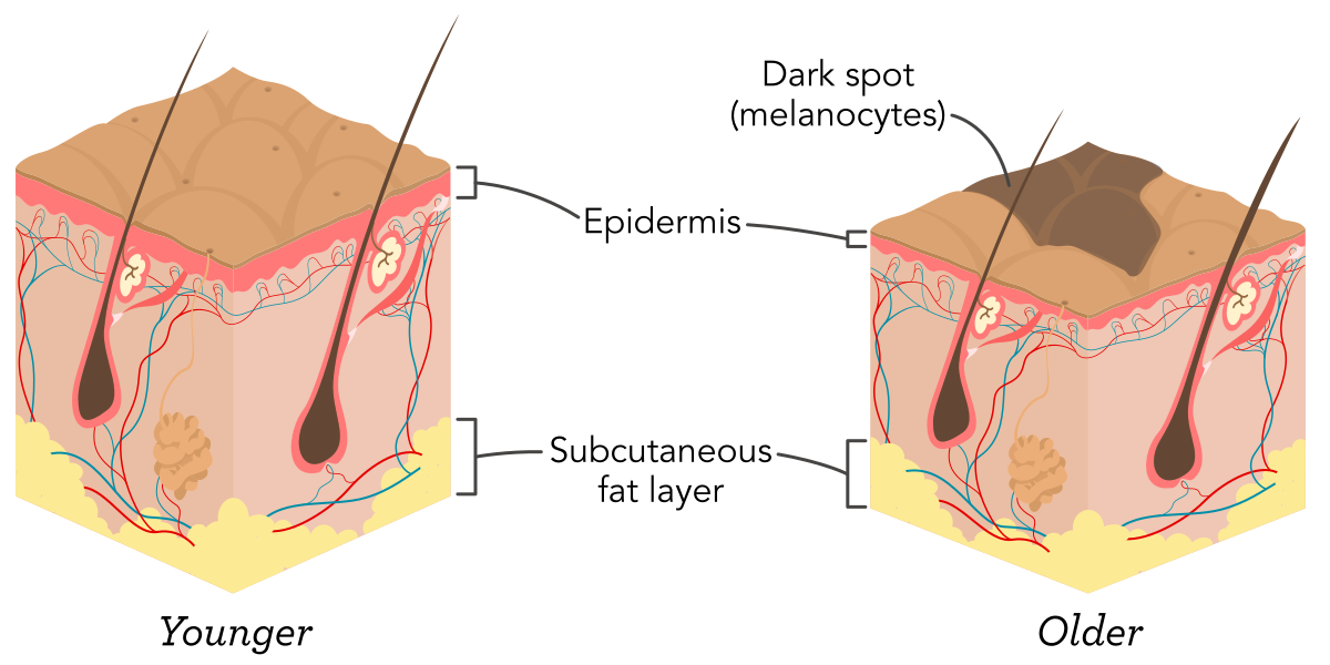
Skin Changes With AgeAs the skin ages, cells grow and repair themselves more slowly, leading to a thinner, more fragile epidermis. Additionally, the subcutaneous fat layer thins, and melanocytes become less active, leading to lighter and darker patches on the skin.
As these age‑related structural changes accumulate, the skin’s ability to protect, repair, and regulate itself becomes progressively less efficient. Reduced cellular turnover, diminished immune surveillance, and thinning of the dermal and subcutaneous layers all contribute to slower healing, increased vulnerability to irritation, and a higher risk of injury or infection. These physiological shifts alter both the appearance and function of aging skin, reinforcing why older adults often present with dryness, fragility, and delayed recovery from even minor insults. Understanding these underlying mechanisms helps nurses anticipate common concerns in older adults and tailor assessments and interventions accordingly.
Use what you have learned about aging and the integumentary system to answer the following question.
A nurse is educating a group of older adults about changes in skin health with aging. Which of the following changes should the nurse include as common in older adults?
Select all that apply.
a
Increased rate of epidermal cell renewal
b
Decreased surface lipids leading to dryness and itching
c
Fewer and less active melanocytes causing patchy skin color
d
Increased subcutaneous fat layer
e
Decline in Langerhans cells reducing immune response
Skin Assessment and Care
Page 1
Skin Assessment and Care
Integumentary assessment is a routine but essential part of nursing practice. A thorough head-to-toe assessment of the integumentary system involves the skin, hair, and nails. Skin especially can reveal early manifestations of systemic illness, circulatory issues, infection, or injury, making it a valuable tool in clinical evaluation.
Nurses must approach skin assessment with attention to detail, using both observation and touch to gather accurate information. A thorough examination includes both subjective and objective data. By learning to assess the skin carefully and consistently, nurses can support early diagnosis, guide treatment decisions, and help prevent complications such as skin breakdown.
Assessment Techniques
Subjective data is gathered by asking the client questions about their skin manifestations, such as itching, burning, tingling, or pain, as these manifestations often occur before visible changes. It also includes asking the client if they have a history of any skin- or hair-related disorders, and if they are currently using any prescription or over-the-counter (OTC) products for their skin, hair, or nails.
Accurate skin assessment is essential for detecting early manifestations of illness or injury, but changes in color and texture can appear differently across skin tones. Historically, most training materials have focused on lighter skin, which can lead to delayed recognition of conditions in clients who have darker tones. Nurses must use inclusive techniques to ensure timely, equitable care for all clients. In some cases, assessment techniques may require the use of tools other than visualization, such as ultrasound or infrared imaging, when changes to skin coloration are difficult to see.
Objective data is gathered by assessing the color, temperature, moisture level, turgor, and any lesions or skin breakdown. Explain to the client what the assessment will involve and make sure there is good lighting available. During visual assessment, natural light is preferred. If natural light is unavailable, always use bright white light for assessment. Inspect high-risk areas like bony prominences, folds, and skin that is under devices such as casts and face masks.
turgor
Compare the appearance of the area being assessed to the surrounding area of skin. Note any changes in color, such as pallor, cyanosis, jaundice, or erythema. Pallor may appear as ashen gray or a yellow-brown hue in more pigmented skin and as a loss of pink in less pigmented skin. To help identify pallor, check highly vascularized areas such as the palms, nail beds, conjunctiva, and buccal mucosa.
pallor
cyanosis
jaundice
Cyanosis is most noticeable in the mucous membranes, such as the lips, tongue, and oral mucosa. It can also be clearly present in the nail beds and soles of the feet. Cyanosis may present as gray, white, or greenish in clients who have deeper skin tones and as blue in clients who have lighter skin tones.
Jaundice is best noted in the changes in scleral color with a yellow discoloration. Other areas to assess include the hard palate, oral mucosa, palms, and soles. Changes in skin color may be more easily observed in lightly pigmented skin and may be less apparent or occur later in highly pigmented skin.
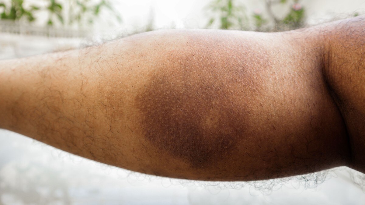
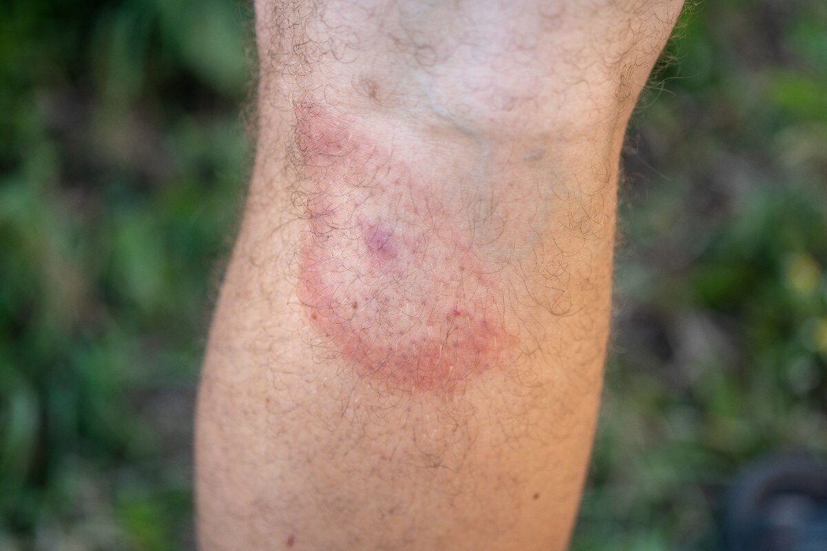
The image following demonstrates the appearance of erythema in clients who have different skin tones.
sclera
© Nomad Rabbi/Shutterstock
© Alexey Androsov/Shutterstock
ERYTHEMA Erythema presents differently on darker skin tones than it does on lighter skin tones.
Assess for ecchymosis, rashes, petechiae, unusual moles, or burns. Assess overall skin integrity and identify any manifestations of breakdown, paying special attention to the skin folds and skin that is under devices such as casts. Assess sacrum and bony prominences, which are identified in the following image. This includes the heels, hips, elbows, and shoulders.
ecchymosis
petechiae
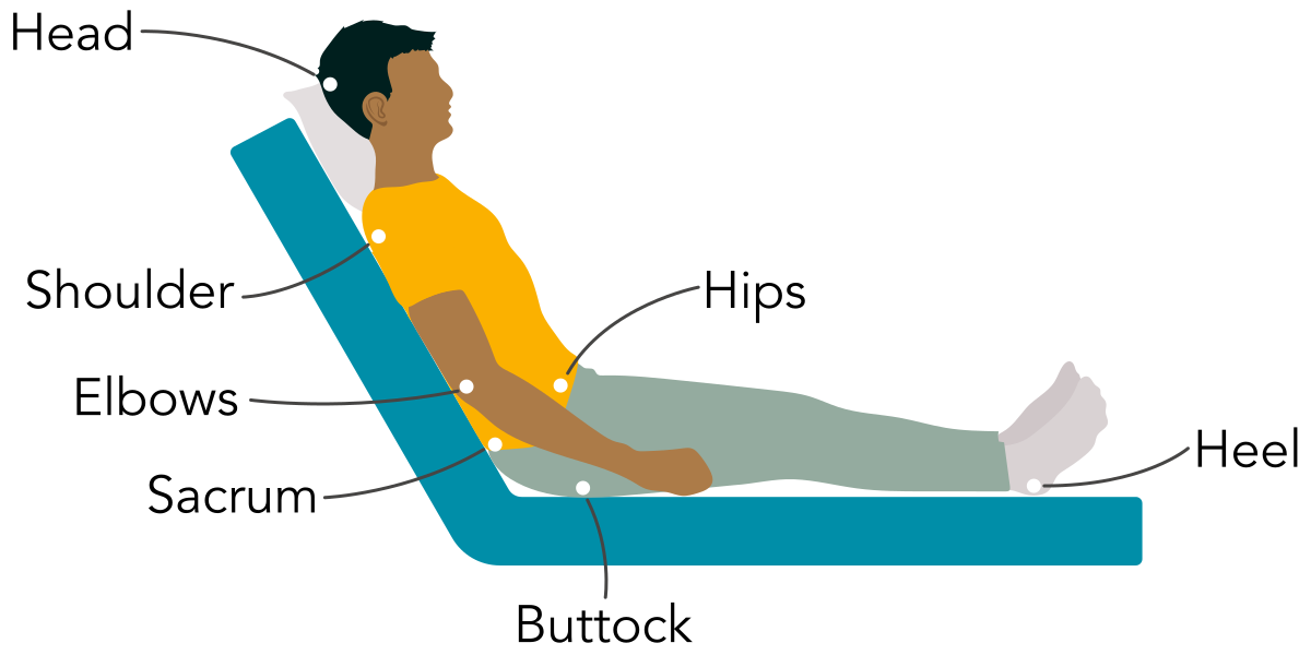
Bony ProminencesSpecial care must be paid to bony prominences. These are areas where the bone is closest to the skin, making the skin especially fragile and prone to breakdown.
Palpation can provide additional information about skin temperature, moisture, texture, and turgor. The dominant hand should be used to check temperature, noting if the skin feels warm, cold, or clammy. Diaphoresis may indicate a fever or systemic illness, while localized warmth may indicate infection or inflammation.
diaphoresis
Nails should be examined for discoloration, cracking, or splitting. While commonly these are minor aesthetic concerns, they could be manifestations of more serious diseases that would require further investigation. Disruption of the nail can cause disability and decreased function in clients. Detailed history and examination are needed for accurate diagnosis. Examine the nails for pitting, which is an unexpected keratinization of the nail matrix that results in deep pits within the nail plate. This is present in conditions such as psoriasis or eczema. Nail shape is another area to assess. Upward curving of the distal nail plate (described as “spoon-like”), could be idiopathic in nature or could be associated with iron deficiency anemia, warranting further investigation. Digital clubbing is the opposite, characterized by increased nail plate curvature that causes the nail to curve downward over the tips of the fingers. In clubbing, the nail typically thickens at the distal fingertip. This is caused by platelet aggregation in the vasculature of the digit, which stimulates the release of growth factors that cause the characteristic clubbed appearance. This manifestation correlates with many systemic conditions.
Use what you have learned about skin assessment to answer the following question.
A nurse is assessing a client who has darker skin for findings of cyanosis. Which of the following areas should the nurse prioritize for inspection to accurately assess for cyanosis?
a
Cheeks and forehead
b
Arms and legs
c
Lips, tongue, and oral mucosa
d
Sclera
Promoting Skin Health
Promoting skin health is a core responsibility in nursing practice. In the hospital, this begins with identifying risk factors for skin breakdown during admission and reassessing regularly throughout the stay. The Braden scale is a widely used, evidence-based clinical assessment tool designed to predict a client’s risk of developing pressure injuries. It evaluates six key risk factors:
Sensory perception
Moisture
Activity
Mobility
Nutrition
Friction and shear
Each area is scored and the total score indicates the client’s pressure injury risk, with a lower score meaning higher risk of pressure injury. The Braden Scale is used routinely in medical settings to guide clinical decision making, such as initiating interventions (repositioning schedules, pressure-relieving devices, moisture management, nutritional support, and more frequent skin monitoring). Braden Scale documentation has been demonstrated to significantly enhance the quality of risk assessments and prevention planning in clients.
Nutrition and hydration play major roles in maintaining skin integrity. Intake and output should be measured and clients should be monitored for manifestations of dietary insufficiency. Skin care routines include gentle cleansing to help avoid infection and use of emollients to keep skin moist and supple. Clients who have incontinence or mobility issues should be protected from excess moisture, and protective barrier products such as ointments, creams, or dressings should be applied as ordered.
emollient
incontinence
Making the Connection: Preventing Pressure Injuries
Pressure injuries happen when constant pressure, friction, or shear forces damage the skin and underlying tissue by cutting off blood supply. Immobility is the greatest risk for pressure injury, but injuries can also result from medical devices (intravenous or respiratory tubing, supportive braces, or indwelling devices) left in place for extended periods of time. This is why it is important to perform a regular, thorough skin assessment, gently checking under and around such equipment for any skin breakdown, and implementing preventative strategies as soon as the devices are placed.
Education is equally important. Nurses must teach clients and family members how to care for their skin at home. This includes checking high-risk areas daily and managing incontinence with protective products. Clients should be educated on adequate hydration and maintaining mobility to prevent pressure injuries. Clear instructions and follow-up support can help prevent complications after discharge. Nurses who understand these strategies and apply them across settings play a critical role in protecting client comfort, safety, and recovery.
Apply what you have learned about skin health promotion to the following question.
A nurse is developing a care plan for a client at risk for skin breakdown. Which of the following interventions should the nurse include to promote skin integrity?
Select all that apply.
a
Schedule regular repositioning to alleviate pressure on vulnerable areas.
b
Apply emollients to keep the skin moist and supple.
c
Limit fluid intake to prevent excess moisture and skin maceration.
d
Use pressure-relieving surfaces to reduce the risk of pressure injuries.
e
Educate the client and family on daily skin checks and incontinence management.
Summary
Page 1
This module detailed the structure and function of the integumentary system, which includes the skin, hair, nails, and associated glands. The skin is the largest organ in the body and serves as a protective barrier against injury, infection, and fluid loss. It also helps regulate temperature, supports sensory perception, and plays a role in vitamin D production. Understanding these functions is essential for accurate assessment and early recognition of changes that may signal health problems.
A clear understanding of underlying pathophysiology allows nurses to provide better care. Disorders such as pressure injuries, skin cancer, and autoimmune issues affect the skin’s ability to protect and heal. Knowing how these conditions develop and impact tissue function helps nurses anticipate complications, choose effective interventions, and teach clients preventive measures, ensuring better overall client outcomes. This knowledge ensures safe, evidence-based care that promotes healing and protects overall health.
Summary Scenario
Think back to the scenario in the overview.
Scenario: Clarence is a 68-year-old client who has type 2 diabetes mellitus. He presents at the clinic for an annual checkup and asks the nurse to assess a wound on his foot that he noticed 2 weeks ago. The nurse notes that there is a deep, open sore on the top of Clarence’s toe. Clarence admits that he often wears tight shoes that rub against his skin. He is worried because the wound seems to be getting worse, not better.
Reflective Questions: What type of skin lesion is Clarence likely experiencing? What factors increase Clarence’s risk of wound development?
Response: After reviewing Clarence’s past medical history, the history of present illness, and performing an assessment, the nurse should identify that Clarence is likely experiencing a foot ulcer related to his diabetes mellitus.
Clarence is at risk for wound development due to several factors. Diabetes mellitus is a condition closely associated with impairments in circulation, which delay wound healing. Neuropathy, which impairs tissue sensation, is also common among clients who have diabetes mellitus. Due to decreased sensation, it is difficult for the client to feel pain as the wound develops, allowing it to progress. Wearing poorly fitting shoes that repeatedly rub against the same place on the skin also puts Clarence at risk for increased skin breakdown.
Clarence should be taught to check his lower legs and feet daily for skin breakdown, and to wear shoes that fit well to prevent pressure injuries. The nurse should also explain the importance of keeping the wound clean and following the provider’s instructions for dressing changes. These steps will help prevent infection and support tissue repair.
Key Takeaways
The integumentary system includes the skin, hair, nails, and glands. It serves as the body's primary defense mechanism.
The skin’s three main skin layers are the epidermis, dermis, and subcutaneous tissue (or hypodermis). These layers work together to maintain overall health and support essential functions.
The skin functions as a dynamic barrier that regulates temperature, prevents infection and fluid loss, and facilitates sensory perception while it continuously renews itself.
The skin hosts specialized immune cells that detect and respond to threats, playing a crucial role in preventing infections and managing immune responses.
Proper nutrition and hydration are essential for maintaining skin health and resilience.
Environmental factors such as UV exposure, pollutants, and irritants can damage the skin, accelerating aging and increasing disease risk.
Skin lesions, which can be primary or secondary, provide important insights into underlying skin conditions by revealing changes in appearance, texture, and progression over time.
Thorough skin assessment and care involve detailed observation, palpation, and inclusive techniques based on skin tone. Skin assessment is essential for early detection of health issues to guide treatment and prevent complications.
Promoting skin health through nutrition, hydration, and education supports overall client well-being.
Lesson References
Learning Resources
Page 1
Guided Notes
With this template, you can easily break down complex concepts into manageable sections while you're working through the content in this module. Use the designated spaces for summarizing each topic and create concise and personalized study notes.
Click to download this file.
Worksheets
Practice, check, and grow. Complete these worksheets to apply what you’ve learned, then use the answer key for self‑review.
Click to download this file.
Click to download this file.