The Immune System
Foreign Antigens Trigger an Immune Response
Antigens are molecules (usually proteins) that can generate an immune response when detected by the body. They are usually found on the surface of cells and are used by the immune system to identify: pathogens (organisms that cause disease), abnormal body cells (e.g. cancerous or pathogen infected cells, which have abnormal antigens on their surface), toxins and cells from other individuals of the same species (e.g organ transplants). There are four main stages in the immune response:
1) Phagocytes Engulf Pathogens
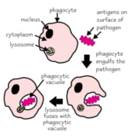
A phagocyte (e.g. a macrophage) is type of white blood cell that carries out phagocytosis (engulfment of pathogens). They’re found in the blood and in tissues and are the first cells to respond to an immune system trigger inside the body. Here’s how they work:
A phagocyte recognises the foreign antigens on a pathogen.
The cytoplasm of the phagocyte moves around the pathogen, engulfing it.
The pathogen is now contained in a phagocytic vacuole (a bubble) in the cytoplasm of the phagocyte.
A lysosome (an organelle that contains enzymes called lysozymes) fuses with the phagocytic vacuole. The lysozymes break down the pathogen.
The phagocyte the presents the pathogen’s antigens — it sticks the antigens on its surface to activate other immune system cells.
2) Phagocytes Activate T-cells
A T-cell (also called a T-lymphocyte) is another type of white blood cell. It has receptor proteins on its surface that bind to complementary antigens presented to it by phagocytes. This activates the T-cell. Different types of T-cells respond in different ways. For example, helper T-cells (Th cells) release chemical signals that activate and stimulate phagocytes and cytotoxic T-cells (Tc cells), which kill abnormal and foreign cells. Th cells also activate B-cells, which secrete antibodies.
3) T-cells Activate B-cells, Which Divide into Plasma Cells
B-cells (also called B-lymphocytes) are also a type of white blood cell. They’re covered with antibodies — proteins that bind antigens to form an antigen-antibody complex. Each B-cells has a different shaped antibody on its membrane, so different ones bind to different shaped antigens.
When the antibody on the surface of a B-cells meets a complementary shaped antigen, it binds to it.
This, together with substances released from helper T-cells, activates the B-cell. This process is called clonal selection.
The activated B-cell divides into plasma cells.
4) Plasma Cells Make More Antibodies to a Specific Antigen
Plasma cells are identical to the B-cell (they’re clones). They secrete loads of antibodies specific to the antigen. These are called monoclonal antibodies. They bind to the antigens on the surface of the pathogen to form lots of antigen-antibody complexes.
An antibody has two binding sites, so can bind to two pathogens at the same time. This means that pathogens become clumped together — this is called agglutination. Phagocytes then bind to the antibodies and phagocytose many pathogens at once. This process leads to the destruction of pathogens carrying this antigen in the body.
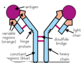
Antibodies are proteins — they’re made up of chains of amino acids. The specificity of an antibody depends on its variable regions, which form the antigen binding sites. Each antibody has a variable region with a unique tertiary structure (due to different amino acid sequences) that’s complementary to on specific antigen. All antibodies have the same constant regions.
The Immune Response Can be Split into Cellular and Humoral
The immune response is split into two — the cellular response and the humoral response.
Cellular — The T-cells and other immune system cells that they interact with, e.g. phagocytes, form the cellular response.
Humoral — B-cells, clonal selection and the production of monoclonal antibodies form the humoral response.
Both types of response are needed to remove a pathogen from the body and the responses interact with each other, e.g. T-cells help to activate B-cells, and antibodies coat pathogens making it easier for phagocytes to engulf them.
The Immune Response for Antigens can be Memorised
The Primary Immune Response
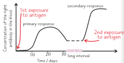
When an antigen enters the body for the first time it activates the immune system. This is called the primary response.
The primary response is slow because there aren’t many B-cells that can make the antibody needed to bind to it.
Eventually the body will produce enough of the right antibody to overcome the infection. Meanwhile the infected person will show symptoms of the disease.
After being exposed to an antigen, both T- and B-cells produce memory cells. These memory cells remain in the body for a long time. Memory T-cells remember the specific antigen and will recognise it a second time round. Memory B-cells record the specific antibodies needed to bind to the antigen.
The person is now immune — their immune system has the ability to respond quickly to a second infection.
The Secondary Immune Response
If the same pathogen enters the body again, the immune system will produce a quicker, stronger immune response — the secondary response.
Clonal selection happens faster. Memory B-cells are activated and divide into plasma cells that produce the right antibody to the antigen. Memory T-cells are activated and divide into the correct type of T-cells to kill the cell carrying the antigen.
The secondary response often gets rid of the pathogen before you begin to show any symptoms (you are immune to the pathogen).