Orbit and Eye Ball
Page 1: Introduction to Orbit and Eyeball
Topic introduction on orbit and eyeball (1 October 2018)
Page 2: Orbit Structure
Shape: Pyramidal shape
Base: Anterior
Apex: Posterior; contains optic canal
Periorbita:
Lined with periosteum, forms the fascial sheath of the eyeball
Continuous with:
Periosteal layer of dura via optic canal and superior orbital fissure
Pericranium via inferior orbital fissure
Page 3: Orbit Wall Bones
Bones:
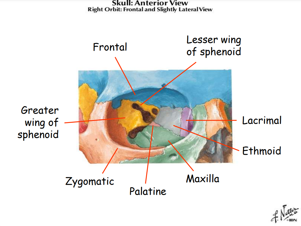
Superior Wall: Frontal bone and lesser wing of sphenoid
Medial Wall: Ethmoid, lacrimal, maxilla, sphenoid
Inferior Wall: Maxilla, zygomatic, palatine
Lateral Wall: Zygomatic bone, greater wing of sphenoid
Apex: Lesser wing of sphenoid
Page 4: Skull - Anterior View
Illustration of right orbit from a frontal and slightly lateral view.
Page 5: Orbital Margin
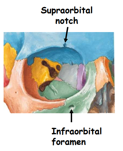
Orbital margin consists of 4 curved sides:
Supraorbital: Frontal bone
Infraorbital: Zygomatic & maxilla
Medial: Lacrimal crests
Lateral: Frontal & zygomatic
Least protected part but offers greatest view
Key features: Infraorbital foramen and supraorbital notch
Page 6: Superior Orbital Fissure
Located between the lesser and greater wings of sphenoid.
Communication: Middle cranial fossa
Nerves and vessels:
Oculomotor nerve, Trochlear nerve, Lacrimal nerve (V1), Frontal nerve (V1), Nasociliary nerve (V1), Abducent nerve
Veins: Superior & inferior ophthalmic veins
Page 7: Other Fissures and Foramina
Inferior orbital fissure: Communicates with pterygopalatine fossa, maxillary nerve, infraorbital nerve & vessels, zygomatic nerve
Optic foramen/canal: Contains optic nerve and ophthalmic artery
Page 8: Anatomy of the Sphenoid Bone
Key structures and foramina associated with sphenoid bone, including:
Foramen spinosum, Foramen ovale, and others.
Locations: Hamulus, pterygopalatine fossa, and choanae (posterior nares)
Page 9: Ethmoidal Foramina
Posterior ethmoidal foramen: Contains posterior ethmoidal artery and nerve
Anterior ethmoidal foramen: Contains anterior ethmoidal artery and nerve; nasociliary nerve (V1)
Page 10: Orbital Fissures and Foramina Overview
Summary of key orbital fissures and foramina:
Optic canal
Superior orbital fissure
Fossa for lacrimal sac
Inferior orbital fissure
Posterior & anterior ethmoidal foramina
Page 11: Fascial Sheath of Eyeball
Tenon’s capsule: Extends from optic nerve to corneoscleral junction; separates eyeball from orbital fat; allows free movement; pierced by tendons of EOM (extraocular muscles).
Check ligaments: Medial and lateral, from muscular sheath (medial & lateral rectus)
Suspensory ligament of the eye: Fascia from inferior and superior oblique muscles.
Page 12: Fascia of Orbit and Eyeball
Horizontal Section: Details components including lateral and medial check ligaments, orbital fat, and fascial sheath of the eyeball.
Page 13: Structure of the Eye
Three layers:
Outer fibrous layer: Sclera and cornea
Middle vascular layer (uveal tract): Iris, ciliary body, choroid
Inner nervous layer: Retina
Page 14: Outer Fibrous Layer
Sclera:
Opaque, white, strong; covers 5/6 of eyeball
Contains lamina cribrosa: weakest point where optic nerve enters
Cornea:
Covers anterior 1/6, first refractive surface, transparent, no blood supply, sensitive to touch (supplied by V1), nourished by aqueous humor, tears, and oxygen
Page 15: Eyeball Cross Section
Elements within cross section include cornea, anterior and posterior chambers, sclera, and lamina cribrosa.
Page 16: Middle Vascular Layer - Iris
Iris:
Colored circular disc on anterior lens surface; central aperture = pupil.
Contains sphincter pupillae and dilator pupillae smooth muscles for contraction and dilation (parasympathetic and sympathetic respectively).
Color determined by melanophore count.
Page 17: Middle Vascular Layer - Ciliary Body and Choroid
Ciliary Body:
Connects choroid to iris with ciliary muscle (smooth muscle).
Ciliary Processes: Folds that secrete aqueous humor.
Choroid: Vascular layer, attaches firm to retina but loosely to sclera.
Page 18: Ciliary Structures
Illustrations showing the suspensory ligament of lens, ciliary processes, and compartments of anterior and posterior chambers.
Page 19: Inner Nervous Layer - Structure
Two layers:
Outer: Retinal pigmented epithelium, fused with choroid.
Inner: Neural layer; posterior 3/4 is retinotopic receptor area; anterior edge forms ora serrata.
Page 20: Cross Section of Inner Nervous Layer
Detailed diagram showing various structures including the iris, ciliary body, and various layers of the retina.
Page 21: Inner Nervous Layer - Fundus
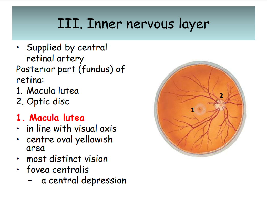
Supplied by central retinal artery:
Key Structures: Macula lutea (distinct vision), optic disc (entry of optic nerve, 'blind spot').
Page 22: Optic Disc Characteristics
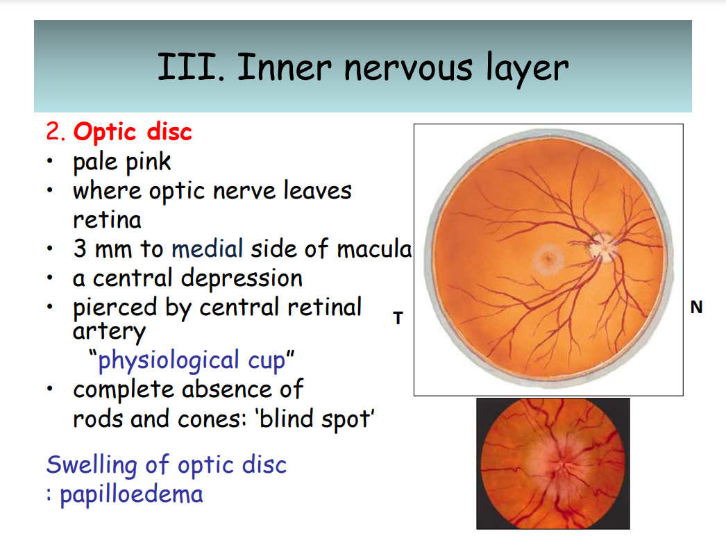
Optic Disc:
Pale pink, location of the optic nerve leaving retina; central depression; absence of rods and cones causes 'blind spot'.
Papilledema indicates swelling of optic disc due to increased CSF pressure.
Page 23: Eyeball Contents
Refractive Media (4 components):
Cornea
Aqueous humor
Lens
Vitreous humor
Page 24: Eyeball Contents - Cornea
Cornea: Transparent, first refractive surface, nourished by aqueous humor and tears, sensitive to touch, no blood supply.
Page 25: Eyeball Contents - Aqueous Humor
Aqueous Humor: Clear fluid in anterior and posterior chambers; secreted from ciliary processes; provides nutrients to cornea and lens.
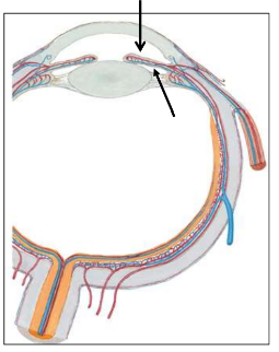
Page 26: Aqueous Humor Drainage
Drainage Path:
ciliary process
posterior chamber
pupil
anterior chamber
canal of Schlemm
Glaucoma: Condition of blockage leading to increased aqueous humor pressure.
Page 27: Contents of the Eyeball - Lens
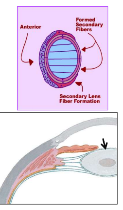
Lens: Biconvex, transparent, posterior to iris, attached to ciliary processes by suspensory ligament; formed by lens fibers.
Page 28: Accommodation of the Eye
Near Object: Ciliary muscle contracts, lens becomes globular.
Far Object: Ciliary muscle relaxes, lens becomes thin.
Page 29: Supporting Structures - Lens
Diagrams showing lens anatomy and formation of secondary lens fibers.
Posterior to iris & anterior to vitreous humor
Transparent, biconcave.
Enclosed in elastic lens capsule.
anterior surface only – cuboidal epithelium
lens fibers – main bulk of lens
Attached to ciliary process by suspensory ligament of lens
Page 30: Cataract Surgery - Phacoemulsification
Steps involved in cataract surgery, showcasing the breaking up and removal of the clouded lens and implantation of an artificial lens.
Page 31: Contents of the Eyeball - Vitreous Humor
Vitreous Humor: Gel-like substance in posterior 4/5 of eyeball, supports lens, holds retina layers in position.
Hyaloid Canal: Narrow channel from optic disc to posterior of lens.
Function of Fluid: Support lens, hold the 2 layers of retina
Page 32: Extraocular Muscles (Anterior View)
Overview of extraocular muscles including superior rectus, superior oblique, lateral rectus, medial rectus, inferior rectus, and inferior oblique.
Page 33: Muscles of the Orbit - Overview
Key muscles include:
Levator palpebrae superioris
Superior, inferior, medial, and lateral rectus muscles
Superior and inferior oblique muscles
Page 34: Function of Orbital Muscles
Levator Palpebrae Superioris: Elevates upper eyelid; innervated by CN III, distal attachment to tarsal plate.
Page 35: Recti Muscles Anatomy
Recti Muscles:
Arise from common tendinous ring.
Attach to sclera on anterior half of eyeball.
Page 36: Oblique Muscles Function
Inferior Oblique: Adduction and elevation (with superior rectus).
Superior Oblique: Abduction and depression (with inferior rectus).
Page 37: Ocular Movement Basics
Center of cornea/pupil as a reference for eye movement.
Movements relate to the anatomical orientation of this 'anterior pole'.
Page 38: Axes of Ocular Movements
Three Axes:
Vertical (Z Axis)
Horizontal (X Axis)
Sagittal (Y Axis)
Page 39: Abductors of the Eye
Muscles involved: Lateral rectus, superior oblique, and inferior oblique.
Page 40: Adductors of the Eye
Muscles involved: Medial rectus, superior rectus, and inferior rectus.
Page 41: Depressors of the Eye
Muscles involved: Inferior rectus and superior oblique.
Page 42: Elevators of the Eye
Muscles involved: Superior rectus and inferior oblique.
Page 43: Intorsion and Extorsion
Intorsion (Medial Rotation): Achieved by superior oblique and superior rectus.
Extorsion (Lateral Rotation): Achieved by inferior oblique and inferior rectus.
Page 44: Orbital Muscles and Nerve Supplies
Overview of muscle functions and their respective nerve supply (CN III, IV, VI).
Example: Levator palpebrae superioris (CN III)
Page 45: Summary of Extraocular Movements
Key muscle actions and their contributions:
Elevation: Superior rectus, inferior oblique
Depression: Inferior rectus, superior oblique
Abduction: Lateral rectus
Adduction: Medial rectus
Page 46: Ocular Muscles and Nervous Supply
Nerves:
Oculomotor (III), Trochlear (IV), and Abducens (VI).
Page 47: Orbit Arteries and Veins
Vascular Supply: Overview of facial vein, superior ophthalmic vein, cavernous sinus, pterygoid plexus, and arteries including the ophthalmic and lacrimal arteries.
Page 48: Lacrimal Apparatus
Components:
Lacrimal gland and ducts
Page 49: Lacrimal Gland Anatomy
Located in lacrimal fossa (superolateral part)
Orbital part- large, superior
Palpebral part- small, inferior
Gland open into superior fornix of conjunctiva by 8-12 ducts
Parasympathetic supply: lacrimal nucleus of CN VII
Sympathetic supply: internal carotid plexus
Page 50: Tear Flow Pathway
Pathway:
Lateral to medial drainage of tears
lacus lacrimalis —> punctum lacrimalis —> canaliculi lacrimalis —> lacrimal sac —> nasolacrimal duct.
Sensory: V1
Motor: pterygopalatine ganglion of facial nerve
Nasolacrimal duct: open into inferior meatus of nose
Page 51: Eyelid Characteristics
Protective function, muscle function, and structures including the tarsal plate and meibomian glands
Common lesions include ptosis due to CN III palsy.
Page 52: Conjunctiva Structure
Has two parts:
Palpebral conjunctiva (covers eyelid)
Bulbar conjunctiva (covers sclera)
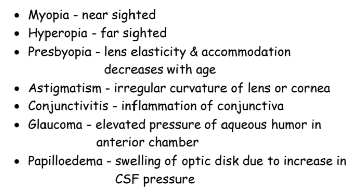
Page 53: Visual Conditions
Page 54: Eye Muscle Testing
Clinical test for eye motion: Following finger in an H pattern isolates muscle pairs for evaluation.
Page 55: Applied Anatomy - Diplopia
Diplopia: paralysis of extraocular muscles (nerve injury related).
Cyclopia: a single orbit & 1 eye.
Coloboma Iridis: cleft in iris.
Medial & lateral strabismus: lesions of cranial nerve in orbit
Page 57: Infection Risks
Cavernous sinus & anterior facial vein are connected by superior opthalmic vein
Allow rapid infection to cranium interior
Infected pimples lead to cavernous sinus thrombosis & meningitis.
Page 58: Cranial Nerve Lesions
Specific symptoms based on which cranial nerve is affected:
Oculomotor: ptosis, diplopia, loss of accommodation.
Trochlear: diplopia when looking down & head tilt.
Abducent: medial strabismus.