BMS165 Topic 5: The Nervous System
Part 1
Nervous System: General Functions
The nervous system is the master control and communication system of the body.
Every thought, action, and emotion reflects its activity
It communicates with body cells using electrical impulses, which are rapid and specific and cause almost immediate responses.
Nervous System: Primary Functions
To carry out its normal role, the nervous system has three overlapping functions:
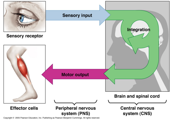
It uses its millions of sensory receptors to monitor changes occurring inside and outside of the body. This information is called sensory input.
It processes and interprets the sensory input and decides what should be done at each moment. This process is called integration.
It then causes a response, or effect, by activating muscles or glands via motor output.
Anatomical Subdivisions
There are two major anatomical subdivisions
Central nervous system (CNS)
Brain and spinal cord enclosed in bony coverings
Peripheral nervous system (PNS)
All nervous system, except the brain and spinal cord, consists of
Nerves: bundles of axons in connective tissue
Ganglia: swellings of cell bodies in a nerve
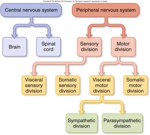
Function Subdivisions of Peripheral Nervous Syste
Sensory (afferent) division (receptors to central nervous system)
visceral sensory and somatic sensory division
Motor (efferent) division (central nervous system to effectors)
somatic motor division
effectors: skeletal muscle
visceral motor division (aka autonomic nervous system)
effectors: cardiac muscle, smooth muscle, glands
sympathetic division (fight or flight)
parasympathetic division (rest and digest)
3 Functions Classes of Neurons
Sensory (afferent) neurons
detect changes in body and external environment
information transmitted into brain or spinal cord
Interneurons (association neurons)
lie between sensory and motor pathways in central nervous system
90% of our neurons are interneurons
process, store and retrieve information
Motor (efferent) neuron
send signals out to muscles and gland cells
organs innervated by motor neurons that carry out responses are called effectors
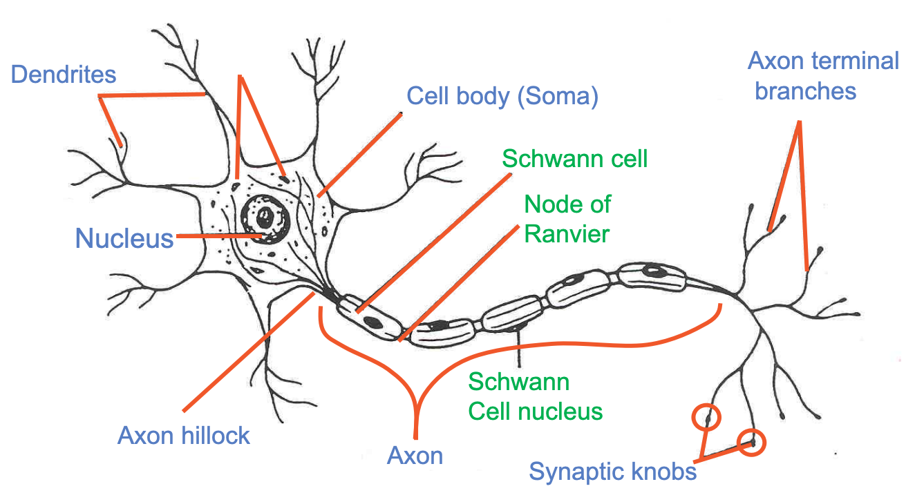
Cell Body
also referred to as Soma
Contains the nucleus and most organelles
grouped in CNS into nuclei
grouped in PNS into ganglia
Axons
Process extending from the outgrowth from the soma called the axon hillock
May be covered in places with a substance called myelin
produced by a cell called a Schwann cell
Conduct the primary signal (action potential)
from initiation in the initial segment, to the terminals where neurotransmitter release occurs
Length from 1mm to 1m
Terminals
The end points of axons
Specialised structures for packaging and releasing neurotransmitters following an action potential
Very closely opposed to special areas (called post-synaptic densities) on the dendrites of their target cells
Terminal + post-synaptic density = a synapse (discussed later)
Dendrites
dendron = tree branch
Receptive area
transmits information (electrical impulses) to cell body
Receives this information from other neurons
Variable in number from 1000s to 1.
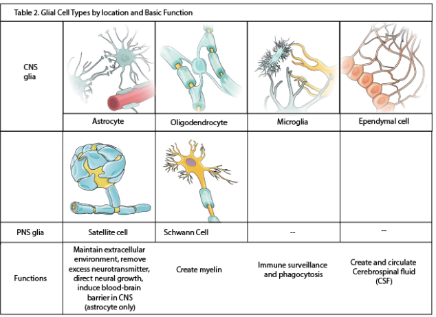
Types of Neuroglial Cells
Even though it is complex, nervous tissue is made up of just two principal kinds of cells
Neurons
Supporting cells
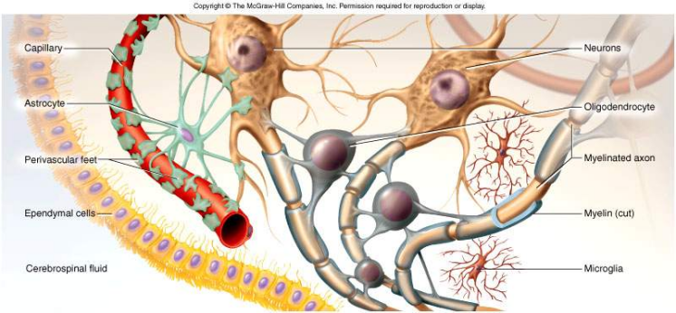
Supporting cells in the nervous system are called neuroglia
Neuroglia are found in both the central nervous system and in the peripheral nervous system
Each type of neuroglia has a specific function
Neurons need neuroglia in order to maintain homeostasis.
Types of Neuroglial Cell
Myelin Sheath
Insulating layer around a nerve fibre
Schwann cells make myelin in the Peripheral Nervous System, and oligodendrocytes make myelin in the Central Nervous System
Formed from wrappings of plasma membrane in a spiral fashion:
20% protein and 80 % lipid (looks white)
The myelin sheath insulates the axon for rapid action potential conduction, as well as separating axons from surrounding extracellular components
All myelination completed by late adolescence
In the Peripheral Nervous System, hundreds of layers wrap axon
The outermost layer of the Schwann cell is called the neurilemma
Covered by basal lamina and endoneurium.
In Central Nervous System - no neurilemma or endoneurium
Oligodendrocytes myelinate several fibres:
Myelination spirals inward with new layers pushed under the older ones
Gaps between myelin segments= nodes of Ranvier
Less nodes of Ranvier in Central Nervous System compared to Peripheral Nervous System.
The role of Myelin
Diameter of fibre and presence of myelin increases the speed (conduction velocity) that electrical signals (action potentials) travel along an axon:
Large fibres have greater conductance and less relative leak and capacitance
Myelin protects large axons
Myelin further reduces leak and capacitance by being an insulator.
Speeds
small, unmyelinated fibres = 0.5 - 2.0 m/sec
small, myelinated fibres = 3 - 15.0 m/sec
large, myelinated fibres = up to 120 m/sec
Multiple Sceloris (MS)
A complex neurodegenerative disease affecting the central nervous system (CNS).
The exact aetiology of the disease is unknown.
Observational research has shown that that it is associated with demyelination inside of the CNS whilst the axons are preserved.
Part 2
Resting Membrane Potential
A neuron that is “resting” has an electrical difference across its plasma membrane.
This means that there are fewer positive ions sitting on the inner surface of the neuron’s plasma membrane than there are on its outer surface.
While the inside of the neuron remains more negative than the outside, the neuron will remain in its resting state.
The electrical difference whilst the neuron is at rest is called the resting membrane potential.
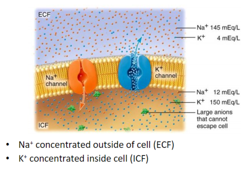
The cations and anions inside and outside of the neuron are different
Electrical Potential and Currents
An electrical potential is the difference in the concentration of charged particles between one point and another
An electrical current is a flow of charged particles from one point to another
Living cells are polarized:
resting membrane potential is -70 mV with a negative charge on the inside of membrane
Ionic Basis of Resting Membrane Potential
At resting state, Na+ (positively charged) is attracted by the electrical force into the -negatively charged cell
Since the concentration of extracellular Na+ is higher, it tends to be pulled into the cell by the concentration force
In the resting state there is almost no Na+ flow.
K+ (positively charged) is attracted into the cell by the electrical force, but repelled outwards by the diffusion force
Protein molecules (negatively charged) tend to be driven out by both the electrical force and the diffusion force
These molecules are too big to pass through the cellular membrane
Cl- (negatively charged) is repelled outward by the electrical force but pulled inward by the diffusion force.
Due to the overall unequal distribution of the charged particles, the inside of the cell is -70 mV relative to outside of the cell. This is the resting potential of the neuron.
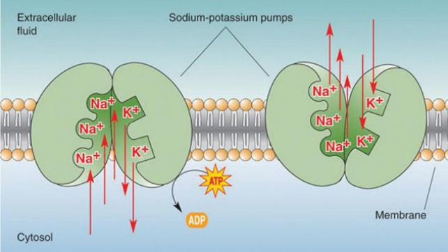
Sodium-potassium Pumps
Local Potentials
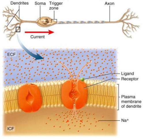
Local disturbances in membrane potential
Occur when a neuron is stimulated by
Chemicals
Light
Heat
Mechanical disturbances
Depolarisation decreases potential across cell membrane due to opening of ligand-gated Na+ channels but not enough to stimulate the voltage gated Na+ channels at trigger zone
Na+ rushes in down concentration and electrical gradients
Na+ diffuses for short distance inside membrane producing a change in voltage called a local potential
Action Potentials
More dramatic change in membrane produced where high density of voltage-gated channels occur
trigger zone up to 500 channels/mm2 (normal is 75)
If threshold potential (-55mV) is reached voltage-gated Na+ channels open (Na+ enters causing depolarisation)
Past 0 mV, Na+ channels close = depolarisation
Slow K+ gates fully open
K+ exits, repolarising the cell
Negative overshoot produces hyperpolarisation
excessive exiting of K+
The Refractory Period
When -55mV is reached , this opens the voltage-gated sodium channels.
Sodium rushes into the neuron then the voltage-gated sodium channels shut. And it doesn’t matter what you do, you cannot open them again.
When they shut, they become deactivated(locked). Recovery from inactivation takes 1 -2 msec. This is the absolute refractory period.
From 2 msec onwards, some of the voltage-gated sodium channels start to come out of inactivation(unlocked) which means that they can be used again. This is the relative refractory period.
Impulse Conduction in Unmyelinated Fibres
Threshold voltage in trigger zone begins impulse
Nerve signal (impulse) - a chain reaction of sequential opening of voltage-gated Na+ channels down entire length of axon
Nerve signal (non-decremental) travels at 2m/sec
Voltage gated sodium channels are distributed evenly along the axon.
As the action potential allows sodium to depolarise a section of axon, the sodium channels adjacent become recruited and propagate the action potential continuously along the axon
The refractory membrane is where the sodium channels become closed (called inactivated) and cannot re-open for a period of time
This keeps the signal moving in one direction (after initiation)
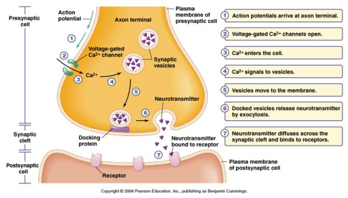
Excitatory Synapse
Nerve signal opens voltage-gated calcium channels in synaptic knob
Triggers release of ACh, which crosses synapse
ACh receptors trigger opening of Na+ channels producing local potential
When voltage reaches -55mV, triggers action potential in postsynaptic neuron
Excitatory and Inhibitory Synapses
There are two kinds of synapses, excitatory and inhibitory.
At excitatory synapses, neurotransmitters induce a depolarizing postsynaptic potential.
Excitatory neurotransmitters bind to protein channels that allow Na+ to enter the next neuron, this makes the post synaptic neuron more positive
At inhibitory synapses, neurotransmitters induce a hyperpolarising post synaptic potential.
Inhibitory neurotransmitters bind to protein channels that allow Cl- to enter the next neuron, this makes the post synaptic neuron more negative.
Stopping Postsynaptic Neuron Stimulation
Diffusion
Some of the released neurotransmitter molecules diffuse away from the synaptic cleft
Enzymatic degradation
Enzymes break down neurotransmitters. Acetylcholinesterase breaks down Ach into acetate and choline with these products having no stimulatory effect
Reuptake of neurotransmitter
Many neurotransmitters are actively transported back into the presynaptic neuron by endocytosis
Part 3
Functions of the Spinal Cord
Conduction
Bundles of fibres (Australian spelling) passing information up and down spinal cord
A fibre is another name for an axon.
Carries information that is afferent (comes in) and efferent (goes out)
Locomotion
Repetitive, coordinated actions of several muscle groups
Central pattern generators are pools of neurons providing control of flexors and extensors (walking)
Reflexes
Involuntary, stereotyped responses to stimuli (remove hand from hot stove)
Involves spinal cord and peripheral nerves, brain informed afterwards
Structure of the Spinal Cord
Cylinder of nervous tissue within the vertebral canal (as thick as a finger)
Vertebral column in an adult extends to L1
Extends through vertebral canal from foramen magnum to L1
31 pairs of spinal nerves arise from cervical, thoracic, lumbar and sacral regions of the cord
each cord segment gives rise to a pair of spinal nerves
Cauda equinae is L2-S5 nerve roots – resembles a horse’s tail
Meninges of the Spinal Cord
There are three fibrous layers that enclose the spinal cord:
Dura mater (outer/superficial layer):
tough collagenous membrane surrounded by epidural space
epidural anaesthesia utilized during childbirth
Arachnoid mater (middle layer):
Pia mater (inner/deep layer):
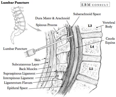
Lumbar Puncture
The entry into the subarachnoid space is commonly described as feeling a "pop" sensation.
The needle insert is then removed, and CSF should begin to drip out.
Anatomy of the Spinal Cord
Central area of grey matter shaped like a butterfly and surrounded by white matter
Grey matter: consists of neuron cell bodies with little myelin and white matter consists of myelinated axons
Pair of posterior (or dorsal) horns
posterior root of spinal nerve is totally sensory fibres
Pair of anterior (or ventral) horns,
anterior root of spinal nerve is totally motor fibres
Connected by grey commissure punctured by a central canal.
Horns: Grey matter of spinal cord: cell bodies
posterior horn: cell bodies of interneurons
anterior horn: cell bodies of motor neurons
Roots: Bundles of fibres connecting spinal cord and spinal nerves
posterior root contains sensory neurons
anterior root contains motor neurons
White matter/columns: Bundles of myelinated axons (fibre tracts) that carry signals up and down, to and from the brainstem
Spinal Cord Tract
White matter of the spinal cord is composed of myelinated axon tracts (or pathways)
If you think of your spinal cord as a tall building, these tracts are like elevators that go straight up to the top of the building.
Tracts can be ascending (go up) or descending (come down from) the brain
Some tracts cross over to the other side of the spinal cord and some do not
Some tracts travel from the brain to the spinal cord and some conduct impulses from one side of the spinal cord to the other
Nerves
A nerve is an enclosed, cable-like bundle of axons (the projections of neurons) in the peripheral nervous system (PNS). A nerve provides a structured pathway that supports the electrochemical nerve impulses transmitted along each of the axons. Each nerve contains many axons that are sometimes referred to as fibers. Within a nerve, each axon is surrounded by a layer of connective tissue called the endoneurium. The axons are bundled together into groups called fascicles. Each fascicle is wrapped in a layer of connective tissue called the perineurium. Finally, the entire nerve is wrapped in a layer of connective tissue called the epineurium.
Ganglia
A ganglion is a cluster of neuron cell bodies of nerves
Posterior root ganglion is sensory cell bodies
Fibres pass through without synapsing
Spinal Nerves
carry information to and from the spinal cord
are named for the region of the cord from which they arise
are formed when ventral root and dorsal roots combine
divides into two branches after formation.
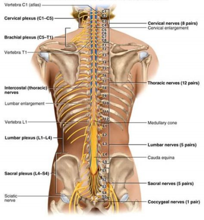
The anterior and posterior branches of each spinal nerve contain both sensory and motor axons, thus damage to a spinal nerve or any of its branches results both in loss of sensation and in paralysis of the area of the body served.
Part 4
Reflexes
Although there are many types of communication between neurons, most of what the body must do every day is programmed as reflexes
Reflexes are rapid, predictable, and involuntary responses to stimuli.
A stimulus is any change that occurs inside or outside of the body.
Reflexes are like one-way streets, once a reflex begins, it always goes in the same direction.
You can think of a reflex as a preprogramed response to a given stimulus.
Somatic and Autonomic Reflexes
The types of reflexes that occur in the body are classed as either somatic or autonomic.
Somatic Reflexes
Involve skeletal muscles and are typically under voluntary control. Examples include the knee-jerk reflex and withdrawal reflex.
Autonomic Reflexes
Involve smooth muscles, cardiac muscles, and glands, and are generally not under voluntary control, such as the regulation of heart rate and digestive processes.
Reflex Arc
Reflexes occur over neural pathways called reflex arcs
All reflex arcs have a minimum of four elements:
A receptor (which reacts to a stimulus)
An effector (the muscle or gland that is stimulated/responds)
A sensory neuron (brings sensory information to the central nervous system)
A motor neuron (delivers information out to the body from the nervous system)
An integration centre (other neurons) are only seen in some reflex arcs.
Patellar Reflex (Somatic Reflex)
Our muscles must not be stretched too far because if they do, the cells making up that muscle may be damaged.
There are a class of reflexes (called stretch reflexes) whose job is to make sure that muscles do not stretch out too far
The patellar reflex (or knee-jerk reflex) is just one of many stretch reflexes
When you hit the tendon with the hammer it causes that muscle to stretch.
This muscle stretch activates sensory neurons in the muscle.
The sensory neurons synapse directly with motor neurons in the spinal cord.
The motor neurons cause the muscle to contract.
Withdrawal Reflex (Somatic Reflex)
A painful stimulus initiates the flexor or withdrawal reflex
This reflex causes automatic withdrawal of the threatened body part form the stimulus.
Salivary Gland Secretion (Autonomic Reflex)
Saliva in the mouth is a biofluid produced mainly by three pairs of major salivary glands
Salivary gland secretion is an autonomic reflex
The volume of saliva secreted is dependent on the intensity and type of taste and on smell, chewing or touch stimulation
Non-Responsive Reflexes
Reflex testing is an important tool in evaluating the condition of the nervous system.
Reflexes that are exaggerated, distorted or absent indicate damage or disease in the nervous system.
Reflex changes often occur before a pathological condition becomes obvious in other ways.