Endocrine
Section 1.1: Endocrine System Anatomy & Physiology
Materials
Endocrine system models
This section introduces you to the anatomy, histology, and physiology of the endocrine organs and hormones. The endocrine system:
Is a diverse group of ductless glands that plays a major role in maintaining homeostasis of multiple physiological variables.
Note: Endocrine glands are different from exocrine glands, which are glands that rely on ducts to release their substance, such as enzymes, tears, sweat, and milk.
Examples of exocrine glands are sweat, salivary, lacrimal, digestive, & mammary glands.
The endocrine system works closely with the other system that maintains homeostasis of multiple physiological variables—the nervous system.
The endocrine system, along with the nervous system, provides means for regulating other organ systems and tissues and maintaining homeostasis.
Although these 2 systems both work toward the same goal of internal communication and coordination, you will notice that the methods by which they do so differ:
The nervous system functions via action potentials (nerve impulses) and releases neurotransmitters that directly affect target cells. The effects are nearly immediate, but they are very short in duration.
In contrast, the endocrine system brings about its effects via the secretion of hormones—chemicals released into the bloodstream that typically act on distant targets. The effects of hormones are not immediate, but they are longer-lasting than those of the nervous system.
In general, hormones regulate the processes of other cells, including inducing the production of enzymes or other hormones, changing the metabolic rate of a cell, and altering permeability of the plasma membrane.
You might think of hormones as the “middle managers” of the body, because they communicate the messages from their “bosses” (the endocrine glands) and tell the “workers” (other cells) what to do.
Some endocrine glands (e.g., the thyroid and anterior pituitary glands) secrete hormones as their primary function. Others, however, secrete hormones as a secondary function, examples of which are the heart (atrial natriuretic peptide), adipose tissue (leptin), the kidneys (erythropoietin), and the stomach (gastrin).
The nervous system is often referred to as our fast response system and tends to have very specific and localized targets of its action.
In contrast, the endocrine system is our slow response system, often taking hours, days, weeks, or longer to produce noticeable physiological changes in the body. Although typically much slower, the endocrine system nevertheless can elicit powerful physiological effects throughout the body and on many organs and tissues simultaneously, as long as the target cells of specific hormones have the necessary receptors.
Endocrine Organs, Hormones, & Functions
Here are the 10 endocrine organs that have hormone secretion as a primary function:
Note: from here on out, students are required to spell out any abbreviated terminologies listed in the lab manual. Abbreviated answered will be considered incorrect. We will inform all of you for any exceptions to this rule.
Hypothalamus
Pituitary gland
Pineal gland
Thyroid gland
Parathyroid glands
Thymus gland
Adrenal glands
Pancreas
Ovaries
Testes.
Let’s take a closer look at each of these organs (note that we discuss the ovaries and testes only briefly here; they are discussed further in Exercises 10 and 11).
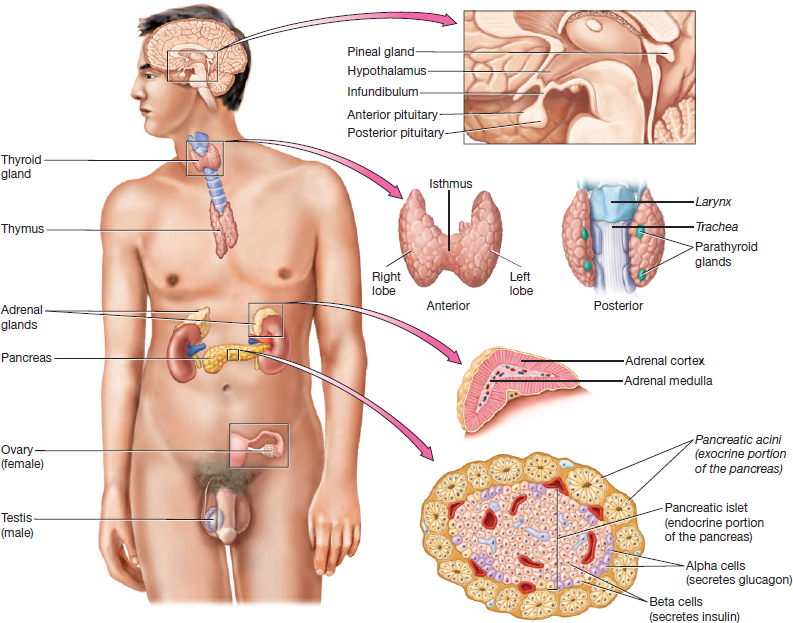
Figure 1.1 Organs and tissues of the endocrine system.
1. Hypothalamus: is the inferior part of the diencephalon and is known as a neuroendocrine organ.
It can be likened as the endocrine system’s chief executive officer (CEO).
It has a close working relationship with the pituitary gland, to which it is attached by a stalk called the infundibulum (in-fun-di-bu-lum).
The hypothalamus produces the following hormones, which will be dependent on whether these hormones interact with the anterior or posterior pituitary glands. Again, these hormones are still produced by the hypothalamus, not by the pituitary gland:
A. Interacts with the Anterior Pituitary:
Inhibiting and Releasing Hormones: are hormones that inhibit and stimulate secretion from the anterior pituitary gland.
In Figure 1.2A, you can see how the hypothalamus communicates with the anterior pituitary gland via a specialized set of blood vessels, called hypothalamic-hypophyseal portal system (HHPS). As shown in the pathway below:
The inhibiting and releasing hormones are synthesized by hypothalamic neurons⇒ then, they enter capillaries in the hypothalamus ⇒ after which they travel through small veins in the infundibulum ⇒ finally, they enter a second capillary bed in the anterior pituitary, where they exit the blood and interact with anterior pituitary cells to influence their functions.
B. Interacts with the Posterior Pituitary:
Oxytocin and Antidiuretic Hormone: These hormones are produced by hypothalamic neurons that extend the length of the infundibulum down into the posterior pituitary gland, where they are stored (Fig. 1.2B).
Oxytocin: Triggers uterine contraction and milk ejection from the mammary glands. Do not confuse its function with prolactin. Oxy- means sharp and -tocin refers to childbirth.
Milk ejection makes sense for this hormone since the birth of the child already happened, therefore it needs the milk ejected from the mother's mammary glands for sustenance.
Antidiuretic Hormone(ADH): Causes water retention from the kidneys. It also assists with blood pressure control. Anti- means against, while diuretic pertains to production of urine.
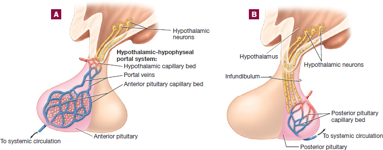
Figure 1.2 Hypothalamus and pituitary gland: (A) hypothalamus and anterior pituitary;
(B) hypothalamus and posterior pituitary.
2. Pituitary Gland: Notice in Figure 1.2 that the pituitary gland is actually made of 2 separate regions:
The anterior pituitary gland or adenohypophysis, is composed of glandular epithelium and secretes a variety of hormones that affect other tissues in the body.
The posterior pituitary or neurohypophysis, is actually composed of nervous tissue rather than glandular tissue.
In response to hypothalamic-releasing hormones, the anterior pituitary gland secretes hormones that stimulate other endocrine and exocrine glands in the body.
Anterior Pituitary Gland: mostly produces hormones known as tropic hormones.
Tropic hormones influence the functions of other endocrine or exocrine glands.
Mnemonic: FLAT-PG
2 reproductive hormones which affects both the testes and ovaries:
Follicle-Stimulating Hormone (FSH), a hormone that will influences and stimulates follicles in the ovary and sustentacular cells in the testes.
Luteinizing Hormone (LH), a hormone that influences and stimulates the oocyte in the ovary and interstitial cells in the testes.
Adrenocorticotropic Hormone (ACTH), which stimulates secretion from the adrenal cortex (a-dre-no-cor-ti-co-tro-pic).
Thyroid-Stimulating Hormone (TSH), which stimulates growth of and secretion from the thyroid gland.
Prolactin, which stimulates milk production from mammary glands. Do not confuse its function with oxytocin's function.
An exception is Growth Hormone (GH), which increases the rate of cell division and protein synthesis in all tissues and has both tropic and non-tropic effects.
Posterior Pituitary Gland: Does not produce any hormones at all and functions merely as a place to store the oxytocin and antidiuretic hormone produced by the hypothalamus.
3. Pineal Gland: Located in the posterior and superior diencephalon.
This neuroendocrine organ secretes the hormone melatonin.
It secretes it in response to decreased light levels.
The hormone acts on the "reticular formation" part of the brainstem to trigger sleep.
4. Thyroid Gland: Located in the anterior-inferior neck superficial to the larynx (not larnyx) or voicebox.
It consists of right and left lobes connected by a thin band of tissue called isthmus.
Microscopically, it is composed of hollow spheres called thyroid follicles (Fig. 1.3).
The cells that line the thyroid follicles are simple cuboidal cells called follicle cells.
The spheres encapsulates a gelatinous, iodine-rich substance called colloid.
The follicle cells respond to TSH from the anterior pituitary by secreting a chemical into the colloid that reacts with iodine to produce 2 different hormones:
Thyroxine (T4): has 4 iodine molecules. Do not confuse with thymosin.
Triiodothyronine (T3): has 3 iodine molecules (tri-i-o-do-thy-ro-nine).
T3 is the most active of the two hormones.
Acts on essentially all cells in the body to increase the metabolic rate, increase protein synthesis, and regulate the heart rate and blood pressure, among other things.
About 10 times as much T4 is produced as T3, and the body converts T4 to T3 when the T3 level in the blood drops.
Between the follicle cells we find another cell type called the parafollicular cells.
These cells produce the hormone calcitonin.
1 of the 2 hormones that play a role in calcium ion homeostasis.
Calcitonin is secreted when calcium ion levels in the blood rise.
It triggers osteoblast activity and bone deposition, thus lowering the blood calcium levels.
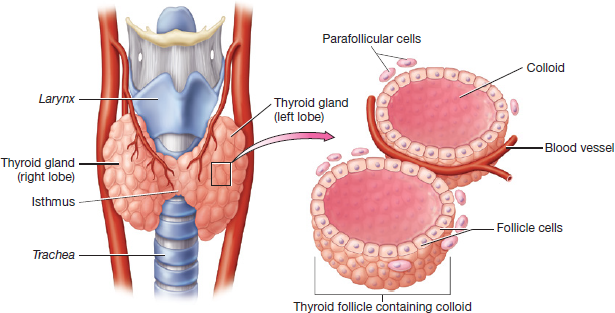
Figure 1.3 Thyroid gland and thyroid follicles.
5. Parathyroid Glands: Refer back to Figure 1.1 where you can see these 4 small glands on the posterior side of the thyroid gland.
They secrete the hormone Parathyroid Hormone (PTH):
Which is the main hormone in the body that maintains calcium ion homeostasis.
PTH is secreted in response to a decreased level of calcium ions in the blood. It triggers:
Osteoclast activity and resorption (losing) of bone tissue.
Increased calcium ion absorption from the intestines.
Increased calcium ion reabsorption (absorbing again) from the kidneys.
They all result in increasing blood calcium levels.
Note: Hormones such as PTH and calcitonin that have opposite actions are called antagonists.
6. Thymus Gland: Sits in the superior mediastinum.
It is large and active in infancy and early childhood, during which time it secretes the hormones thymosin and thymopoietin.
Both of these hormones stimulate the development of T lymphocytes within the thymus gland.
In adults most of the thymic tissue is gradually replaced by fat and other connective tissue.
7. Adrenal Gland: As the name implies, the adrenal glands sit atop the superior pole of each kidney (ad- means next to, while renal refers to kidney).
Like the pituitary gland, the adrenal gland is actually composed of 2 separate structures:
Outer adrenal cortex
Inner adrenal medulla
The glands are surrounded by a thin layer of connective tissue - adrenal capsule (Fig. 1.4).
Adrenal Cortex: The superficial region of the adrenal gland consists of glandular tissue.
It secretes steroid hormones (derived from cholesterol), in response to stimulation by the adrenocorticotropic hormone produced by the anterior pituitary gland and other factors.
It is made up of 3 sections/zones:
Mnemonic: Zona GFR
Note: pay attention to the words "type" of steroid hormone vs "examples" of these types of steroid hormones.
Zona Glomerulosa: The outermost zone of the adrenal cortex, it secretes a type of steroid hormone called mineralocorticoids.
A specific example of mineralocorticoid is aldosterone, which regulates fluid, electrolyte, and acid-base homeostasis.
Mnemonic:GMA (Glomerulosa, Mineralocorticoid, Aldosterone)
Zona Fasciculata: The middle zone of the adrenal cortex, it secretes a type of steroid hormone called glucocorticoids.
A specific example of glucocorticoid is cortisol, which regulates the stress response, blood glucose, fluid homeostasis, and inflammation.
Mnemonic: FGC (Fasciculata, Glucocorticoid, Cortisol)
Zona Reticularis: The innermost zone of the adrenal cortex, it secretes a type of steroid hormone called gonadocorticoids and also glucocorticoids.
Examples of gonadocorticoid are androgens and estrogen, which affect the gonads and other tissues.
Androgens control many aspects of male development and reproductive physiology.
Adrenal Medulla: The deep region or core of the adrenal gland.
It consists of modified postsynaptic sympathetic neurons that secrete the hormone adrenal catecholamines which are derived from amino acids, examples are:
Epinephrine and norepinephrine which are secreted into the blood in response to sympathetic stimulation (fight or flight).
The adrenal catecholamines have the same effects on target cells as the neuronal catecholamines released by sympathetic neurons, such as:
Dilation of bronchioles; increase in the rate and force of contraction of the heart; constriction of blood vessels serving the skin and abdominal viscera; dilation of the pupil; and more (these all makes sense since it is preparing the body for fight or flight).
Adrenal catecholamines also:
Prolongs the sympathetic response, as the effects of neuronal catecholamines are terminated after only a few seconds.
Are able act on target cells that are not innervated by sympathetic neurons.
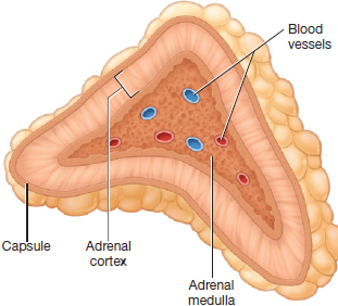
Figure 1.4 Adrenal gland.
8. Pancreas: An elongated and spongy gland located below and behind the stomach. It has both endocrine and exocrine functions.
Exocrine Functions: Are carried out by a group of cells called pancreatic acini or acinar cells, which occupies about 99% of the gland and release pancreatic juices into pancreatic ducts.
These pancreatic juices contribute to neutralizing hydrochloric acid, digesting protein, fat, and starches.
Endocrine Functions: Are carried out by these small round islands called pancreatic islets, which are embedded within the pancreatic acini (Fig. 1.5).
The cells within the pancreatic islets secrete the hormones insulin and glucagon, which play a major role in regulating blood glucose levels.
Insulin: produced by cells called beta (β) cells.
It triggers the uptake of glucose by cells, which decreases the concentration of glucose in the blood.
Glucagon: produced by alpha (α) cells, is insulin’s antagonist.
It triggers the release of stored glucose from the liver and the production of new glucose, which increases the concentration of glucose in the blood.
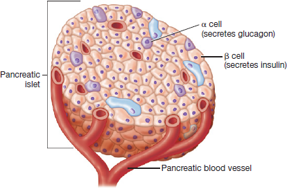
Figure 1.5 Pancreatic islet.
9. Testes: Anatomically speaking, these are the male reproductive organs located in the scrotum that produce sperm cells (male gametes).
Cells within the testes called interstitial cells produce a steroid hormone called testosterone.
This hormone promotes the production of sperm cells and the development of male secondary sex characteristics such as a deeper voice, greater bone and muscle mass, and facial hair.
10. Ovaries: Anatomically speaking, these are the female reproductive organs, located in the pelvic cavity that produce oocytes (female gametes).
The ovaries produce steroid hormones called estrogen and progesterone.
Estrogen play a role in the development of oocytes, female secondary sex characteristics such as breasts and subcutaneous fat stores, and a variety of other processes.
Progesterone has a number of effects that prepare the body for pregnancy.