Bio Unit 8 - DNA
DNA is the same in all organisms
DNA Discovery
1869 - Friedrich Miescher
studied Leukocytes - white blood cells
discovery named “Nuclein” —> Deoxyribonucleic Acid (DNA)
Frederick Griffith’s Experiment
Streptococcus Pneumoniae bacteria
goal: create pneumonia vaccine
Two strains of bacteria:
R Strain - Nonvirulent: rough appearance in dish
mouse lives
S Strain - Virulent: smooth appearance in dish
Smooth capsule [coating] protects bacteria from immune systems
mouse died
2 Trials:
Heat (killed smooth strain bacteria)
mice lives, no disease
Mixed Rough strain with heat-killed Smooth strain
killed mice
living smooth strain bacteria in mouse
Conclusion: Rough strain bacteria took “Transforming Principle” from heat-killed Smooth bacteria = Virulents
Breakdown:
(deadly) Smooth strain bacteria killed by excess heat
Cell membrane ruptures = DNA released
(harmless) Rough strain makes up deadly DNA
DNA incorporated into genome
Genome: entire set of DNA instructions in cell
Transforms into deadly strain
debate over what was transforming the material
Oswald Avery’s Experiment
1944 - Avery repeats Griffith’s experiment with 2 added enzymes
Added Enzymes that destroyed carbohydrates, lipids, and proteins to heat-killed Smooth strain
transformation still occured
Added Enzymes that destroyed nucleic acids to heat-killed Smooth strain
transformation didn’t occur
Conclusion: DNA = the genetic material
Hershey-Chase Experiment
1952 - Alfred Hershey and Martha Chase
used Bacteriophage to prove if DNA or proteins = the genetic materials
Bacteriophage: viruses that infect bacteria and use the host cell to replicate itself
Protein Coat + DNA
Experiment:
protein coat - red radioactive sulfur
virus DNA - blue radioactive phosphorus
Results:
infected cells only contained the blue radioactive phosphorus (DNA)
Conclusion:
Virus injects DNA to replicate
Avery results confirmed
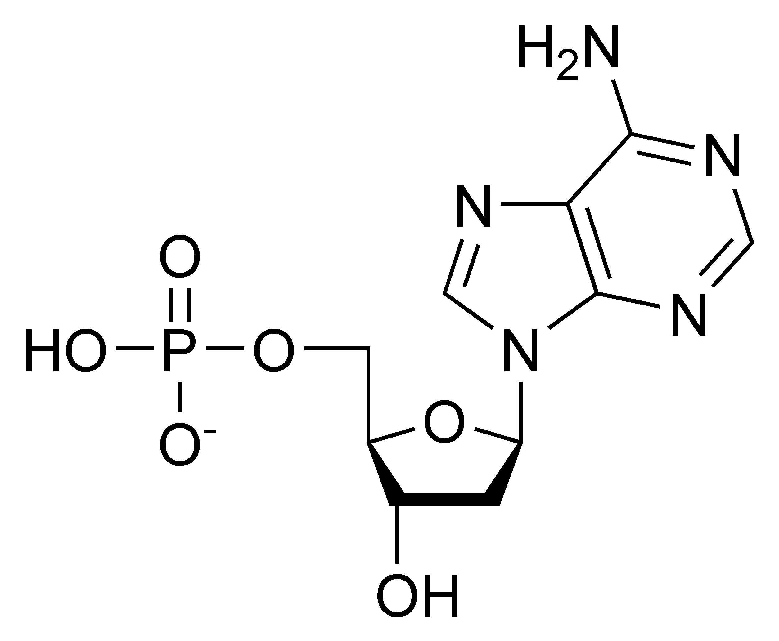
Nucleotide Structure
1929 - Phoebus Levene discovers Deoxyribose
first to describe nucleotide structure
Each nucleotide contains:
Phosphate Group:
Deoxyribose Sugar: 5 -carbon sugar
Nitrogenous Base: make up ladder rungs
Adenine (A)
Guanine (G)
Cytosine (C)
Thymine (T)
Purines - Double-ring nitrogenous bases
Adenine
Guanine
Pyrimidines - Single-ring nitrogenous bases
Cytosine
Thymine
Uracil (RNA only)
proposed Tetranucleotide Hypothesis (wrong)
DNA contains equal amounts of each nitrogenous base
4 Nucleotides bonded together
Base Pairing
1949 - Erwin Chargaff analyzed percentages of each base in DNA
A = T
C = G
Hydrogen bonds hold bases together (weak)
3 bonds between G & C
2 bonds between A & T
Rosalind Franklin
1952 - 1st photographed 3D structure of DNA using x-ray diffraction
Photo 51
James Watson and Francis Crick
1953 - stole Franklin’s photo to build the 1st correct 3D model of DNA
DNA = double stranded double helix
got credit for it
DNA Structure
2 Repeating Strands of Nucleotides
strands supported by a Repeating Phosphate-sugar backbone
bases paired in the middle (rungs)
Antiparallel - strands run in opposite directions
5’ to 3’ and 3’ to 5’
# of carbon atoms in deoxyribose = strand running direction
count clockwise from carbon after central oxygen
oxygen in central ring
5’ : free phosphate group (circle)
3’ : Un-bonded sugar
Function of DNA
stores genetic information
copies itself (same DNA of same cells)
can express genetic information
\\
Nucleotide
DNA Replication
process where the cell copies itself
each new cell has an identical copy
cells must divide because large cells don’t have enough DNA to give instructions to the cell
hereditary information is contained in Genes
Parent Strand —> 2 Replicated Strands
Semiconservative Replication
2 Replicated Strands: Each with 1 old strand and 1 new strand
old strand = template for new strand to be created
Origin of Replication
Eukaryotic cells have multiple origins (speeds up)
Replication Bubble (formed here)
site of replication
Replication forks
at either ends of bubble
move in opposite directions —> bubble grows with forks
continues until DNA is copied
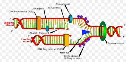
Replication Enzymes
Topoisomerase: unwinds DNA double helix
stops DNA from snapping
Helicase: unzips DNA strands by breaking hydrogen bonds between nitrogenous bases
Single Strand Binding Proteins (SSBs): keep the 2 seperated strands apart
(DNA naturally wants to rejoin)
Parent Strand: original DNA strand/template for new strand
DNA Polymerase: adds the DNA Nucleotides to the new strand
adds complementary bases together
only adds nucleotides from 5’ to 3’ direction
creates leading and lagging strands
Leading Strand: made continuously towards replication fork (5’ to 3’)
Lagging Strand: made in fragments away from replication fork
Okazaki Fragments: small segments of DNA created through lagging strand
DNA Ligase: Bonds Okazaki Fragments together to make 1 continuous DNA strand
DNA Primase: adds a short strand of RNA
RNA Primer: shows DNA Polymerase where to start adding nucleotides