Muscle Tissue Types
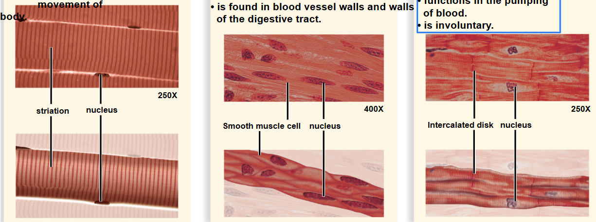
Smooth – involuntary muscle found in hollow organs and vessels. Spindle-shaped cells, each with a single nucleus. NO striations
Cardiac – involuntary muscle found in the heart. Branching Striated Cells, Single nucleus.
Skeletal – voluntary muscle that is attached to the skeleton. Has striated cells with multiple nuclei.
Structure and Organization of muscle tissue
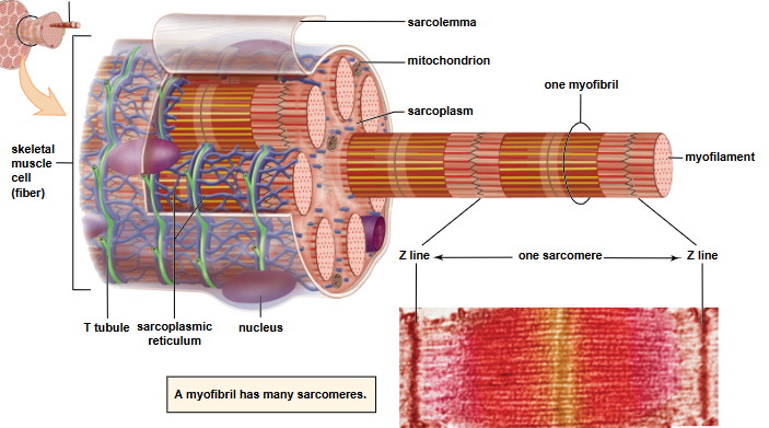
Sarcomere: functional contractile unit extending from Z line to Z line; contains overlapping actin (thin) and myosin (thick) filaments that slide during contraction (filament lengths remain the same; overlap changes).
Muscle fibers are arranged in bundles called fascicles.
Myofibrils are bundles of myofilaments that run the length of a fiber.
Myofilaments are proteins (actin and myosin) that are arranged in repeating units.
Sarcolemma: the plasma membrane. It’s the fine transparent tubular sheath which envelops the fibers of skeletal muscles.
Sarcoplasm: The cytoplasm
Muscle Contraction Mechanics
Sliding filament model: during contraction the actin and myosin filaments slide past each other — filament lengths do not shorten; sarcomere shortens
⭐What is required for the muscle to contract?
Nerve impulses travel down a motor neuron to a neuromuscular junction.
Acetylcholine (ACH) is released from the neuron and binds to the muscle fiber.
This binding stimulates the fiber causing calcium to be released from the sarcoplasmic reticulum.
Released calcium combines with troponin, a molecule associated with actin.
This causes the tropomyosin threads around actin to shift and expose myosin binding sites.
Myosin heads bind to these sites forming cross-bridges.
ATP binds to the myosin heads and is used for energy to pull the actin filaments towards the center of the sarcomere – contraction now occurs.