Tooth development
1. Introduction to Tooth Development
Tooth development is similar to the development of other organs such as the lung, kidney, and limbs. It results from epithelial-mesenchymal interactions, with similar molecular mediators involved across different organs.
Teeth form through a sequential series of inductive signals between epithelium and neural crest-derived mesenchyme. These interactions lead to the differentiation of specialized structures like incisors, canines, premolars, and molars.
In some species, teeth can be formed from both endoderm and ectoderm or from ectoderm alone.
2. Primary Epithelial Band
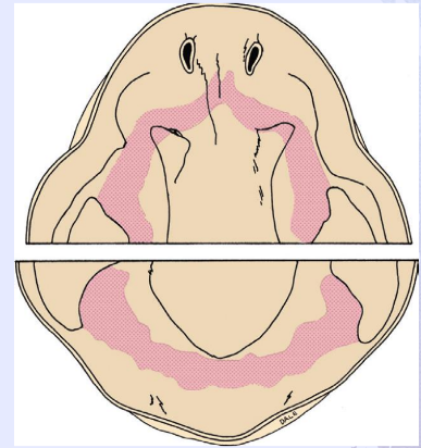
By 37 days of development, a continuous band of thickened epithelium, roughly horseshoe-shaped, forms the primary epithelial band in the positions of the future dental arches.
This band gives rise to two subdivisions:
Dental Lamina (responsible for tooth development).
Vestibular Lamina (responsible for forming the vestibule between lips and teeth).
A key event in tooth initiation is the formation of placodes within the primary epithelial band, which initiate the formation of different tooth families.
3. Ectomesenchyme in Tooth Development
Ectomesenchyme is a form of mesenchyme derived from neural crest cells. It forms the tissues of the neck and cranium and is responsible for the development of dental tissues through interactions with the oral epithelium.
Initially, up to 12 days, the first arch epithelium can form tooth-like structures when combined with neural crest cells. This potential is transferred to neural crest cells as development progresses.
4. Stages of Tooth Development
Tooth development proceeds in distinct stages, though they form part of a continuous process, making it challenging to draw precise lines between stages.
Primary Epithelial Band Formation (37 days of development):
The primary epithelial band thickens in the location of the future dental arches.
Bud Stage (Week 6-8 in Utero):
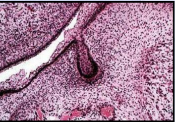
Characterized by the rounded growth of the epithelium into the underlying mesenchyme.
Dental lamina forms buds called the enamel organ, surrounded by condensed ectomesenchyme (future dental papilla).
The dental sac/follicle encircles both the enamel organ and the dental papilla, contributing to the formation of cementum and periodontal ligament.
Enamel Organ will eventually form the enamel.
• Dental Papilla will form dentin and pulp.
• Dental Sac / Follicle will give rise to cementum & the periodontal ligament
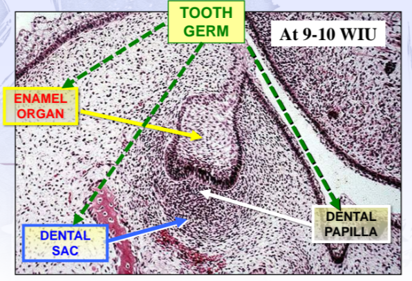
Cap Stage (Week 9-10 in Utero):
The developing enamel organ undergoes unequal growth, leading to a concave inner surface.
Layers of the enamel organ during this stage:
Outer Enamel Epithelium (OEE): Single cuboidal cell layer on the convex surface.
Inner Enamel Epithelium (IEE): Columnar cells on the concave surface, which later become ameloblasts (enamel-forming cells). The IEE will eventually become ameloblasts that will form the enamel of the tooth crown by differentiating into tall columnar cell
Stellate Reticulum (SR): Star-shaped cells connected by desmosomes.to keep space for the developing enamel.
Stratum intermedium (SI) : They are rich in alkaline phosphatase enzyme essential for enamel mineralization
Enamel Knot: Signaling center that organizes cusp development.
Bell Stage (Week 14-18 in Utero):
This stage sees an increase in the size and complexity of the enamel organ. It is divided into early (before hard tissue formation) and late/advanced (marked by the formation of the first layer of dentin) stages.
Histodifferentiation (formation of ameloblasts and odontoblasts) and morphodifferentiation (final crown shape) occur.
The cervical loop (meeting point of OEE and IEE) becomes the future site for root formation.
The enamel organ begins to break away from the dental lamina, as mesenchymal invasion occurs. This gives rise to the successional lamina, which is responsible for the development of permanent teeth.
5. Dental Lamina and Fate
The dental lamina serves as the primordium for the deciduous teeth and loses contact with the oral epithelium after its role is complete.
It breaks up through mesenchymal invasion and can give rise to epithelial remnants called epithelial pearls or cell rests of Serre. These remnants can sometimes form odontogenic cysts or even supernumerary teeth (extra teeth).
Permanent teeth develop from the successional lamina, which forms during fetal development and continues after birth for some teeth (e.g., molars).
6. Late-Bell Stage and Tissue Differentiation
Dentin formation by odontoblasts marks the start of the late bell stage.
The following changes occur:
Outer Enamel Epithelium: Develops capillary loops for nutrient exchange.
Inner Enamel Epithelium: Differentiates into ameloblasts under the influence of newly formed dentin, which triggers reciprocal induction.
Stellate Reticulum: Shrinks as the formed enamel occupies the space.
Stratum Intermedium: Provides support for ameloblasts during enamel mineralization, with high levels of alkaline phosphatase for enamel mineralization.
The dental lamina eventually disintegrates and loses its connection with the developing teeth.