Untitled Flashcard Set
What are the Functions of the Skeletal System?
🌸Support
Bearing the weight of the body
🌸Protection
Encasing essential organs
Ex: ribcage protects the heart and lungs from injury
🌸Movement
Joints provide movement for bones
🌸Storage
Storage of minerals to be released into the bloodstream
Storage of fat in yellow bone marrow
🌸Manufacturing
Production of red & white blood cells from red bone marrow
Called hemapotoiesis
Types of Bones
Bones are a sold matric of living cells and fibers surrounded by calcium deposits
Bones are classfied by their shape:
Flat Bones
Irregular Bones
Long Bones
Short Bones
Sesamoid Bones
Long Bones
Long bones have 2 bacis regions:
Diaphysis: Shaft, long part of bond
Epiphyses: End of bone
Between these layers is a thin layer of internal cartilage called the epiphyseal plate (growth plate)
The ends of the epiphyses are covered with an external layer of cartilage called articular cartilage which provides smooth movement of joints and cushion from shock
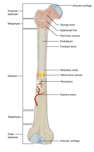
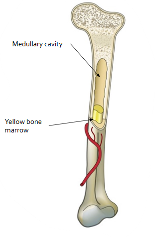
In the diaphysis of the long bone, a hollow medullary cavity is found
Red bone marrow fills this cavity in young people. Age causes the red marrow to be replaced with fatty yellow bone marrow.
It is within the bone marrow that new blood cells are produced (called hematopoiesis)
Spongy & Compact Bone
The outer layer of the bone is made of tough connective tissue called periosteum. It is the location of muscle attatchment and bone repair.
Beneath the periosteum is a thick layer of compact bone.
At the ends of long bones, the spongy bone layer is beneath the compact bone.
How do Spingy and Compact Bone Differ?
Spongy Bone
Spongy bone is a lattice of trabeculae (“little beams“) that are found along lines of stress for perfect resistance from compression
Between the trabeculae are spaces filled with marrow or blood vessels
Compact Bone
Compact bone is arranged in cylinders called osteons
Osteons are arranged in concentric cricles called lamellae
These lamellae surround a central (or Harversian) canal that contains blood vessels and nerves
The central canals are connected by perforating (Volkmann’s) canals running perpendiculary
What are the Types and Functions of Speacialized Bone Cells?
Osteocytes = mature bone cells make up the majority of the bone structure
Osteocytes are connected with tentacle-like canaliculi
Canaliculu connect all bone cells, allowing them to receive nutrients and remove wastes
Osteoclasts & osteoblasts line the Haversian canals canals and the surface of the compact & spongy bone
Osteoclasts break down bone
Osteoblasts produce new bone
How is Bone Formed?
An embryo’s skeleton is made of cartilage
Near the third month of embryo development, osteoblasts begin to secrete mineral deposits that replace the cartilage. The osteoblasts then mature into osteocytes.
This process of incorporating calcium & minerals into cartilage to become bone is known as ossification.
As a child grows, tall columns of chondrocytes (cartilage cells) at the epiphyseal plate divide and then deteriorate as the matrix around them calcifies. These cells are then known as osteoblasts, which form spongy bone.
Osteoclasts secrete acid to enlarge the medullary cavity, as the bone grows so that marrow is available for all cells.
What is the Composition of Bone?
Organic
35% Osteoid (made of ground substance & collagen)
Provides the flexibility & tensile strength requiered to keep bones from constantly breaking
Lack of collagen causes “Brittle Bone Disease“
Inorganic
65% Mineral Salts
Crystaline salts made of hydroxyapatites
Ca10(PO4)(OH)2
Provides bone strength & hardness
Lack of hydroxyapatite causes “Rickets“