Unit 4, Topic 1: Mitosis and Asexual Reproduction
1. Limits to Cell Growth
A. Cells divide rather than continuing to grow larger because..
The larger a cell becomes, the more demands the cell places on its DNA
If the cell grows too large, it will have trouble moving enough nutrients and waste across its cell membrane
B. Problem #1: Our DNA has its limits
All of the information that a cell needs to function is stored in the DNA of the cell
DNA is packaged into chromosomes. A chromosome consists of one very long linear DNA molecule consisting of thousands of genes
Each gene is the instruction for making a particular protein that the cell needs
The cell is constantly making copies of these genes and sending the copies (in the form of RNA) out to the ribosomes
When the cell is small, the information stored in the cell’s DNA is adequate to meet the needs of the cell
As the cell grows too large, there is an “information crisis”. The DNA cannot keep up with the demands of running a larger cell
C. Problem #2: A Growing Cell Needs More Food
A cell must take in a constant inflow of food, oxygen, and water across the membrane
Waste products must constantly be crossing the membrane to leave the cell.
A larger cell requires much more food, oxygen, and water. Larger cells generate more waste.
As the cell grows, the volume of the cell increases much more rapidly than the surface area of the cell membrane
When the cell gets too large, the membrane surface area is not adequate to transport the large quantities of food and water in, and waste products out
2. Cell division
Cell division is the process by which cellular material is divided between two new daughter cells
1 Mother cell →2 Daughter cells. The daughter cells are identical to each other and the mother cell
Each daughter is half the size of the parent cell, but immediately begins growing
A typical human cell has about 2 meters of DNA. Before the cell can divide, all of this DNA must be copied and then the two copies separated so that each daughter cell ends up with a complete set of DNA
Each species has a characteristic number of chromosomes in each cell nucleus; humans have 23 pairs/46 total
3. Chromosomes During Eukaryotic Cell Division
A. Each cell must first copy its chromosomes before cell division occurs
B. Each daughter cell gets a complete copy of that information
C. Cell division occurs in two main stages:
Mitosis - Division of the nucleus
Cytokinesis - Division of the cytoplasm
D. The chromosomes are not visible except during cell division. At the beginning of cell division, the chromosomes condense into compact, visible structures that are easily seen with a microscope
E. Well before cell division takes place, each chromosome is replicated or copies
F. At the beginning of cell division, each chromosome consists of two identical “sister chromatids.” These chromatids are connected at an area called a centromere.
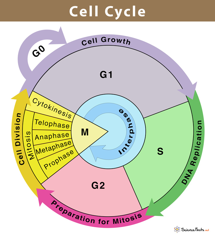
4. The Cell Cycle
The cell cycle is: the series of events cells go through as they grow and divide
The cel cycle is the life of the cell from the time it is first formed from a dividing parent cell until its own division into two cells
During the cell cycle:
Cell grows
Cell prepares for division
Cell divides into 2 daughter cells
The cycle consists of give major phases
G1, S (synthesis), G2, M (mitosis), Cytokenesis
Before a cell can begin mitosis and actually divide, it must:
Form duplicates of chromosomes
PRoduce organelles for daughter cells
These preparations occur during the G1, S, G2 phases of the cell cycle
Collectively known as interphase
G1 phase
Cell doubles in size
Enzymes, organelles, and other molecules double in number
S phase
DNA replication
G2 phase
Cell assembles special structures for cell division
When interphase in complete, the cell is ready to begin the process of cell division
Interphase
Of the cell cycle, interphase accounts for 90% of the time
Nucleus is cell defined and bounded by the nuclear membrane
Outside of the nucleus are two centrioles. Their function is to organize the microtubules into a spindle
They will move apart as spindle microtubules grow out of them
G1 phase is a period of intense biochemical activity. The cell doubles in size, and the enzymes, organelles, and other molecules double in number.
The chromosomes have duplicated during the S phase and they appear as a jumbled mass of fibers, not yet condensed.
In the G2 phase, the cell assembles the special structures needed for cell division
5. Mitosis
Early prophase
The chromosomes coil and thicken and become distinct from one another. The chromosomes are now visible
The nucleolus disappears/disintegrates
The chromosomes are doubled throughout their length
Each half of the double chromosome is a chromatid
The chromatids are connected by a centromere
The centrioles separate and start moving to opposite ends of the cell. A spindle made of microtubules begins to form
Late prophase
The nuclear membrane fragments and mictrotubules invade the nuclear area. The spindle is completely formed
The spindle is a structure that will help to separate the chromosomes. During prophase the pairs of chromatids become attached to the fibers of the spindle
The centrioles have moved to the opposite poles, forming the spindle as they go
Metaphase
The centrioles are at opposite poles
The spindle fibers will push and pull chromosomes
The chromosomes line up at the center of the cell
Each chromosome is connected to a spindle fiber at its centromere
Anaphase
The centromeres divide and chromatids move to opposite sides of the cell
The mictrotubules begin to shorten and pull chromatids apart to the centrioles
By the end of anaphase, the two ends of the cell have equivalent and complete sets of chromosomes
Telophase
Nuclear membrane begins to form
Nucleolus returns
Cell begins to pinch in (clevage furrow, in animals only)
Reults: 2 identical cells
6. Cytokinesis
A. At the end of mitosis, two nuclei have been formed. Each nucleus has an identical set of chromosomes
B. Cytokinesis is the division of cytoplasm
C. Cytokinesis usually occurs at the same time as telophase
D. In animal cels, a cleavage furrow pinches the cell membrane inward until the cell si pinched into two separate cells. each new cell contains its own nucleus, cytoplasm, and organelles
E. In plants, it is not possible for the cell to pinch inward because of the rigid cell wall. In plants, a cell plate forms midway between the two nuclei. The cell plate continues to form across the cell until two separate cells have been formed
7. The Importance of the Cell Division Process
A. Results of Mitosis
In unicellular plants and animals: results in new offspring by asexual reproduction
In multicellular organisms: results in the growth & repair of the organism
B. Importance of Mitosis
The two new cells are exact duplicates
This ensures that new cells will be able to carry on the same function as the parent cell
8. Regulation of the Cell Cycle
A. The frequency of cell division varies with the type of cell. Skin cells divide frequently throughout our lives. Liver cells maintain the ability to divide but only do so on rare occasions—say, to repair a wound. The most specialized cells, such as muscle and nerve cells, do not divide at all
B. Controls on Cell Division
When cells come into contact with other cells, they respond by not growing
When an injury, like a cut in the skin, occurs, the cells at the edge of the injuriy begin to divide rapidly
When the healing process nears completion, the rate of cell division slows down
C. Cell Cycle Regulators
There are many proteins found on the inside and the outside of the cell that regulate cell division
Some of the proteins are responsible for starting/stopping cell division
Other proteins seem to speed up/slow down the cell division process
These proteins send out signals that prevent excessive cell growth. This keeps the tissues of the body from disrupting one another
D. Unconftrolled Cell Growth
If the cells in a tissue grow uncontrollably, the consequences may be severe
Cancer cells do not respond to the signals that regulate the growth of cells. Cancer cells divide uncontrollably and form masses of cells called tumors. These tumors can damage the surrounding healthy tissues by stealing their nutrients.
All cancers have one thing in common: the protein regulators that control the cell cycle have failed at their job
Chromosomes | Other | |
Interphase (G1, S, G2) |
|
|
Early Prophase |
|
|
Late Prophase |
|
|
Metaphase |
|
|
Anaphase |
|
|
Telophase |
Identical sets |
|
Cytokenesis |
|
|
Cancerous cells vs. Healthy cells
What is cancer?
Cancer is when what cell process in uncontrolled?: Mitosis
Normal cells of the body spend most of their time in which phase of mitosis: Interphase
How are cancer cells different from regular cells?
Spend less time in interphase
Have uncontrolled mitosis - they do not stop division after a certain point
Grow faster than normal cells
They can break away from groups of cells and travel to other places in the body
They have a different physical structure - often with parts that allow them to grip and grab onto other cells and tissues
What is one way that cancer cells are physically different than normal cells?
Many have structures that allowed them to grip onto other cells
What are some causes of cancer?
You can end up dying from cancer because the cancerous cells take the nutrients meant for the healthy cells, preventing healthy cells from getting what they need for proper function. This can lead to tissue dying, and eventually death.
Causes:
Chemicals - many chemicals cause mutations, leading to problems with mitosis
UV radiation - when tanning or exposed to the sun, you absorb UV light. UV light causes DNA to be mutated, which can alter the cell mitosis cycle in the future.
Viruses - an example is the HPV virus which is known to cause cervical cancer in females if they are exposed
X-rays - they radiate cells, causing mutations
Genetics - some people are born with genes that are more likely to mutate over time or have cancerous properties. If cancer runs in the family, then you are likely to have a higher risk of getting cancer.
All of these increase the likelihood of cells becoming cancerous, meaning they may not repair and follow a proper cell division cycle
What is a tumor?
A tumor is defined as a cluster of cells going through uncontrolled mitosis
There are two types of tumor. Benign or malignant.
Benign: tumor cells that do NOT migrate to other parts of the body and do not harm other parts of the body
Malignant - these tumors have cells that can migrate to other parts of the body, potentially causing the cancer cells to spread in the body
Metastasis - when a tumor or cancer cells have spread from their original source
How is cancer treated?
In order to determine if a tumor is cancerous, a doctor will take a biopsy of it.
Biopsy - a doctor removes a sample of the cells from the tumor to analyze under a microscope to determine the nature of the cells
If a tumor is deemed cancerous, there can be multiple treatments.
Surgery - often, the doctors will remove the tumor and attempt to remove the cancer from the body and reduce the chance of it spreading
Chemotherapy - To kill cancer cells in the body, doctors can inject powerful drugs into the body. This is usually done as an injection of drugs into the blood. These drugs kill cells quickly and aggressively. These drugs will also kill many normal cells in the body. So, this therapy can make people very weak and frail.
Radiation treatment - If the tumor is cancerous, you can direct radiation at the tumor. The radiation will kill the cells in the tumor, causing the tumor to shrink and, hopefully, die.