Mechanical Traction
Functional Unit of the Spine
• TWO adjacent vertebrae & the intervening IVD
• Motion segment of the spine
• Anterior – WB
• Posterior – Mov’t
Pain Sensitive
• VB
• IVD
• Outer 1/3 AF
• ALL
• PLL
• Facet
• NR
• Muscle
Pain Tolerance
• Nucleus Pulposus
• Outer 2/3
Injury to the MOTION SEGMENT:
• Spondylosis- Degeneration of the intervertebral disc
• Spondylolysis- Defect in the pars interarticularis or the arch of the vertebra
• Spondylolisthesis- Forward displacement of one vertebra over other
• Retrolisthesis- backward displacement of one vertebra on another
Annulus Fibrosus
• Outer laminated portion the disc
• Contains 20 concentric, collarlike rings of collagenous fibers that criss-cross each other to increase their strength & accommodate torsion movements
• Three Zones
• Outer Zone – Sharpey’s Fibers
Nucleus Pulposus
• At birth, hydrophilic mucoid tissue
• Gradually replaced by fibrocartilage
• Water-binding capacity of the disc decreases with age, and degenerative changes (spondylosis)
• Initially, the disc contains approximately 85% to 90% water
• Decreases to 65% with age
• Adheres to the vertebral body by means of the cartilaginous end plate
• Avascular, with only the periphery receiving a blood supply
• Receives nutrition by diffusion, primarily through the cartilaginous end plate
• Until the age of 8 years, IVD have some vascularity, decreases with age
Aneural
• Posterior periphery of the AF – nerve supply?
• Direct vertical pressure on the disc can cause the disc to push fluid into VB
• ↑ pressure on the IVD = Schmorl's nodules
• Herniations of the NP into the VB
• Normally, an adult is 1 to 2 cm (0.4 to 0.8 inch) taller in the morning than in the evening (20% diurnal variation)
Herniation
• Localized displacement of disc material beyond the normal margins of the disc,
• Defined circumferentially by the outer edges of the ring apophyses of the vertebral body end plates &, by the end plates above and below the disc space
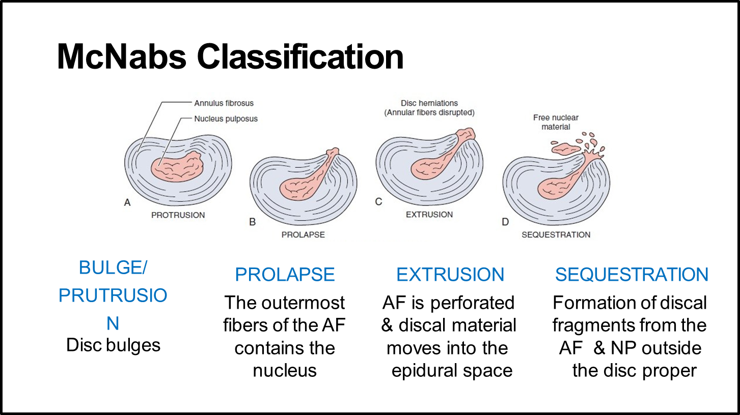
IVD Herniation
McNab’s Classification
(BPES)
• Bulging
• Prolapsed
• Extrusion
• Sequestration
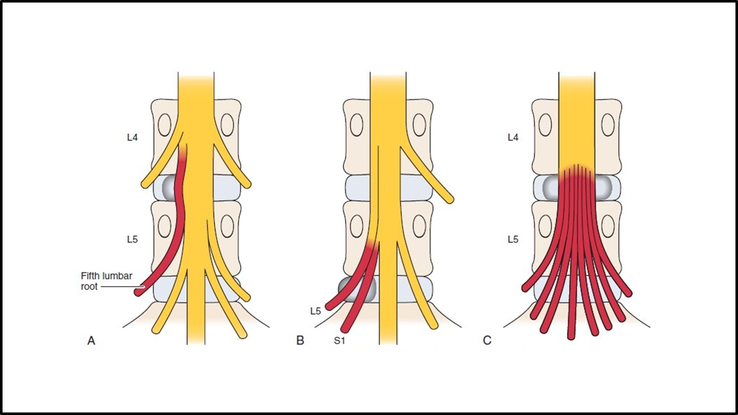
IVD Herniation can lead to…
• Pressure on the spinal cord itself (upper lumbar spine) leading to a Myelopathy
• Pressure on the cauda equina leading to Cauda Equina Syndrome
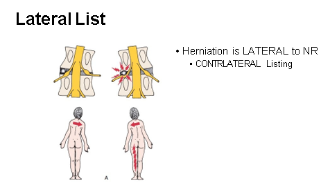
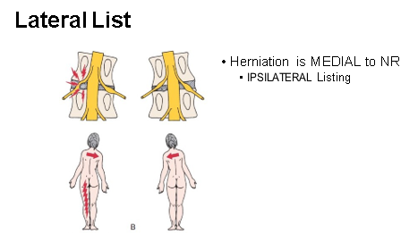
• Pressure on the nerve roots (most common)
L5-S1
• MC site of problem in the spine
• Greater WB
• COG passes → ↓ shearing force
• Transition of mobile segment (L5) to fused or stable (S1) segment
• Greater angle compared with other vertebrae
• Large amount of mov’t
• The herniations from the fragments of the nucleus pulposus into the vertebral body is called:
• Schmorl's nodules
Mechanical Effects
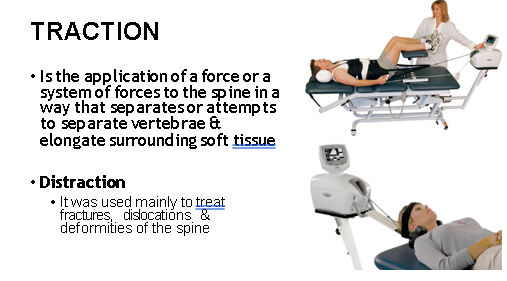
• Stretching of muscles and ligaments
• Separation of apophyseal joints
Effect on spinal movement
• Distraction of the vertebral bodies with enlargement of IVF
• 1-2 mm separation per IV space
• ↓ pain, paresthesia while traction is applied
Effects on bone
WOLFF’S LAW
• Bone changes in response to compressive or distractive loads
• Intermittent traction with a rhythmic on & off load cycle provides distraction load & promotes movement
Effects on ligaments
Viscoelactic Property
• Property of ligaments that allow them to resist shear forces & return to their original form following the removal of a deforming load
• Slow loading rates allow the ligament to lengthen as it absorbs the force of the load
Effects on Articular Facet Joints
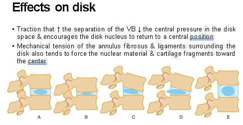
• Meniscoid structures, synovial fringes, or osteochondral fragments (calcified bone chips) impinged between joint surfaces are released → ↓ symptoms when joint surfaces are separated
Effects on Muscular System
• ↓ EMG activity of Erector Spinae = muscle relxation
• ↑ BF, activation of muscle proprioceptors, Gate Control Theory
Effects of Nerves
• Pressure on nerves or roots from bulging disk material, irritated facet joints, bony spurs, or narrowed foramen size causes the neurologic malfunctioning often associated with spinal pain
• ↑ blood’s circulation to the nerve, ↓ edema and allowing the nerve to return to normal functioning
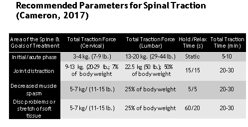
Parameters
• Position:
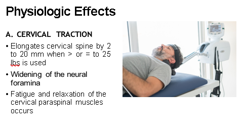
• 0-5 cervical fl: IV space at C1-C5
• 25-30 flexion: for C5-C7
• 0 : disc dysfunction
Force:
• 7% of the body weight
• 10-15 lbs. initial tx.
• 25-30 lbs : elongation of the spine occur
Treatment time:
• 5 – 10 min HNP
• 10-30 min for other conditions
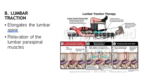
Parameters
Position:
• 45-60 of hip flexion : IV space at L5-S1
• 75-90 of hip flexion: at L3-L4
Force:
• ½ of body weight : male
• 1/3 of body weight : female
Treatment time:
• 5-10 min for HNP
• 10-30 min for other condition
Major Parameters for the Application of Traction
• Positioning
• Weight to be used
• Duration
• Continuous vs. intermittent application
• Concomitant modalities to be given
INDICATIONS
• Relief of pain from disc herniation
• Radiculopathies
• Patient immobilization as in the treatment of acute disc herniation
CONTRAINDICATIONS
• Inadequate expertise
• Cervical ligament instability
• Underlying tumor or malignancy
• Severe osteopenia
• Vertebro-basilar arterial disease
• Cord compression & infections of the spine
• Old age
• Pregnancy
• RA
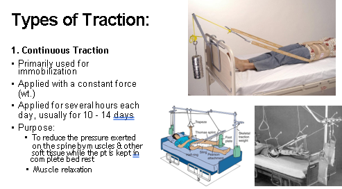
2. Sustained Traction
• Is also applied continuously but usually for no longer than 45 - 60 min
• > greater forces can be applied
• Purpose:
• Relaxation of muscles
• Stretching of soft tissue
• separation of bony surfaces
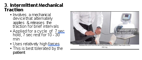
Purposes:
• To separates bony surfaces
• Mobilize the jts
• Stretch soft tissue & relax the muscles around the jt.
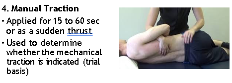
Level-specific Manual Traction
L3 – L4
L4 – L5
L5 – S1
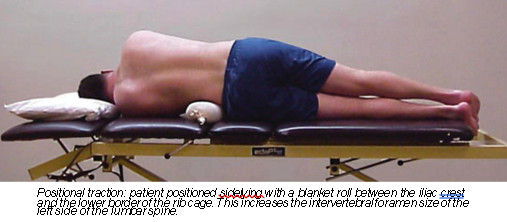
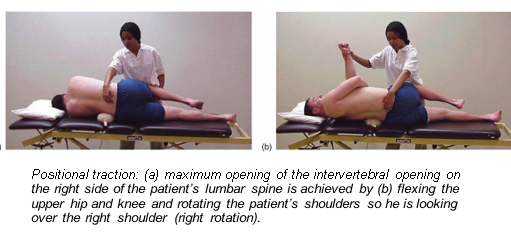
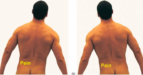
5. Positional Traction
• Applied by positioning the patient in a way that will affect the relationship of the bony surfaces in the area treated
• Duration of treatment is 5 - 30 min
• Can be done unilaterally / bilaterally
• Purpose:
• To alleviate pressure on an entrapped nerve
• Relax the muscle in spasm
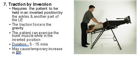
6. Gravity - assisted Traction
• Patient is placed in a position that favors distraction
• Duration : 10 - 30 mins
• Permits the gravity to help the overall distraction of targeted tissue
Mechanical Traction
General Parameters:
1. Body position: prone, supine, hip position, bilateral, or unilateral direction of pull
2. Force used
3. Intermittent traction: traction time and rest time
4. Sustained traction
5. Duration of treatment
6. Progressive steps
7. Regressive steps