Femur
Femur: Anatomy and Clinical Relevance
I. Overview of the Femur
Location: The femur is the longest, strongest, and heaviest bone in the body.
Function: The femur supports the weight of the body, facilitates movement of the lower limbs, and is a key bone in walking, running, and standing.
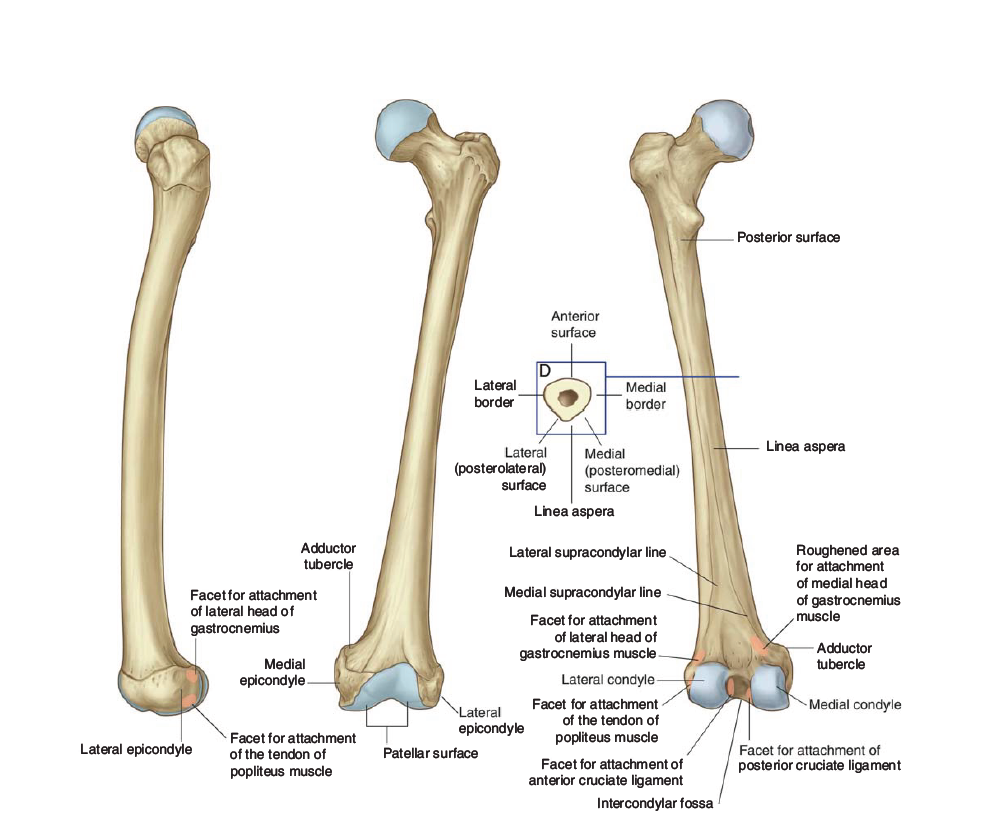
II. Structure of the Femur
The femur consists of the following parts:
Head:
Spherical structure that fits into the acetabulum of the pelvis to form the hip joint.
Fovea Capitis: A small depression on the head where the ligamentum teres attaches.
Blood Vessel: The artery to the head of the femur, a branch of the obturator artery, supplies blood to the femoral head through this fovea.
Neck:
Constricted portion between the femoral head and shaft. The neck is a common site of fractures, particularly in elderly individuals.
Greater Trochanter:
Large bony prominence located laterally at the junction of the neck and shaft.
Muscle Attachments:
Gluteus medius and minimus (abductors of the hip).
Piriformis (lateral rotator of the hip).
Obturator internus and gemelli muscles (lateral rotators of the hip).
Lesser Trochanter:
Smaller prominence located medially and posteriorly on the femur.
Muscle Attachment:
Iliopsoas muscle (primary hip flexor).
Shaft (Diaphysis):
The long cylindrical part of the femur. The shaft is slightly curved for strength and flexibility.
Muscle Attachments:
Adductors (adductor longus, adductor magnus) (medial side).
Vastus medialis, vastus lateralis, and rectus femoris (quadriceps group) (anterior side).
Medial and Lateral Condyles:
Located at the distal end of the femur, these condyles articulate with the tibia to form the knee joint.
Muscle Attachments:
Gastrocnemius muscle (posterior side) attaches to the femoral condyles, contributing to knee flexion and plantarflexion.
Patellar Surface:
The smooth surface on the anterior side of the femur where the patella (kneecap) articulates.
Intercondylar Fossa:
A deep notch between the condyles, which houses the cruciate ligaments of the knee joint.
III. Function of the Femur
Support and Load Transmission:
Transmits the body's weight from the hip to the knee joint during standing, walking, and running.
Movement:
Provides attachment sites for muscles that enable movement of the hip and knee (flexion, extension, rotation).
Shock Absorption:
The curved shape of the femur helps absorb shock and distribute forces during locomotion.
Red Blood Cell Production:
The femur contains bone marrow, which produces red blood cells.
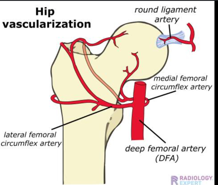
IV. Blood Supply to the Femur
Arteries Supplying the Femur:
Profunda Femoris Artery (Deep Femoral Artery):
A major branch of the femoral artery that supplies the femur and its muscles.
Perforating branches of the profunda femoris artery supply the femoral shaft and surrounding muscles.
Medial and Lateral Circumflex Femoral Arteries:
Branches of the profunda femoris artery, supplying the hip joint and head of the femur.
Artery to the Head of the Femur:
This small artery arises from the obturator artery and supplies the femoral head through the fovea capitis. This is the main blood supply to the femoral head, especially important in young individuals.
A loss of blood supply to the femoral head (e.g., due to fracture) can result in avascular necrosis.
V. Clinical Significance
Femoral Fractures:
Neck of the Femur Fractures: Common in elderly people due to falls. These fractures can disrupt blood supply to the head, leading to avascular necrosis.
Diaphyseal Fractures: May occur from high-impact trauma and often require surgical intervention.
Avascular Necrosis of the Femoral Head:
Occurs when the blood supply to the femoral head is compromised, leading to bone death. It can be caused by fractures, dislocations, or certain conditions like corticosteroid use or sickle cell disease.
Osteoarthritis of the Hip Joint:
Degeneration of the hip joint often involves the femoral head and acetabulum. This leads to pain and limited range of motion, especially in older adults.
Legg-Calvé-Perthes Disease:
A condition in children where blood supply to the femoral head is temporarily interrupted, leading to necrosis and deformity of the femoral head.
VI. Summary
The femur is a strong bone that plays a critical role in weight-bearing, movement, and stability.
It serves as the attachment site for major muscles that control the hip and knee.
Blood supply to the femoral head, through the artery to the head of the femur, is crucial for its health and function.
Femoral fractures and conditions like avascular necrosis highlight the importance of maintaining the femur’s blood supply and integrity.
Tips for Remembering:
Trochanters: Remember the "Greater Trochanter" as the site for the Gluteus Medius and the Lesser Trochanter as the site for the Iliopsoas.
Visualize the artery to the head of the femur running through the fovea capitis for understanding blood supply.
Remember:
The femur is involved In the knee joint.
Articulations:
Patellofemoral articulation- articulation of the patella with the patellar surface of the femur.
Tibiofemoral articulation- articulation of the medial and lateral condyle of the tibia that of the femur.
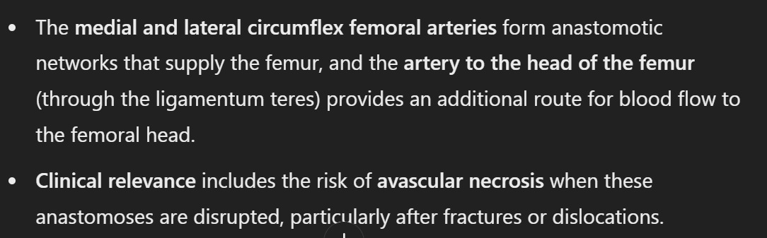
ANASTOMESES