LAB EXAM 2
Pipeting
withdrawing liquid:
press pipet stopper to the FIRST stop
use first stop ONLY
dip tip into liquid
let go of pipet stopper to pull up liquid
pull pipet tip out of liquid
check that liquid has been sucked up
dispensing liquid:
place pipet tip into container
press down on pipet stopper to the SECOND stop
pull pipet out of tube before releasing finger from stopper
1918 Flu Video. Be able to:
describe similarities and differences between the 1918 flu pandemic and the COVID-19 pandemic
similarities:
virus spread fast
many deaths
confused doctors
overflow of hospitals
understaffed hospitals
mutated virus
lack of awareness
population denial
transcontinental travel
virus was asymptomatic at first
differences:
war was going on, resulted in parades, and work at factories
science is more advanced now and we know about viruses
communication is done easier now via social media
successful virus with COVID-19
PCR ribotyping. Be able to:
explain the how ribotyping works
PCR-ribotyping: identification of organisms based on differences in the rRNA gene
STEPS:
PCR: PCR is used to copy rRNA gene
create enough material for detection
Digest: restriction enzyme detect and cut DNA in places where the sequence differs between species
This creates DNA fragments of different sizes depending on the species.
gel electrophoresis: view the different sizes of DNA to determine the species
DNA is placed into a gel and an electric current is applied.
Smaller fragments move faster and farther than larger ones.
Final Result (Ribotyping Pattern)
GOAL: detect differences in rRNA gene sequence to make identification
Different species or strains will have different band patterns.
By comparing the pattern to known samples, you can identify the bacteria.
describe how to set up a PCR experiment and how it works
PCR: Polymerase Chain Reaction
PCR function: makes many copies of DNA
used to amplify rRNA genes - makes many copes of rRNA gene
Setting up a PCR experiment:
you combine several key components in a tube and keep them cold (on ice):
Primers (forward and reverse): short (20 nucleotides) pieces of DNA designated by scientist to bind to either side of region to be copied
Two roles of Primers:
defining what regions of DNA is copied
provide 3’ end for building DNA
DNA polymerase: to build DNA
dNTPs: the enzyme and building blocks used to make new DNA
template: DNA you are copying
Bacteria: a small amount of bacterial cells
How PCR works:
Tubes are loaded into thermocycler that brings the samples through specific temperature cycles:
thermocycler: machine that heats and cools tubes over and over again
95°C: DNA opens
56°C: Primers bind to DNA
72°C: DNA polymerase builds DNA
explain what a restriction enzyme does
restriction enzymes: cut DNA at specific sequences of nucleotides
recognize specific sequence of DNA and cuts them
CANNOT be controlled to cut specific sequence we want
found naturally in bacteria
explain how gel electrophoresis works
Gel electrophoresis: technique used to separate DNA by size and for visualizing DNA
STEPS:
DNA is mixed with stain and a heavy fluid (glycerol), then loaded into well of gel
glycerol makes DNA heavy enough to sink to bottom of well
gel is placed in buffer and a current is applied across the buffer chambers so that one end is negative and other end is positive
negative electrode is placed on the side nearest to samples
positive electrode placed on the opposite side
DNA move through gel towards positive side (since DNA is negatively charged)
small molecules fit through the holes better large molecules, so small molecules will travel faster
results in separation of DNA molecules by size
DNA can be seen because of stain
size can be determined by running a DNA ladder made up of mixtures of DNA fragments of known size along sample DNA
interpret gel electrophoresis data, including ribotyping data
You compare your sample bands to the ladder
helps estimate the size (in base pairs) of each band in your sample
The number and size of bands depend on the species
compare your sample’s pattern to known/reference patterns
CRISPR. Be able to:
explain the purpose of CRISPR for bacteria in nature
CRISPR in bacteria protects bacteria from:
viral infection
defend against bacteriophages
explain how scientists use CRISPR to edit DNA
CRISPR in genome editing allows scientist to cut anywhere they choose (vs. Molecular cloning that can only cut at specific restrictive enzyme cut sites)
guide RNA (gRNA) targets cas9 to cut the DNA where DNA / gRNA sequence match
once the DNA is cut, a gene deletion, insertion, or other modification is formed
describe the molecular players of CRISPR as used in genetic engineering (cas9, gRNA, donor DNA, directed homology repair)
cas9: used to cut DNA where gRNA binds
a bacterial endonuclease that forms a double strand break (cut) in DNA at a specific site within a larget recognition sequence or target site
gRNA: guide DNA, binds the DNA and directs cas9
used to target Cas9 in cutting DNA in a desired location
gRNA is complementary to lac z gene sequence
donor DNA: piece of DNA with homology (ends are same) to lacZ
DNA that is patched into the cut of DNA, using bacterial repair machinery
include the desired sequence flanked on both sides by “homology arms” that match the sequence upstream and downstream of the cut
have homology arms that are sequences that match parts if destination DNA
homology arms help donor DNA line up correctly
directed homology repair: DNA repair enzymes insert donor DNA into lacZ because of the homology, repairing the cut made by cas9
DNA repair enzymes turned on in presence of arabinose
homologous recombination, enzymes patch the break using donor DNA
explain how lacZ was disrupted in the CRISPR lab
LacZ gene codes for enzyme b-gal
if b-gal is expressed by bacteria in presence of X-gal, bacteria colonies turns blue
CRISPR-Cas9 is used to cut the bacterial DNA within the lacZ gene
provide cells with large quantities of donor DNA, which includes an insert with a stop codon that will disrupt the gene function
causes colonies to be white instead of blue

interpret results similar to those from the CRISPR lab
lacZ intact = blue colonies
lacZ disrupted = white colonies
LacZ cut but did not get repaired = bacteria die, no colonies
example:
Tube A:
has E. coli that were grown with NO arabinose
arabinose sugar is a nutrient E.coli needs to express the enzymes needed for DNA repair
plasmid included had:
donor DNA
did NOT have guide RNA
DNA repair did not occur (due to no guide RNA telling cas9 where to cut)
results in blue colonies
colonies stay blue because there was no arabinose and guide RNA was missing, preventing cas9 from knowing where to cut
Tube B:
has E. coli that were grown with NO arabinose
plasmid included has:
donor DNA
guide RNA
DNA repair did not occur (due to no arabinose)
results in no colonies, due to bacteria dying
bacteria dies because cas9 cut the lacZ gene but do not repair it due to no arabinose
Tube C:
has E. coli that were grown with arabinose
plasmid included has:
donor DNA
did NOT have guide RNA
DNA repair did NOT occur
results in blue colonies
colonies stay blue because there was no arabinose and guide RNA was missing, preventing cas9 from knowing where to cut
Tube D:
has E. coli that were grown with arabinose
plasmid included has:
donor DNA
guide RNA
DNA repair occurred
results in white colonies
lacZ gene was disrupted
RT-PCR. Be able to:
explain how RT-PCR is used in COVID-19 testing, including the enzymes and process of RT-PCR
RT-PCR: reverse transcription polymerase chain reaction
STEPS:
obtain sample, containing cells, bacteria, SARS-CoV-2 virus
extract all DNA from sample
use reverse transcriptase enzyme to copy RNA into complementary DNA (cRNA)
reverse transcriptase comes from retrovirus
PCR and DNA polymerase and primer specific the SARS-CoV-2 N-gene and conjugated to fluorescence molecule
N-gene is the gene detected in SARS-CoV-2 gene
add a fluorescent probe allows amount of target DNA be detected
fluorescence = positive result
no fluorescence = negative result
Immunoassay Rapid Tests and ELISA. Be able to:
explain how each rapid test works (Staph, Strep, and Covid)
rapid tests: antibodies conjugated to detect antigens in patient sample
Staph Test: uses agglutination assay that uses antibodies conjugated to blue latex beads to detect S. aureus antigen
Indirect Agglutination Immunoassays test: add latex beads to detect pathogen or antibody in sample
assay is specific for S. aureus because antibodies is specific to S. aureus antigen
Strep Test: uses lateral flow immunoassay that use antibodies conjugated to colored beads to detect S. pyogenes antigen OR used to detect SARS-CoV-2 antigen
STEPS:
apply fluid sample to designated location on test strips
if positive test, antigen will be included in sample
if negative test, antigen will not be present
bead-conjugated antibody is located in designated location to apply sample
if positive test, bead-conjugated antibody α antigen
antigen will bind to antibodies that are attached to beads
if negative, there is no antigen to bind to bead-conjugated antibody
fluid goes through lateral flow down through test strip
test strips have stripes:
first stripe has antibodies adhered to stripe
first stripe = antibody α antigen
if positive test, antibody α antigen α bead-conjugated antibodies
antigen that are bound to bead-conjugated antibodies binds to antibodies located in first stripe
causes beads to gather in stripe, changing color of stripe
if negative result, antibody do not have anything to stick to so first stripe does not change color
second stripe has antibodies adhered to stripe
second stripe = antibody α antibody
if positive test, antibody α bead-conjugated antibody α antigen
constant region of bead-conjugated antibodies that have an antigen bound to it will stick to antibody that is bound to stripe
causes beads to gather in stripe, changing color of stripe
if negative test, antibody α bead-conjugated antibody
constant region of bead-conjugated antibodies bind antibody that is bound to stripe (even if does not have antigen attached)
causes beads to gather in stripe, changing color of stripe
used as control
Covid Test: uses lateral flow immunoassay that use antiboides conjugated to reddish-gold beads to detect SARS-CoV-2 antigen
interpret rapid test results
Staph Test:
Positive: blue clumps on test card
Negative: no blue clumps
Staph Test:
Positive: two red lines on test strip
Negative: one red line on test strip
explain how an ELISA works
ELISA (Enzyme-Linked Immunosorbent Assay): detect antigens or antibodies using enzyme-linked antibodies that yield a color change upon substrate reaction.
involve antibody is conjugated to an enzyme
if positive test, enzyme reacts with substrate, causing a color change
Types include:
Direct ELISA: Detects antigens from sample
detects current illness
Steps:
in 2 wells, add patient sample to both
in well with antigens: molecules, proteins and antigens from sample stick to plastic in well
in well with no antigens: just proteins and molecules stick to well
wash wells
washes off anything that didn’t stick
add enzyme-conjugated antibody
well with antigen: enzyme-conjugated antibody α antigen
α: indicates what the antibodies bind
enzyme-conjugated antibodies bind to antigen
well with no antigens: enzyme-conjugated antibodies float around
wash again
in well with antigens: antibodies stay, as they are bonded to antigens that are stuck to well
in well with no antigens: antibodies get washed away
add substrate
in well with antigens:
positive result = substrate causes color change
in well with no antigens:
negative result = no color change

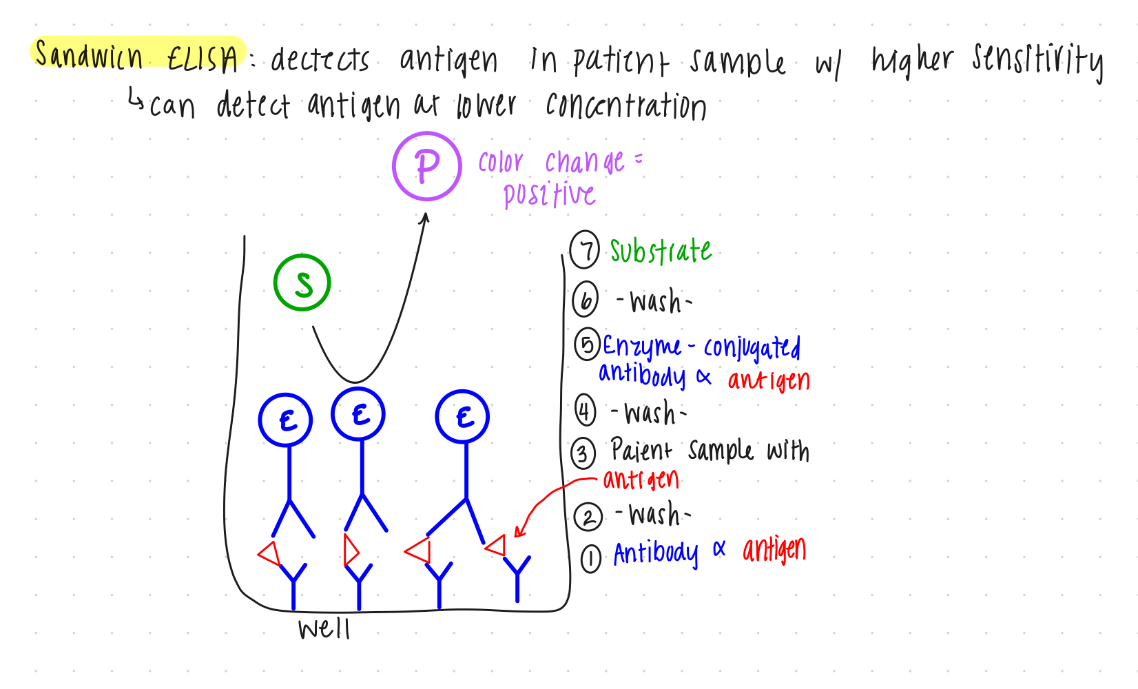
Sandwich ELISA: detects antigen in patient sample with higher sensitivity
can detect antigen at lower concentration
detects current illness
STEPS:
add antibodies to well
antibodies stick to bottom of well
wash
washes antibodies that did not stick to well
add patient sample containing antigen
antibodies α antigen
antigen bind to antibodies that are stuck to well
wash
washes away anything that isn’t antigen
add enzyme-conjugated antibody
if antigen is present, enzyme-conjugated antibody α antigen
antibody binds to antigen that is bound to antibody
forms antibody-antigen-antibody sandwich
wash
antibodies stay, as they are bonded to antigens that are bound to antibody that is bound to well
add substrate
positive result = substrate causes changes color
INDIRECT ELISA USED IN THIS LAB
Indirect ELISA: Detects antibodies in patient samples
detects current illness OR might have antibodies for illness
STEPS:
add antigens that are generated from lab
antigens stick to bottom of the well
wash
washes antigen that did not stick to well
add patient sample with antibodies
antibody α antigen
antibodies bind to antigens that are stuck to well
wash
add enzyme-conjugated antibodies
enzyme-conjugated antibodies α antibodies
enzyme-conjugated antibodies bind against the constant region of antibodies bound to antigen
wash
add substrate
positive result = substrate causes changes color

interpret the results of an ELISA
positive: change to purple color
negative: did not change color
White Blood Cells. Be able to:
identify unknown white blood cells under the microscope
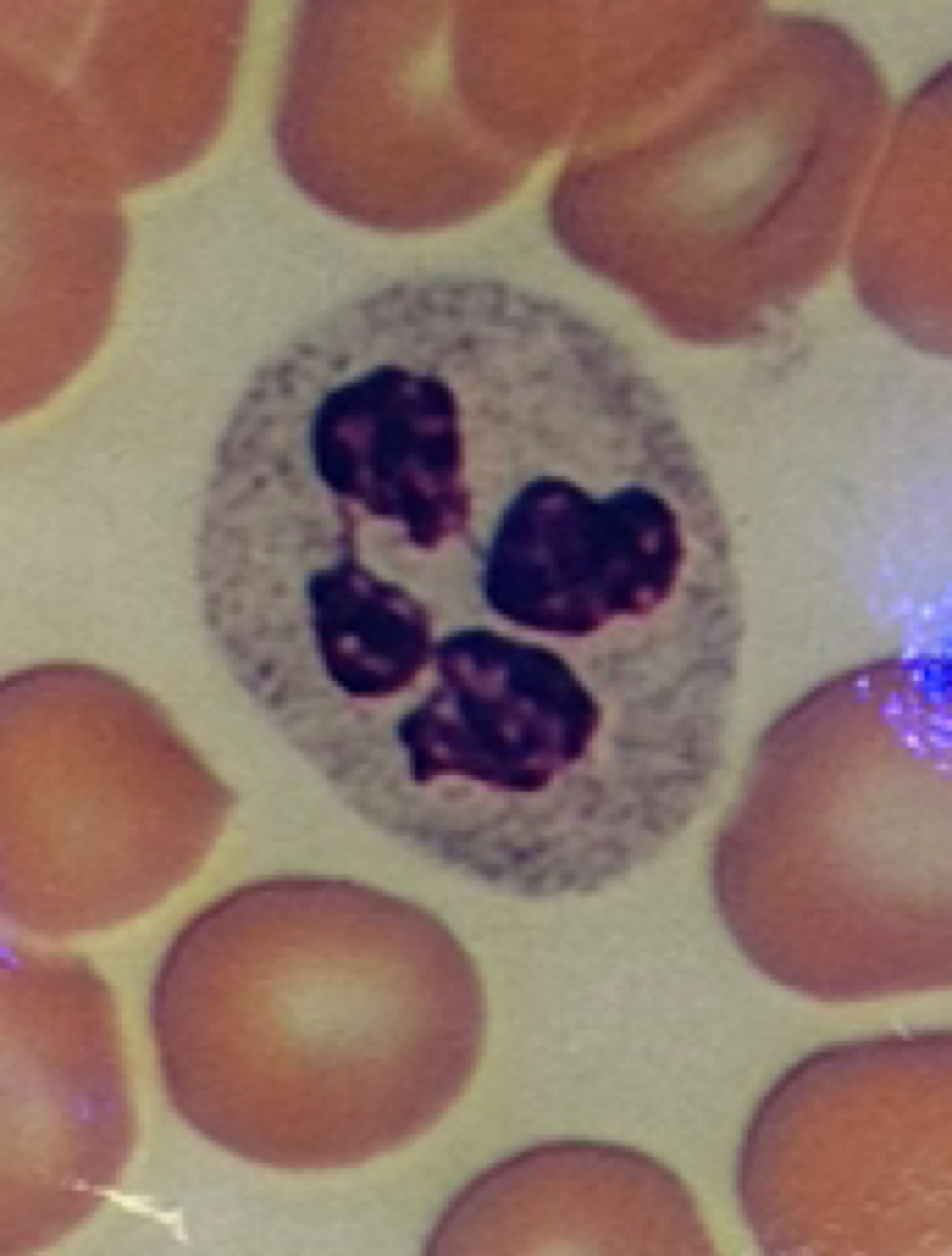
Neutrophils:
bigger than RBCs
multi-lobed nucleus
granules
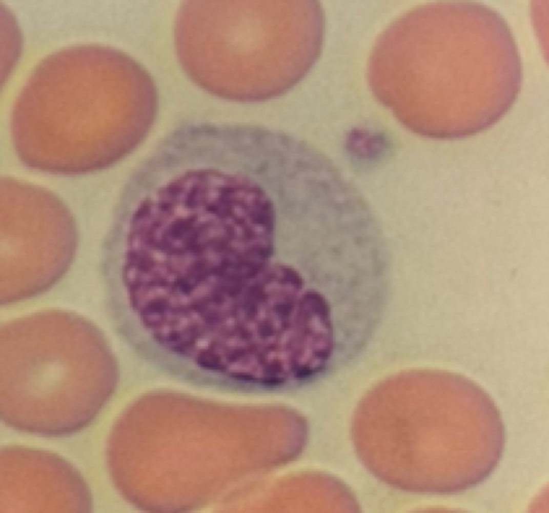
Lymphocytes:
only a little bigger than RBCs
No granules
spherical nucleus

Monocytes:
largest WBC
No granules
kidney-bean shaped nucleus
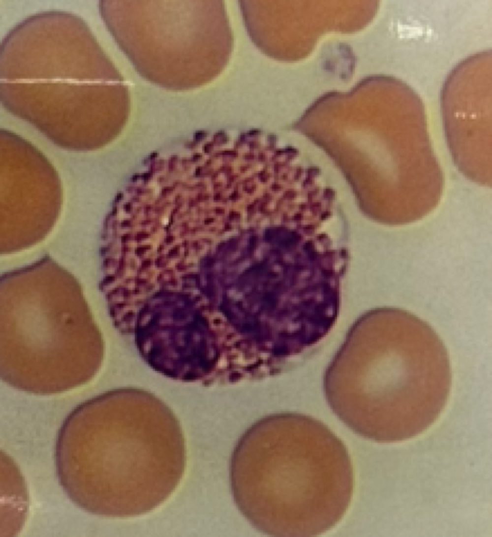
Eosinophils:
bigger than RBCs
two lobes in nucleus
acidophilic, pink granules
Basophils:
Much bigger than RBCs
two-lobed nucleus
deep blue granules
no cytoplasm

name the order of frequency of each white blood cell (from most frequent to least frequent)
Order of Frequency of white blood cell:
Neutrophils, Lymphocytes, Monocytes, Eosinophils, Basophils
Neutrophils: most abundant
Lymphocytes: 2nd most abundant
Monocytes: 3rd most abundant
Eosinophils: 2nd least abundant
Basophils: least abundant
Memory device: Never Let Monkeys Eat Bananas
state that some health conditions may disrupt the frequency of white blood cells in blood
Calling the Shots Vaccine Documentary. Be able to:
name the concerns people have about vaccination
fears about getting hurt
misinformation from influential people advising against vaccines
fraudulent studies
individualism - concern about individual child over community
hearing emotional stores of harm
side effects of vaccines
distrust due to medical history of harm
heard that vaccines cause autism
religious / cultural beliefs or moral grounds
personal ethical grounds - personal objective to any medical intervention
political grounds - idea that mandatory vaccinations are violation of individual liberties
explain why measles cases have increased in America
growing numbers of un-vaccinated individuals has lead to outbreak of measles
less herd immunity
state what type of cancer can be prevented by a vaccine
cervical cancer, HPV
state if there is scientific evidence of a link between vaccines and autism
NO - studies have been debunked as fraudulent
Simulated Epidemic
define incidence, prevalence, and index case
Incidence: number of new cases of a particular time period
Prevalence: the total number of cases at a particular time point
Index Case: first known case of a particular disease, which presumably is the source of new infections
calculate incidence and prevalence
Incidence = (number of new cases / total population) x K
does not include number of cases that existed prior to a specified time period
K = a power of 10, used to turn a decimal into a whole number
bring the incidence number above 1 so it can be reported as a whole number of cases per some number of people.
E.g. if incidence is calculated as 0.125, a K of 1000 might be used and incidence would be expressed as 125 cases per 1000 people rather than 0.125 cases per person.
Prevalence = (number of total cases at a time point / total population) x K
Pathogens. Be able to:
define pathogen and parasite
pathogen: microbe that causes disease
parasite: eukaryotic pathogens that cause disease themselves or act as a vector in spreading disease caused by microbial pathogens
include fungi, protozoans, helminths (worms), and animals
identify the example pathogens under the microscope or from an image
recall the information about scientific name, common name, classification, and associated disease from the pathogen chart
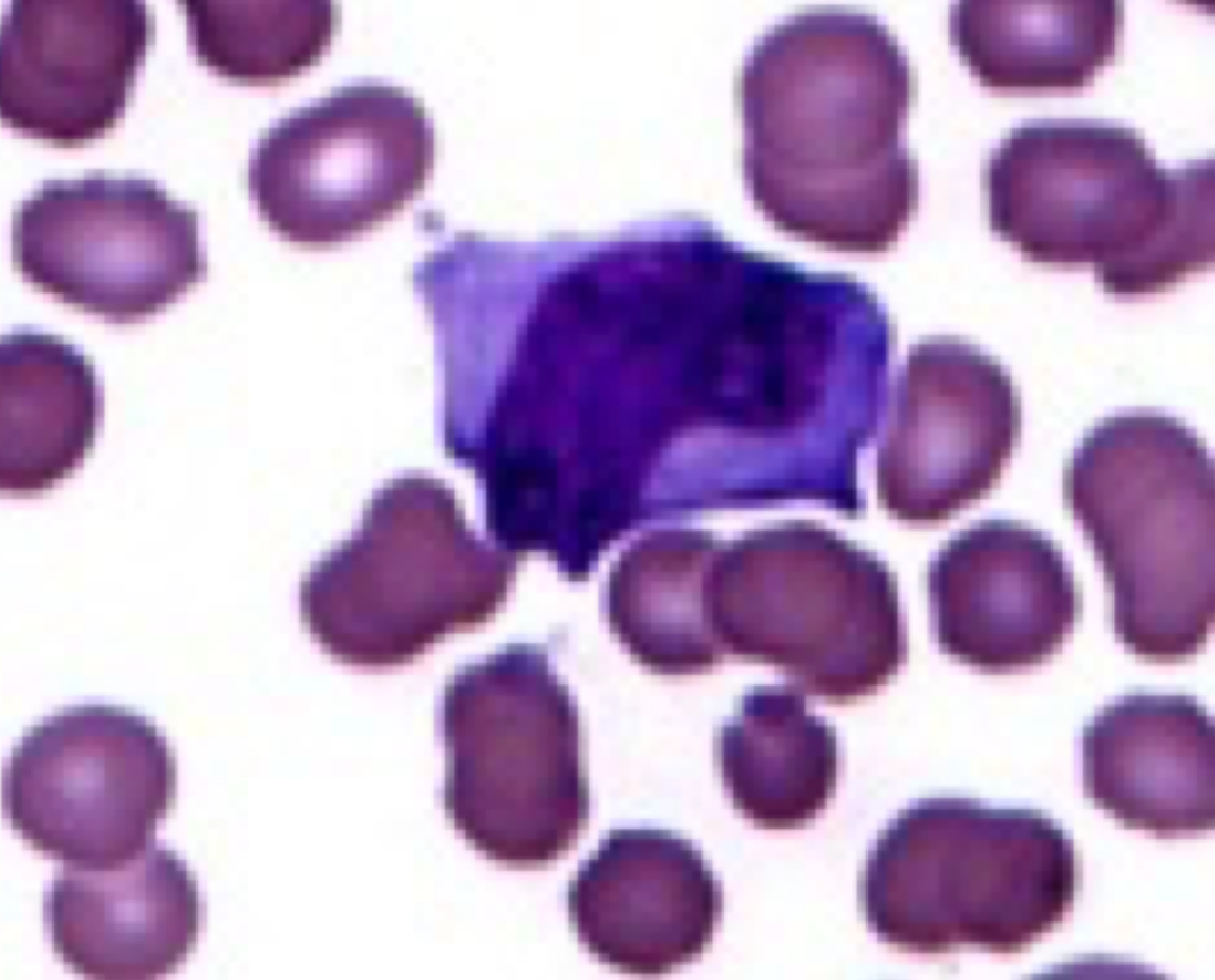
Epstein-Barr Virus
no common name
classification: dsDNA virus
associated disease: infectious mononucleosis (mono)
under microscope: looks like melting lymphocytes, downey WBCs
surrounded by red blood cells
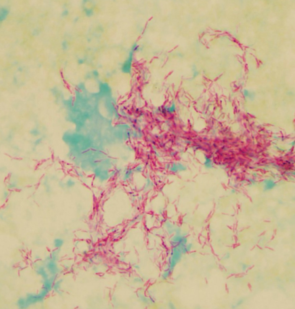
Mycobacterium tuberculosis
No common name
Classification: Acid fast bacillus
Associated disease: tuberculosis
under microscope: looks like pink bacilli (rod) in clusters or in a line
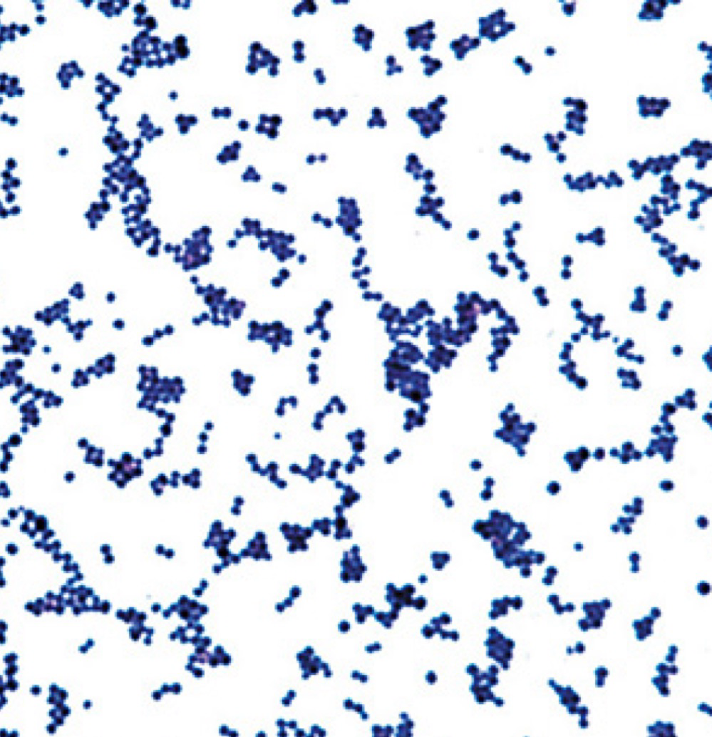
Staphylococcus aureus
no common name
classification: gram positive Staphylococcus bacteria
associated disease: staph infection
under microscope: looks like purple cocci (round) in clusters
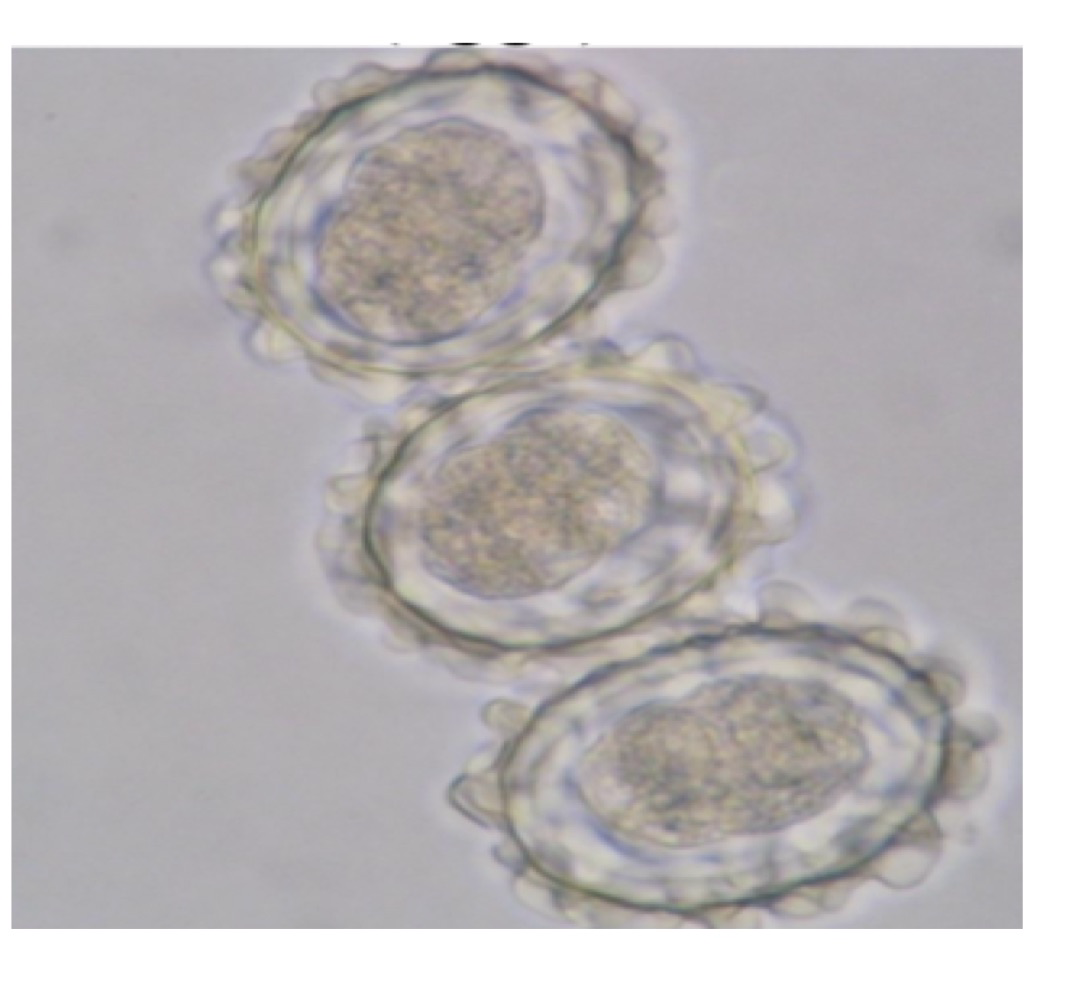
Ascaris lumbricoides
Common name: Round worm
Classification: nematode helminth
Associated disease: ascariasis
under microscope: looks like a big egg with ring and dark center, some have bumps on surface
they are eukaryotes so eggs are big
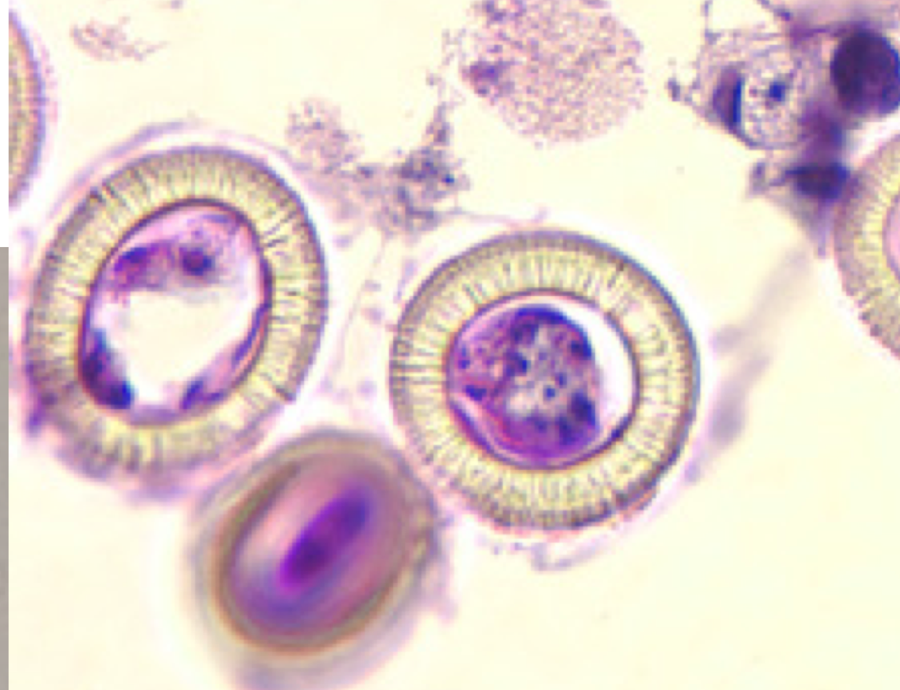
Taenia solium
common name: tapeworm
classification: cestode helminth
Associated disease: taeniasis
under microscope: looks like egg with thick rings, rings contain lines
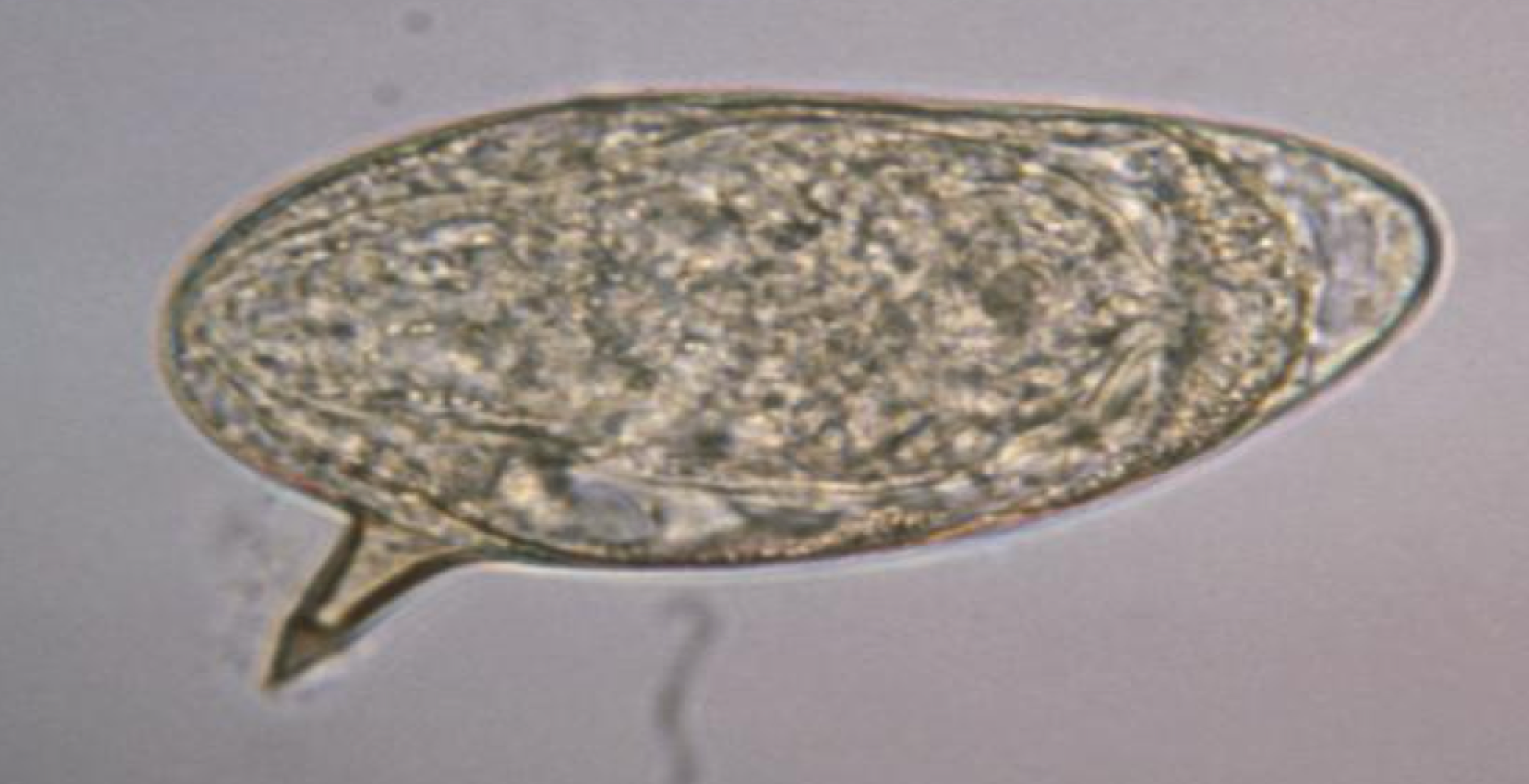
Schistosoma mansoni
no common name
classification: Trematode helminth
associated disease: schistosomiasis
under microscope: looks like oval with spine (sharp edge) coming out of it, looks like a word bubble
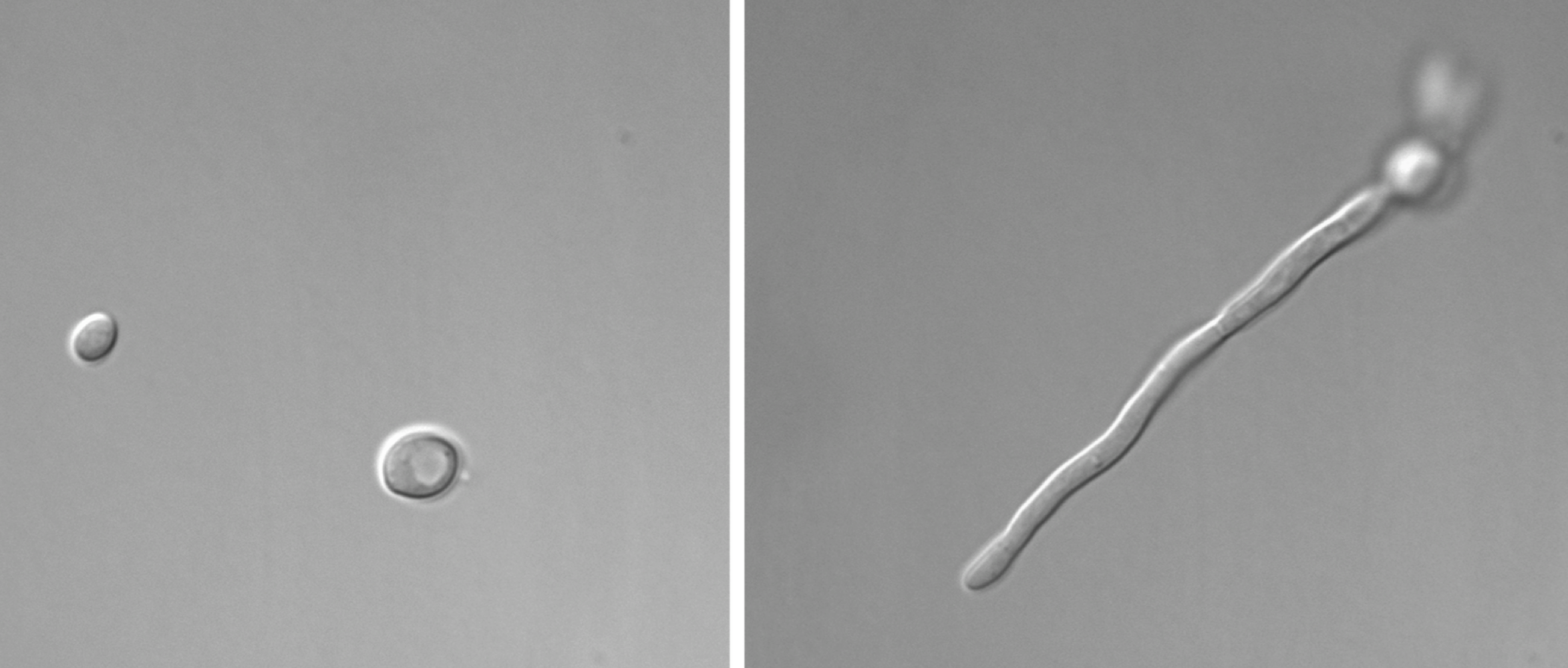
Candida albicans
common name: yeast
classification: Fungus
associated disease: candidiasis (yeast infection)
under microscope: looks like clear dots, others look like clear hyphae
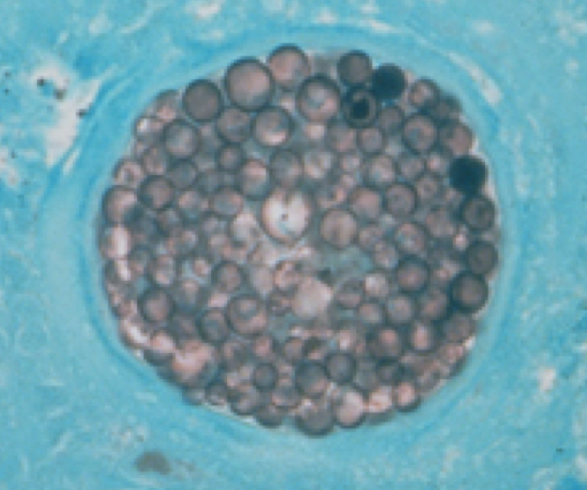
Coccidioides immitis
no common name
classification: fungi
associated disease: valley fever (coccidioidomycosis)
under microscope: looks like eggs clustered together in a circle, blue background
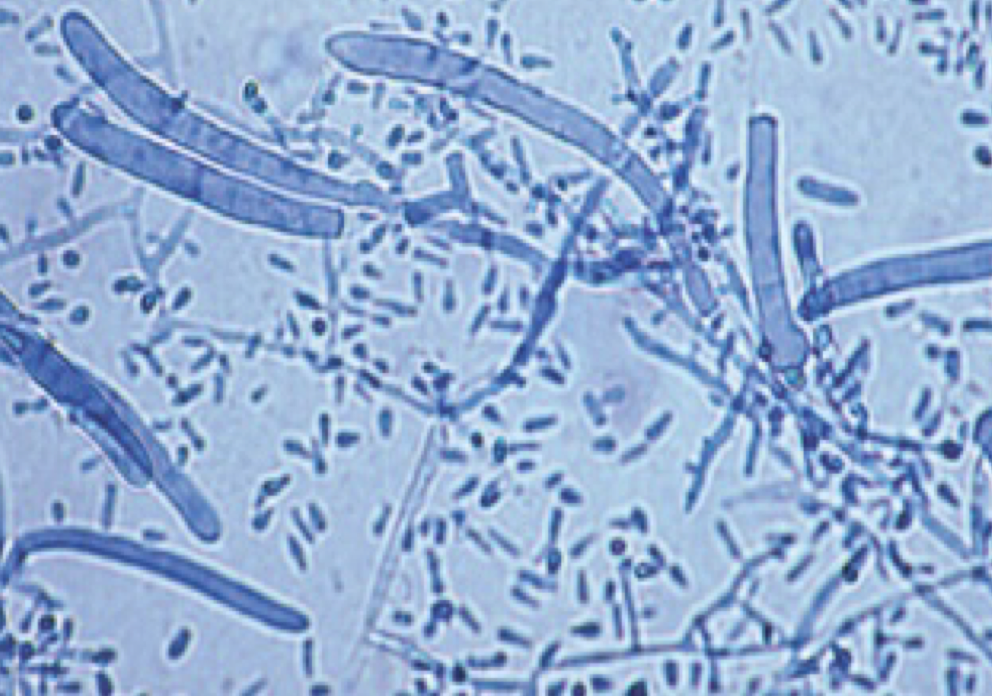
Trichophyton rubrum
no common name
classification: fungi
associated disease: ringworm, athlete’s foot, jock itch (tineas)
under microscope: looks like bacilli (rods) with different sized hyphae
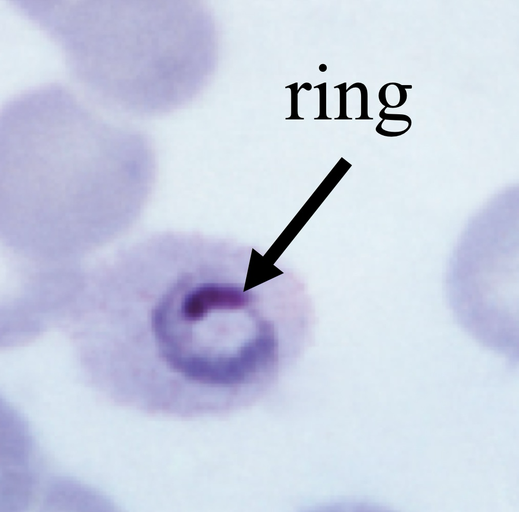
Plasmodium spp.
no common name
classification: protozoan
common disease: malaria
under microscope: looks like red blood cell with a ring in it, surrounded by red blood cells
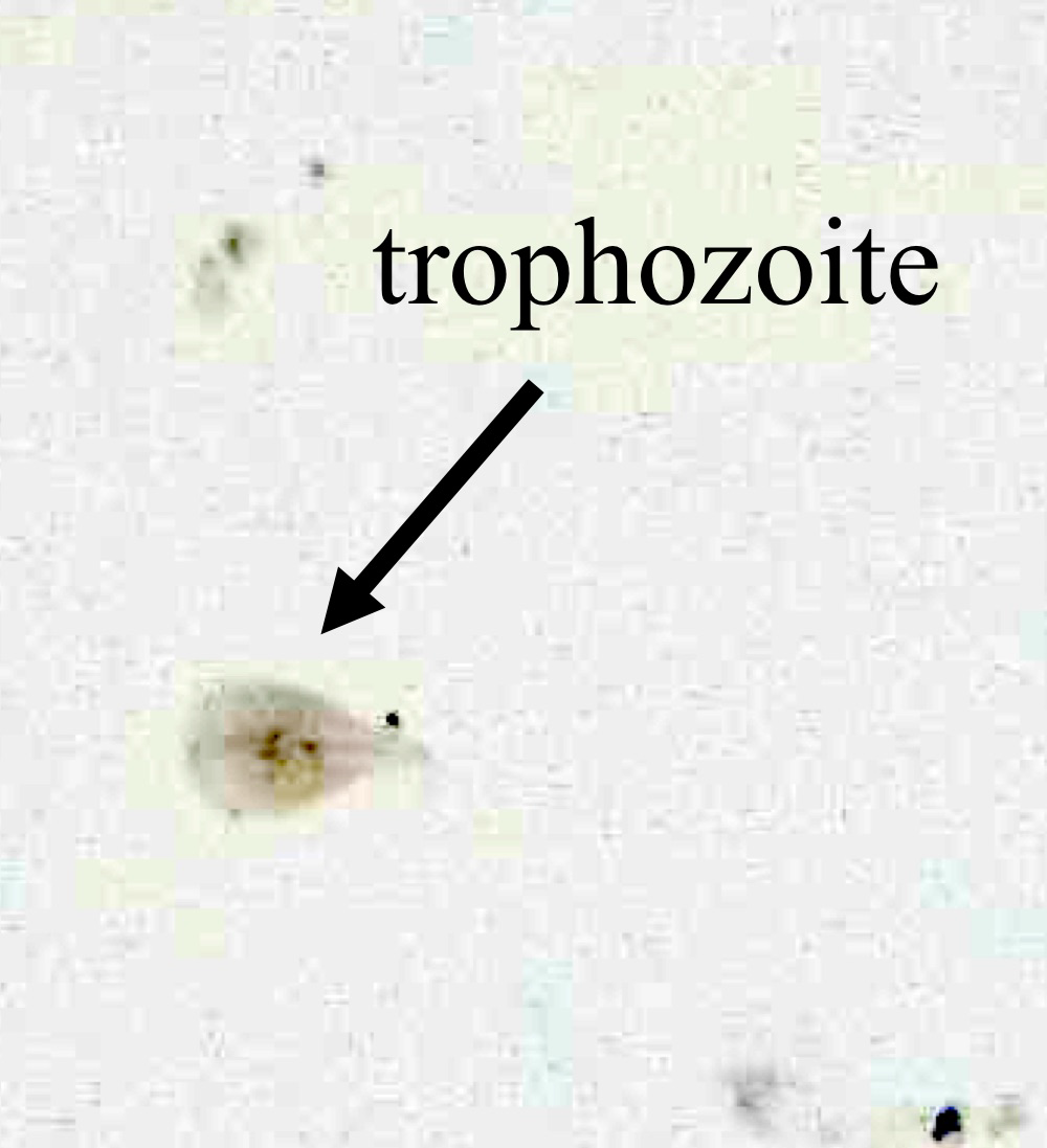
Giardia spp.
no common name
classification: protozoan
common disease: giardiasis
under microscope: looks like pale tear-drop shaped shell
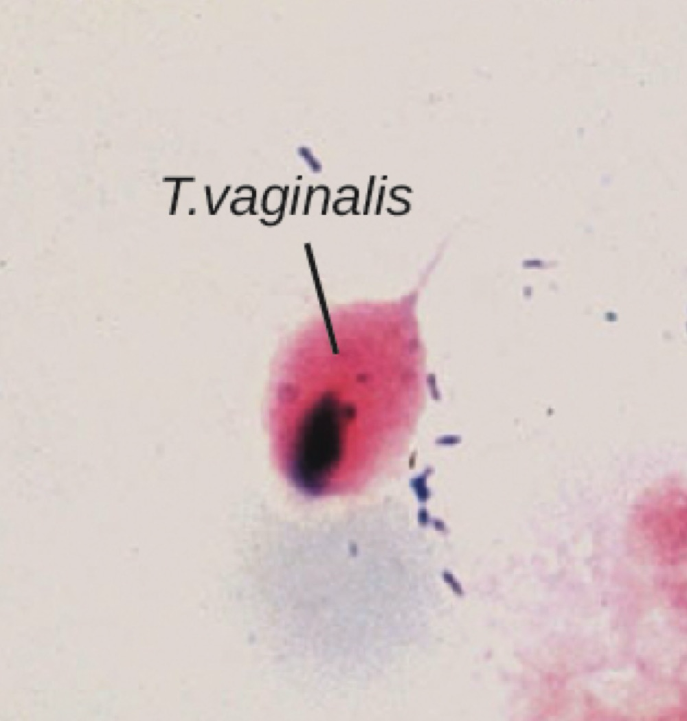
Trichomonas vaginalis
no common name
classification: protozoan
associated disease: trichomoniasis
under microscope: looks like pink and purple ovals with long flagellum coming off of cell
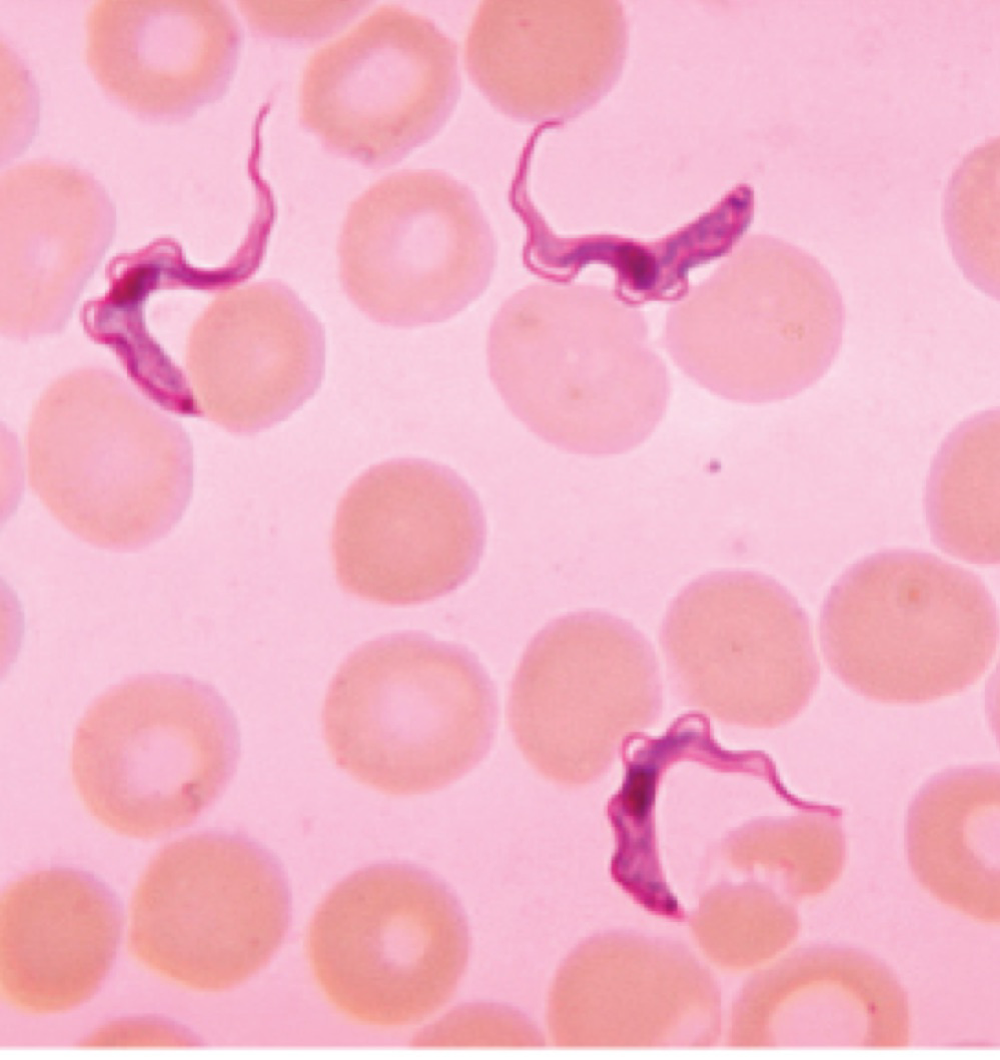
Trypanosoma spp.
No common name
Classification: Protozoan
associated disease: trypanosomiasis, chaga’s disease
under microscope: looks like squiggly looking cells surrounded by red blood cells
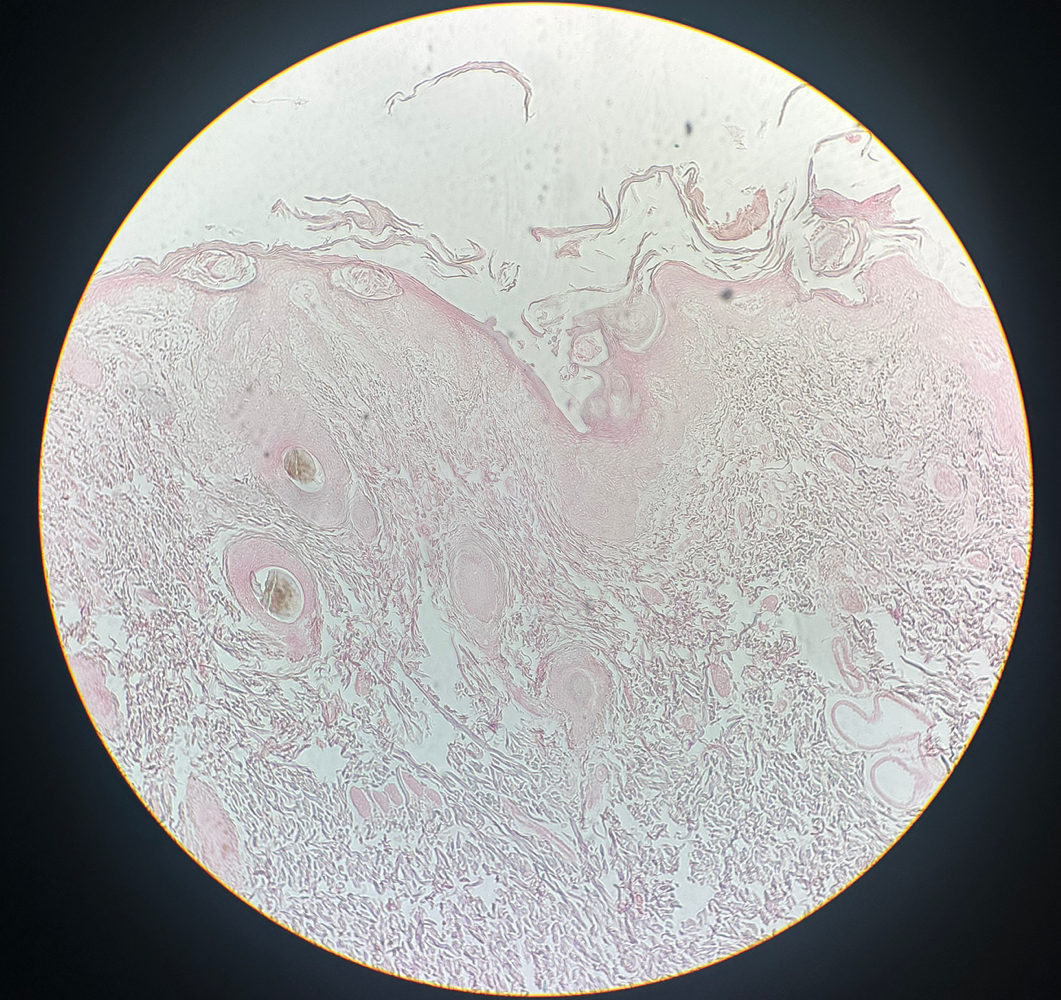
Sarcoptes scabiei var. hominis
common name: mite
classification: animal (pathogen)
associated disease: scabies
under microscope: looks like skin slide with mites and eggs under first layers OR a round, translucent bug
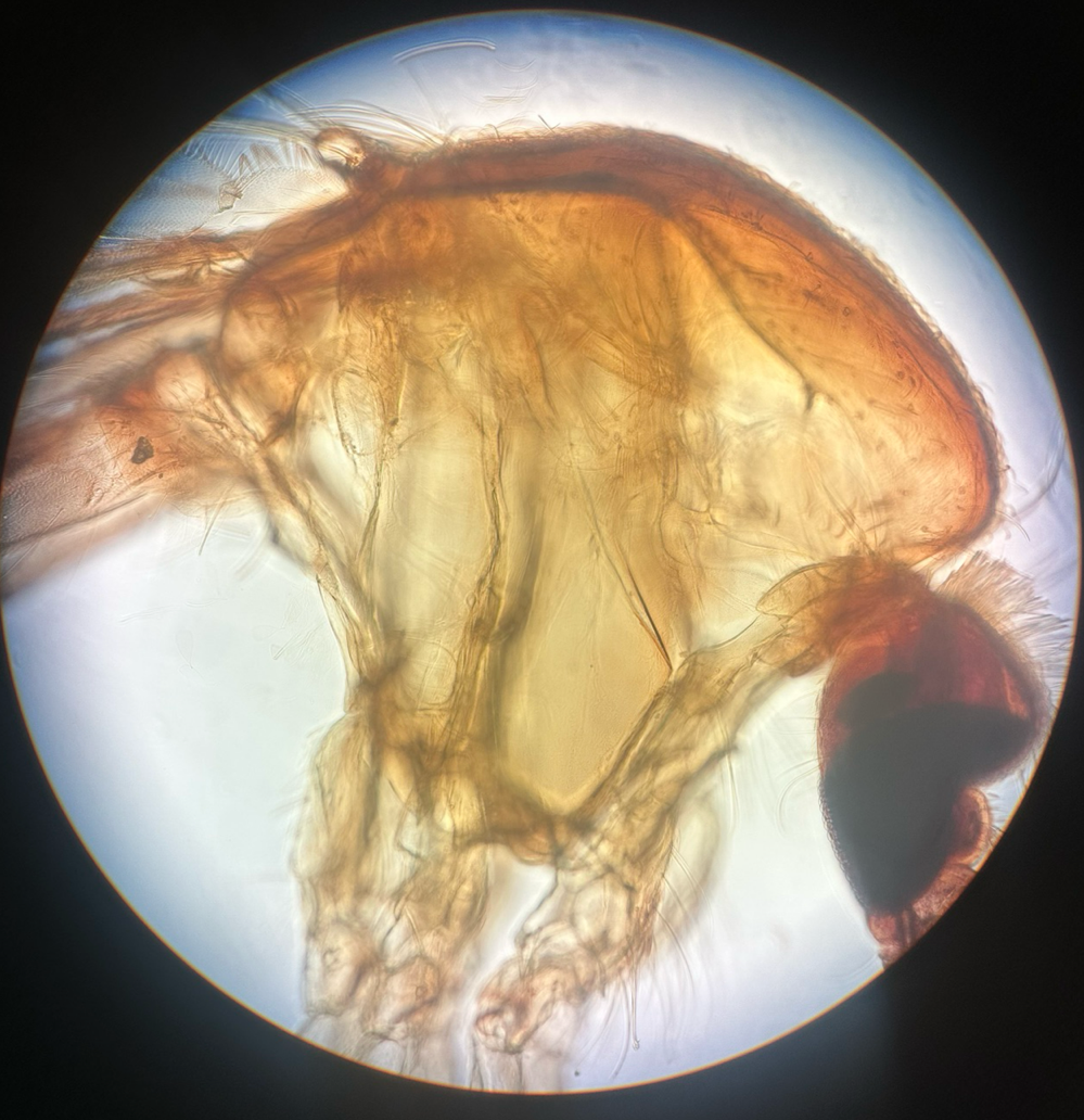
Anopholes spp.
common name: mosquito
classification: animal (biological vector)
associated disease: malaria (and many others)
under microscope: looks like a mosquito with long legs
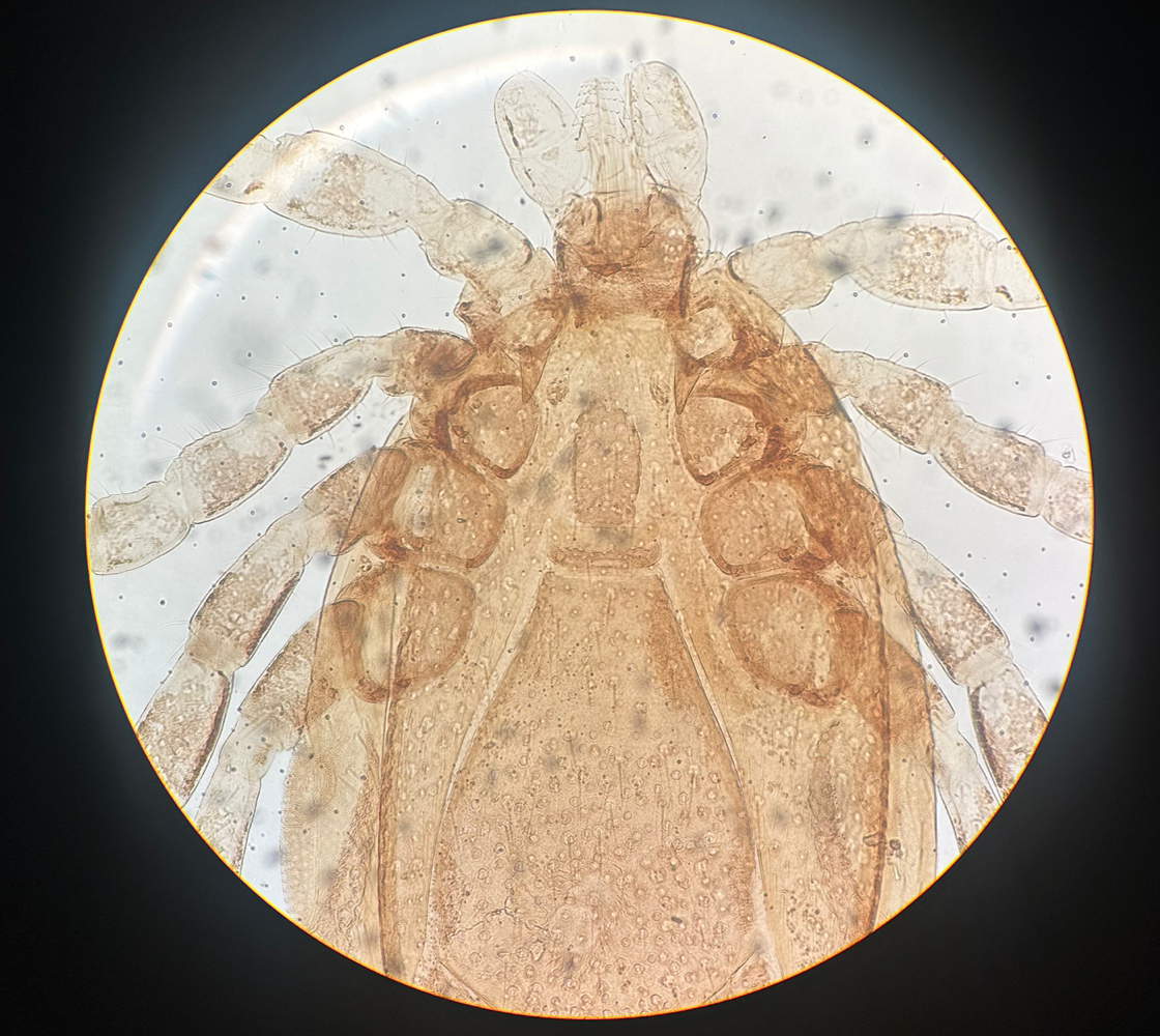
Ixodes spp.
common name: tick
classification: animal (biological vector)
common disease: Lyme disease (any many other)
under microscope: looks like yellow, oval shaped bug
Throat Culture. Be able to:
recognize and interpret alpha-, beta-, and gamma-hemolysis
alpha-hemolysis: reduction of hemoglobin in red blood cells (but not lysed)
appears as a greenish clearing around the colony
looks translucent with darker hue in the background
beta-hemolysis: lyse red blood cells
appears as translucent clearing around the colony
looks completely translucent (vs alpha-hemolysis that has a darker hue in the background)
gamma-hemolysis: no clearing, lack of hemolysis OR reduction of hemoglobin
appears as no clearing around the colony
looks like dark colones
state which pattern of hemolysis is associated with pathogens
beta-hemolysis is associated with pathogens, as they lyse red blood cells, releasing nutrients to make the available for the pathogen
Simulated UTI. Be able to:
define cystitis, pyelonephritis, glomerulonephritis, clean-catch
cystitis: bacterial infection of the bladder
pyelonephritis: infection of one or both of the kidneys
glomerulonephritis: inflammation of the glomeruli of the nephrons
clean-catch: washing labia and urethral opening / penis, then release small amount of urine, then resume urination filling container to collect specimen
goal of a clean catch is to get rid of bacteria in urethra
explain the benefit of a semi-quantitative streak method in UTI detection
cheap and fast
interpret urine culture results
nutrient agar is used:
if culture results on nutrient agar plate is equal or greater than 105 CFU/mL = positive for a UTI
if culture results on nutrient agar plate is less than 104 CFU/mL = negative for a UTI
explain how MacConkey agar is selective and differential
selective for gram-negative bacteria
allows growth of only gram-negative bacteria
differential for lactose fermentations
lactose fermenters turn colonies pink
pink growth aids in UTI confirmation
example: E. Coli is gram negative lactose fermentor
Snyder Test
explain how bacteria contribute to tooth decay
layer of proteins and carbohydrates forms when clean teeth come in contact with saliva
microbes are attracted to this food source and form a biofilm called plaque
sucrose from food is broken down by bacteria in the mouth to glucose and fructose
glucose used to make dextran, which is part of the extracellular matrix of plaque
fructose is fermented, producing organic acids
acids dissolve the mineral in the teeth
low pH results in net demineralization of tooth enamel and susceptibility to caries
name the purpose of the pH indicator in the Snyder test
bromocresol green pH indicator is used to determine susceptibility of dental caries
acid production causes bromocresol green to turn from green to yellow, which indicates a decrease in pH and an increased risk of dental caries.
interpret Snyder test results
green = negative, neutral pH
yellow = strongly positive, acidic pH
higher risk of dental caries
Tiny Earth
explain where most antibiotics come from
most antibiotics come from microbes, used to out compete each other
explain why microbes make antibiotics
microbes continuously compete with each other for space and resources
they have evolved the ability to produce antibiotics that harm competitors and increase the chances of their own survival
design and implement experiments to test antibiotic activity
tiny earth spread/ patch assay
This method involves spreading various microbial cultures on an agar plate, followed by putting different bacteria strains at specific locations on the plate to observe for zones of inhibition
if zone of inhibition forms, that specific microbe makes antibiotics
