Anatomy of the Cerebellum
Page 5: Cerebellum: Location
Posterior cranial fossa
Tentorium cerebelli
Fourth ventricle
Page 6: Cerebellum Functions
Maintenance of equilibrium (balance)
Coordination of voluntary movement
Provides precise timing and appropriate patterns of skeletal muscle contraction
Control muscle tone and posture
Learning & memory of motor tasks
Page 7, 8, 9: Cerebellum Connection with Brainstem
Cerebellum is connected to posterior brainstem
Attached to superior, middle & inferior cerebellar peduncle
Page 10: External Features
Cerebellum consists of 2 cerebellar hemispheres, joined in midline by vermis.
Surface displays alternating parallel elevations called folia and grooves known as sulci to increase surface area.
Vermis
Cerebellar hemisphere
Folia & sulcus
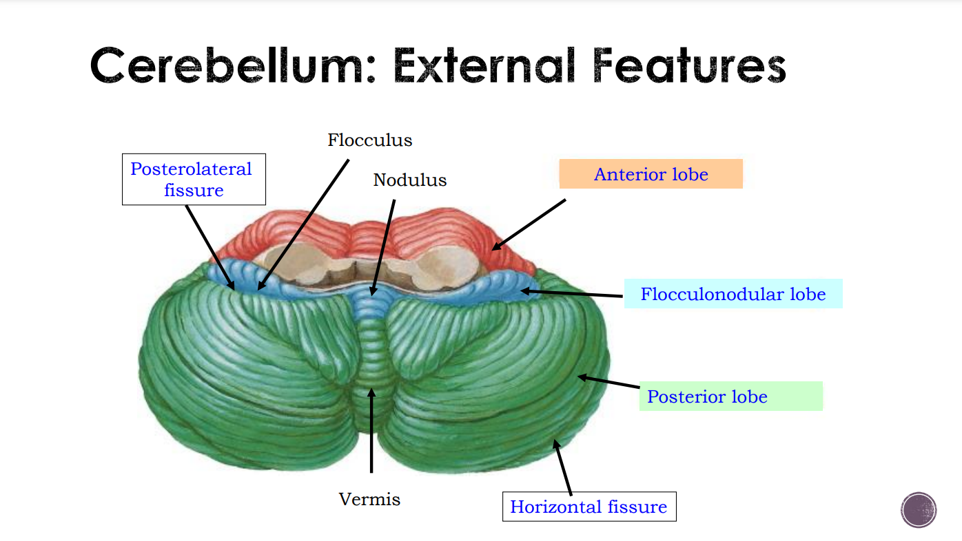
Page 13: Cerebellum Fissures
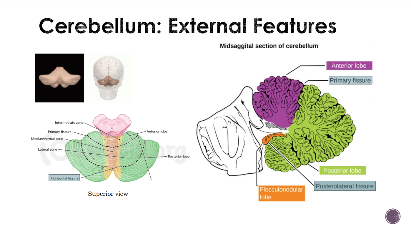
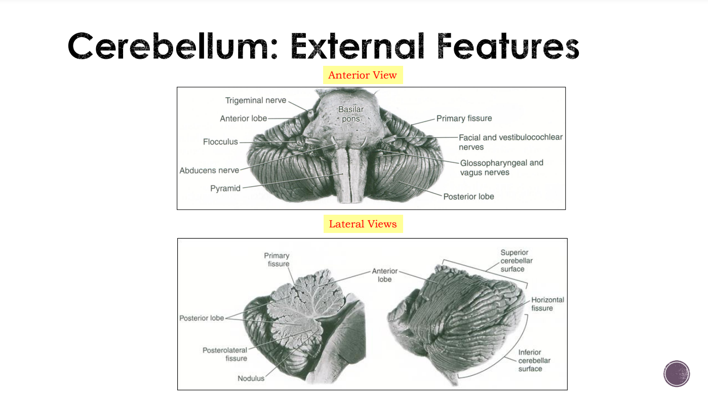
Primary Fissure: V-shaped fissure separating the anterior and posterior lobes.
Uvulonodular (posterolateral) Fissure: separating the flocculonodular lobe and the posterior lobe.
Horizontal Fissure: margin separating the superior and inferior surfaces of the cerebellum.
Page 14: External Features
Page 18
Cerebellum: External Features
Page 19
Cerebellum has 3 lobes:
Anterior lobe
Posterior (middle) lobe
Flocculonodular lobe (composed of left/right flocculi and nodulus)
Lobes can be further divided into lobules
Page 20
Cerebellum is composed of outer cortex (gray matter) and inner white matter
Embedded in white matter are deep cerebellar nuclei (also gray matter):
Dentate (most lateral)
Emboliform
Globose
Fastigial (most medial)
Emboliform & globose nuclei are collectively known as interposed nuclei
Page 21: Cerebellum Internal Features
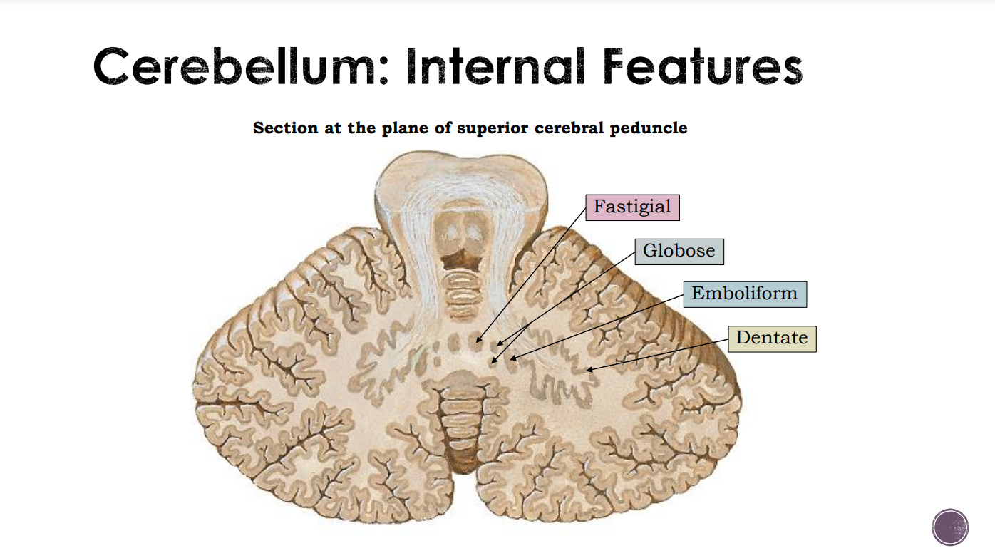
Page 22: Deep Nuclei / Intracerebellar Nuclei
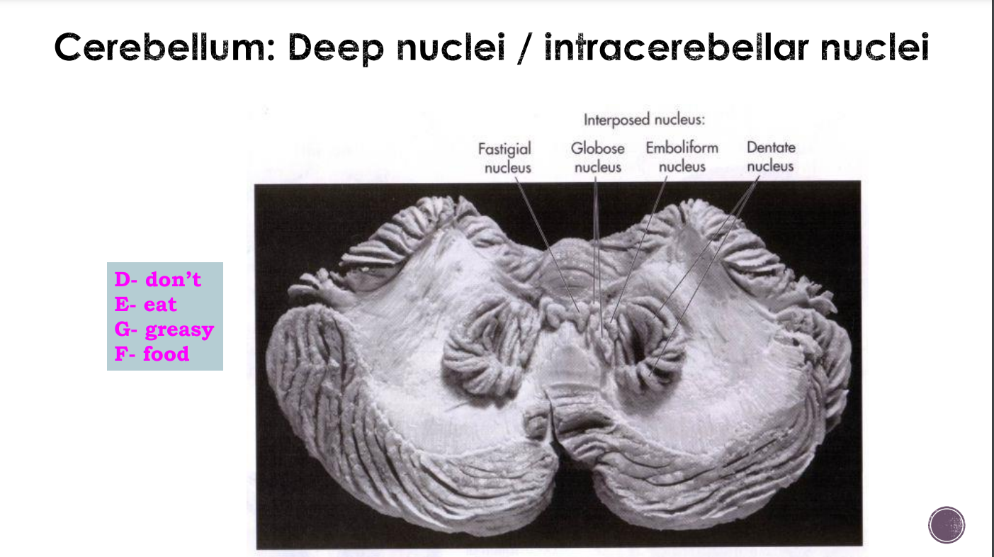
Page 23: Deep Nuclei/Intracerebellar Nuclei
Deep cerebellar nuclei are gray matter embedded deep in the white matter of the cerebellum
They are the main source of cerebellar output
Page 24: Deep Nuclei Input
Each cerebellar nucleus receives input from specific areas of cerebellar cortex:
Fastigial nucleus – receives input from the cortex of flocculonodular lobe and vermis
Globose & emboliform nuclei – receive input from the paravermal zone
Dentate nucleus – receives input from the lateral hemisphere of cerebellum
Page 25: Cerebellum Subdivisions
Anatomic subdivision
Lobe subdivision
ant, post & flocculonodular lobes
Longitudinal zone
vermis, paravermis, lateral hemisphere.
Functional area
Vestibulocerebellum, spinocerebellum, cerebrocerebellum
Phylogenic subdivision
Archicerebellum, paleocerebellum, neocerebellum
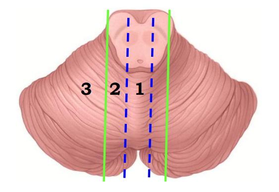
Page 26: Longitudinal Zones
Cerebellum can be divided into 3 longitudinal zones (based on functional & connections)
Vermis
Paravermal zone (Intermediate zone) - medial strip of cerebellar hemisphere adjacent to vermis
Lateral hemispheric zone - lateral part of cerebellar hemisphere
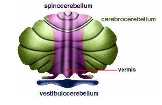
Page 27: Functional Areas
Cerebrocerebellum
Corresponds to lateral zone of cerebellar hemisphere
Spinocerebellum
Corresponds to vermis & paravermal zone
Vestibulocerebellum
Corresponds to flocculonodular lobe
Page 28
Cerebrocerebellum:
The largest division, formed by the lateral hemispheres
Involved in planning movements and motor learning
Receives inputs from the cerebral cortex and pontine nuclei
Sends outputs to the thalamus and red nucleus
Regulates coordination of muscle activation and is important in visually guided movements.
Spinocerebellum:
Comprised of the vermis and intermediate zone of the cerebellar hemispheres
Involved in regulating body movements by allowing for error correction; also receives proprioceptive information.
Vestibulocerebellum:
Functional equivalent to the flocculonodular lobe
Involved in controlling balance and ocular reflexes
Receives inputs from the vestibular system
Sends outputs back to the vestibular nuclei
Page 29: Phylogenic (Evolutionary) Subdivision
The division of the cerebellum into three major parts or regions based on its evolutionary development helps understand how the cerebellum has evolved in different species to serve varied functions.
Page 31: Cerebellar White Matter
Arbor vitae is the white matter present in the cerebellum of the brain. It has a tree-shaped structure.
Page 32: Cerebellar White Matter
White matter fibres:
Intrinsic
Connect different regions of the cerebellum.
Afferent
Most abundant fibers.
Enter mainly through middle & inferior cerebellar peduncle.
Fibers are from cerebrum, spinal cord, and inner ear.
Efferent
Output of the cerebellum
Axon of the Purkinje cells of the cerebellar cortex
Majority will synapse with deep cerebellar nuclei (DCN)
Fibers from DCN leave the cerebellum via superior cerebellar peduncle
Page 33: Corticopontocerebellar pathway
Fibers from cerebral cortex
Descend through corona radiata & internal capsule.
Synapse with pontine nuclei
Fibers from pontine nuclei (transverse fibers of the pons)
Cross midline; enter opposite side of cerebellum via middle cerebellar peduncle.
Page 34: Cerebro-olivocerebellar pathway
Fibers from cerebral cortex
Descend through corona radiata & internal capsule.
Synapse with olivary nuclei
Fibers from olivary nuclei
Cross midline; enter opposite side of cerebellum via inferior cerebellar peduncle.
Page 35: Cerebroreticulocerebellar pathway
Fibers from cerebral cortex (sensorimotor area)
Descend through corona radiata
Synapse with Reticular formation
Fibers from Reticular formation (reticulocerebellar fibres)
Enter the same side of cerebellum via inferior & middle cerebellar peduncles.
Page 36: Cerebellar Afferent Fibers (from Spinal Cord)
Anterior spinocerebellar tract: upper & lower limbs
Posterior spinocerebellar tract: trunk & lower limbs
Cuneocerebellar tract: upper limbs and upper thorax
All receive information from muscle spindle, joint, and tendon
Page 37: Cerebellar Afferent Fibers (from Inner Ear)
Vestibular nuclei receive information from inner ear ; concerning motion from the inner ear.
Page 38: Cerebellar Efferent Fibers
1. Purkinje Cell Axons:
Principal output neurons of the cerebellar cortex
Transmit processed information from the cerebellar cortex to other parts of the brain, primarily the deep cerebellar nuclei and vestibular nuclei
Modulate and coordinate motor commands, contributing to motor control and coordination.
2. Deep Cerebellar Nuclei Output:
Deep cerebellar nuclei, including fastigial, interposed, and dentate nuclei, serve as efferent fibers.
They receive input from Purkinje cells and project to various parts of the brain, including the thalamus and brainstem
Essential for transmitting refined motor commands and playing a key role in motor planning and execution.
Page 39: Blood Supply
Superior Cerebellar artery, a branch of basilar artery
Anterior Inferior Cerebellar artery, a branch of basilar artery
Posterior Inferior Cerebellar artery, a branch of vertebral artery
Page 40
Each cerebellar hemisphere is connected by nervous pathways principally with the same side of the body
A lesion in one cerebellar hemisphere produces signs and symptoms limited to that same side of the body.
Page 41
Clinical Signs:
Dysdiadochokinesis - impairment in performing rapid alternating movements.
Ataxia - unsteady gait, difficulties with balance, and issues with fine motor skills.
Nystagmus - involuntary, rhythmic movement of the eyes.
Intention tremor
Scanning dysarthria - slow, monotonous speech.
Heel-shin test positivity – unable to produce smoothly and accurately moving their heel down their shin, typically due to a loss of coordination.