3. Intraocular tumours: Retina 2
Overview of Retinal Tumours
Topic: Retinoblastoma and Astrocytic Hamartoma
Retinoblastoma
Definition and Origin
A malignant congenital tumour derived from neural elements of the embryonic retina.
Epidemiology
Diagnosis Age: Affected cells are only present in the first few years of life; most cases diagnosed prior to age 3.
Incidence Rate: Rare, affecting 1 in 18,000 live births.
Prevalence: Represents 3% of all childhood cancers.
Significance: Most common primary intraocular malignancy in childhood.
Hereditary Nature: Bilateral cases generally hereditary, linked to a mutation of the RPE1 gene on chromosome 13.
Inheritance Pattern: Inherited in approximately 40% of cases in an autosomal dominant (AD) manner with variable penetrance.
Clinical Presentation
Timing of Presentation:
Presents within the first year if bilateral.
Presents around 2 years of age if unilateral.
Common Signs:
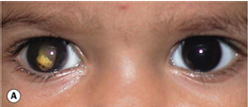
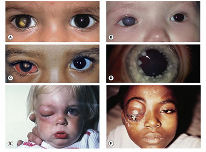
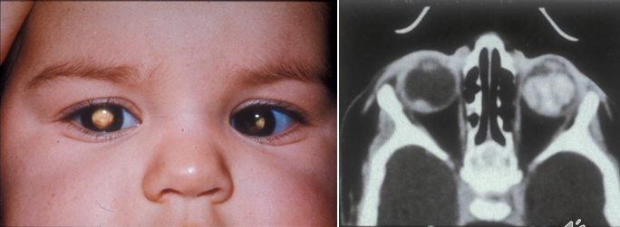
Leukocoria: 60% of cases presenting sign.
Strabismus: Presenting sign in 20% of cases.
Additional Presentations:
Secondary glaucoma.
Diffuse retinoblastoma may invade anterior segment leading to red eye from tumour-induced uveitis.
Iris nodules may be associated with pseudo-hypopyon.
Orbital inflammation; orbital invasion may cause proptosis.
Differential Diagnosis for Leukocoria
Possible Causes:
Toxocara granuloma.
Persistent hyperplastic primary vitreous.
Coats disease.
Retinopathy of prematurity.
Congenital cataract.
Note: Any child presenting with strabismus must have a dilated examination to rule out retinoblastoma.
Tumour Characteristics
Appearance of Early Tumours:
May appear as flat or round white lesions.
Intraretinal tumour often presents as homogenous, dome-shaped white lesions; can become irregular and may show white calcific flecks.
Endophytic tumours project into the vitreous as white masses.
Exophytic tumours present as subretinal, multi-lobed white masses.
Possible Overlying retinal detachment (RD).
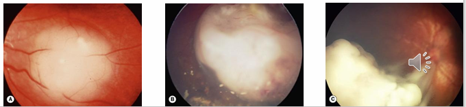
Growth Patterns of Tumours
Types of Growth:
Endophytic: Growth into vitreous.
Exophytic: Growth into subretinal space leading to retinal detachment.
Optic nerve invasion: Tumour may spread along optic nerve to the subarachnoid space of the brain.
Diffuse infiltration of retina.
Metastatic spread: Can extend to regional lymph nodes, lungs, brain, and bones.
Risk Factors for Metastatic Disease
Increased Risk:
Advanced tumour stage.
Retrolaminar optic nerve involvement.
Choroidal invasion.
Anterior chamber (AC) involvement.
Orbital spread.
Repeat occurrences after treatment.
Management and Treatment
Importance of Early Diagnosis: Correlates with a higher chance of preserving vision, salvaging the eye, and preserving life.
Treatment Options Include:
Laser treatment for small tumours.
Chemotherapy in conjunction with local treatments.
Use of radioactive plaques.
Enucleation as a common treatment for large tumours.
Mortality Rate: Overall mortality rate is about 10%; this escalates to 65% in patients with optic nerve involvement beyond the point of surgical transection.
Genetic counselling is necessary, and screening for at-risk family members is recommended.
Patients with heritable retinoblastoma also have a predisposition to non-ocular cancers such as pineoblastoma, osteosarcoma, soft tissue sarcoma, and melanoma.
Astrocytic Hamartoma (Astrocytoma)
Definition and Nature
Composition: Hamartomas consist of tissue that is normal for the location but is malformed or mal-arranged.
Origin: Astrocytic hamartomas arise from astrocytes in the optic nerve head (ONH) and nerve fibre layer (NFL).
Characteristics:
May be bilateral and show multiple lesions in the same eye.
Can occur anywhere in the retina but predominantly found in ONH.
Related Conditions:
Often associated with Tuberous sclerosis and neurofibromatosis.
May also occur as isolated findings in otherwise healthy patients.
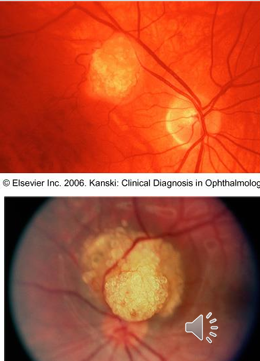
Appearance:
Can present as flat, dirty-white translucent oval lesions.
May also appear as calcified, whitish-yellow, multiple lobed, mulberry-like lesions.
fish egg appearance.
Size Range: From half-disc diameter (DD) to several DD in size.
Symptoms: Generally asymptomatic, though visual acuity (VA) may be decreased, and possible visual field defects due to compression of axons on ONH.
Prognosis: Benign (non-life-threatening). Won’t metastasise. Can lead to changes in VF due to compression of nerve fibres.
Cavernous Retinal Haemangioma
Definition: A rare, congenital vascular mass in the retina.
Characteristics:
Typically unilateral.
Commonly diagnosed in the 2nd to 3rd decades of life, often presenting with vitreous haemorrhage.
Generally a coincidental finding.
Referral needed - can cause bleeding.
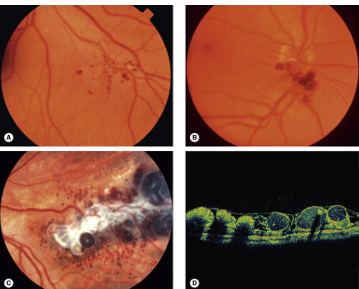
Appearance: Clusters of aneurysm-like formations resembling a bunch of grapes in the peripheral retina.