General Microbiology: Methods of Investigating Microorganisms
Culturing Microorganisms
Culturing microorganisms is a fundamental practice in microbiology to grow and study microbes under controlled conditions.
Studying Microbiology
Microbiologists use 5 basic techniques to grow, examine and characterize microorganisms in the lab.
Inoculation
This step involves introducing a tiny sample of microorganism (the inoculum) into a suitable growth medium (inoculation~liquid or solid). The medium provides the necessary nutrients for the microorganism to grow. The inoculum can come from various sources, such as clinical samples, environmental swabs, or previously cultured organisms.
Incubation
Once inoculated, the sample is placed in an incubator to promote growth. The temperature, oxygen levels, and humidity are controlled to match the conditions required for the specific microorganism. For example, many human pathogens are incubated at 37°C to mimic human body temperature.
Isolation
If multiple species of microorganisms are present, isolation is done to separate them. Streaking techniques on agar plates are commonly used to obtain individual colonies. Each colony arises from a single microorganism, leading to a pure culture that can be further studied.
Inspection
After incubation, the growth of the microorganisms is examined. This involves looking at the physical appearance of colonies, their color, shape, and size.
Identification
Finally, the microorganisms are identified using a variety of methods.
Identification of Organisms Measures
Phenotypic Methods
These methods focus on the observable traits of microorganisms, including their behavior on different types of media, morphology, and biochemical activities.
Media
Enriched media: Contains extra nutrients to support the growth of fastidious organisms (e.g., blood agar).
Selective media: Promotes the growth of specific organisms while inhibiting others (e.g., MacConkey agar for Gram-negative bacteria).
Differential media: Allows differentiation between species based on their biochemical characteristics (e.g., lactose fermentation on MacConkey agar).
Characteristic media: Identifies organisms based on their specific enzymatic activities (e.g., Simmons Citrate agar).
Direct Microscopic Examination
Observation of microorganisms under a microscope, often using stains like Gram stain or acid-fast stain to differentiate bacteria based on their cell wall properties.
Staining
Specific stains like Gram staining or acid-fast staining highlight structural differences, allowing for initial classification (Gram-positive vs. Gram-negative).
Biochemical Tests
Rapid Tests
Bacteriophage Typing
Flow Cytometry
Immunological Methods
These methods detect and identify microorganisms based on their interaction with the immune system, specifically antibodies or antigens.
Precipitation: Detects the presence of antigens by mixing them with antibodies in solution, leading to visible clumping (precipitate).
Agglutination Reactions: Used to identify the group of antigens for streptococci. (GBS- Group B Streptococcus)
Fluorescent Antibodies
ELISA (Enzyme-Linked Immunosorbent Assay)
Immunoblot (Western Blot)
Genotypic Methods
These methods rely on analyzing the genetic material (DNA or RNA) of microorganisms to identify them.
Nucleic Acid Probes
PCR (Polymerase Chain Reaction): Polymerase Chain Reaction (PCR) is a powerful technique used to amplify specific DNA sequences, making millions of copies from a small sample. The process involves three main steps: denaturation (separating DNA strands with heat), annealing (binding short primers t the target DNA), and extension (synthesizing new DNA strands with a DNA polymerase enzyme):
Nucleic Acid Sequencing:
RFLP (Restriction Fragment Length Polymorphism)
Plasmid Fingerprinting: Identifies microorganisms by analyzing their plasmid DNA, which can carry unique genetic markers specific to certain bacterial strains.
Successful Microbe Identification
The successful identification of microbe depends on:
Using the proper aseptic techniques.
Correctly obtaining the specimen.
Correctly handling the specimen
Quickly transporting the specimen to the lab.
Once the specimen reaches the lab it is cultured and identified.
Specimen Collection
Successful identification depends on how the specimen is collected, handled and stored. The general aseptic procedures be used including sterile sample containers and sampling methods to prevent contamination of the specimen. After identifying a microorganism from a clinical sample, susceptibility testing is performed to determine which antimicrobial agents (e.g., antibiotics, antifungals) are most effective at controlling or killing the pathogen.
Phenotypic Method
Microscopic morphology
Include a viewing the organism under a microscope to observe a combination of cell shape, size, Gram’s stain, acid fast reaction, special structures e.g. endospores, granule, flagella and capsule can be used to give an initial presumptive identification.
Macroscopic morphology
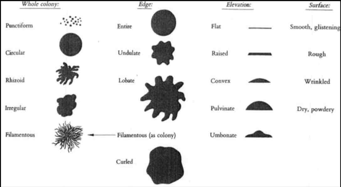
Observing traits that can be accessed with the naked eye e.g. appearance of colony including texture, shape, pigment, speed of growth and growth pattern in broth.
Physiology/Biochemical characteristic
Focus on the metabolic and enzymatic capabilities of bacteria, allowing microbiologists to distinguish between species based on their biochemical activities.
Immunological Methods
Immunological methods involve the interaction of a microbial antigen with an antibody (produced by the host immune system). So, it uses antibodies to identify the specific antigen.
Precipitation Reactions
Precipitation occurs when soluble antigens (small molecules like proteins) react with antibodies to form a visible, insoluble complex. This complex becomes large enough to form a precipitate that can be seen with the naked eye or under a microscope.
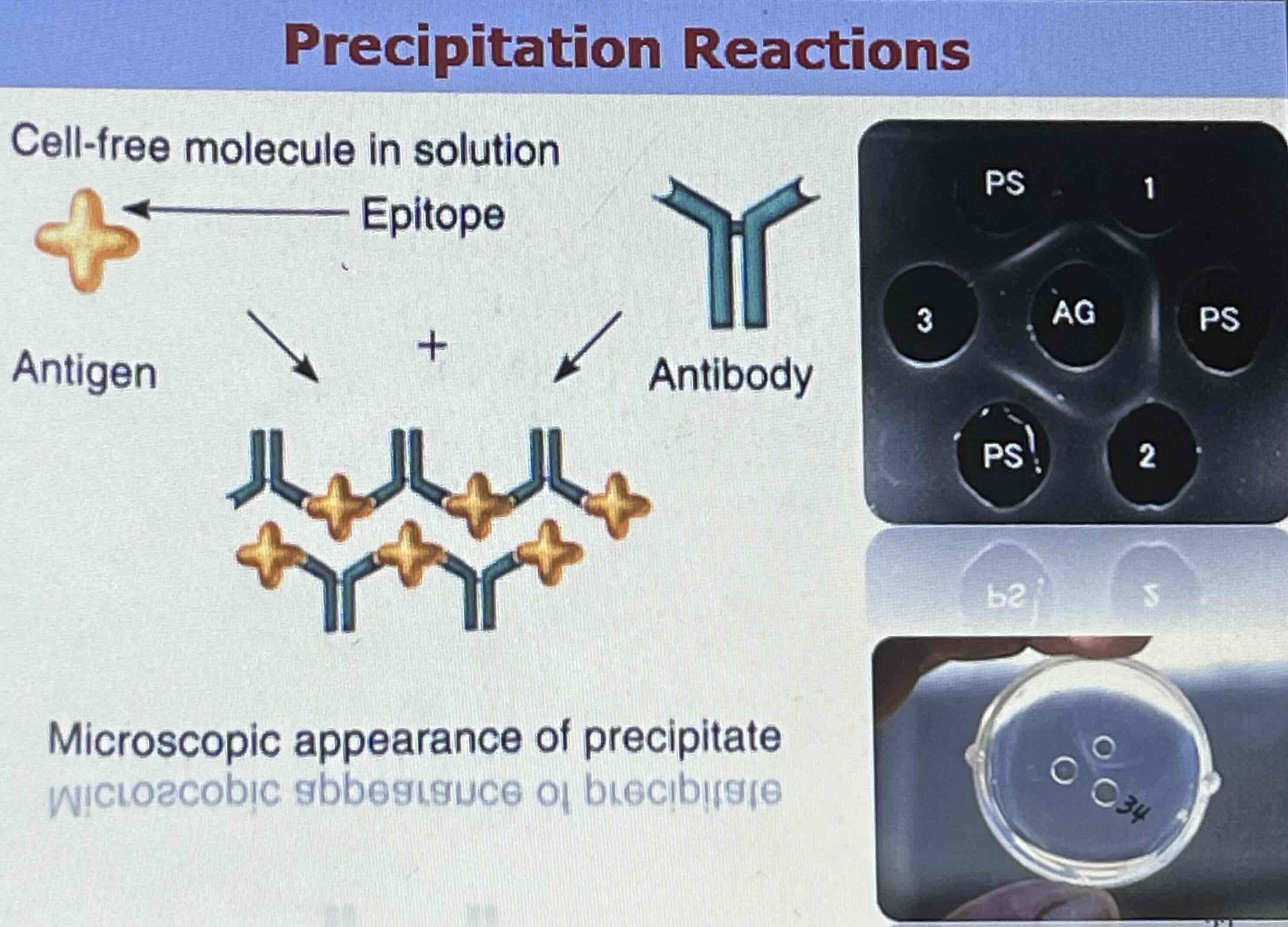 How it works: When the concentration of antigen and antibody is optimal, they bind in solution to form lattice-like structures that settle out of solution as a precipitate.
Agglutination Reactions
Agglutination occurs when particulate antigens (such as whole bacterial cells, red blood cells, or latex beads) react with antibodies, causing the particles to clump together into visible masses.
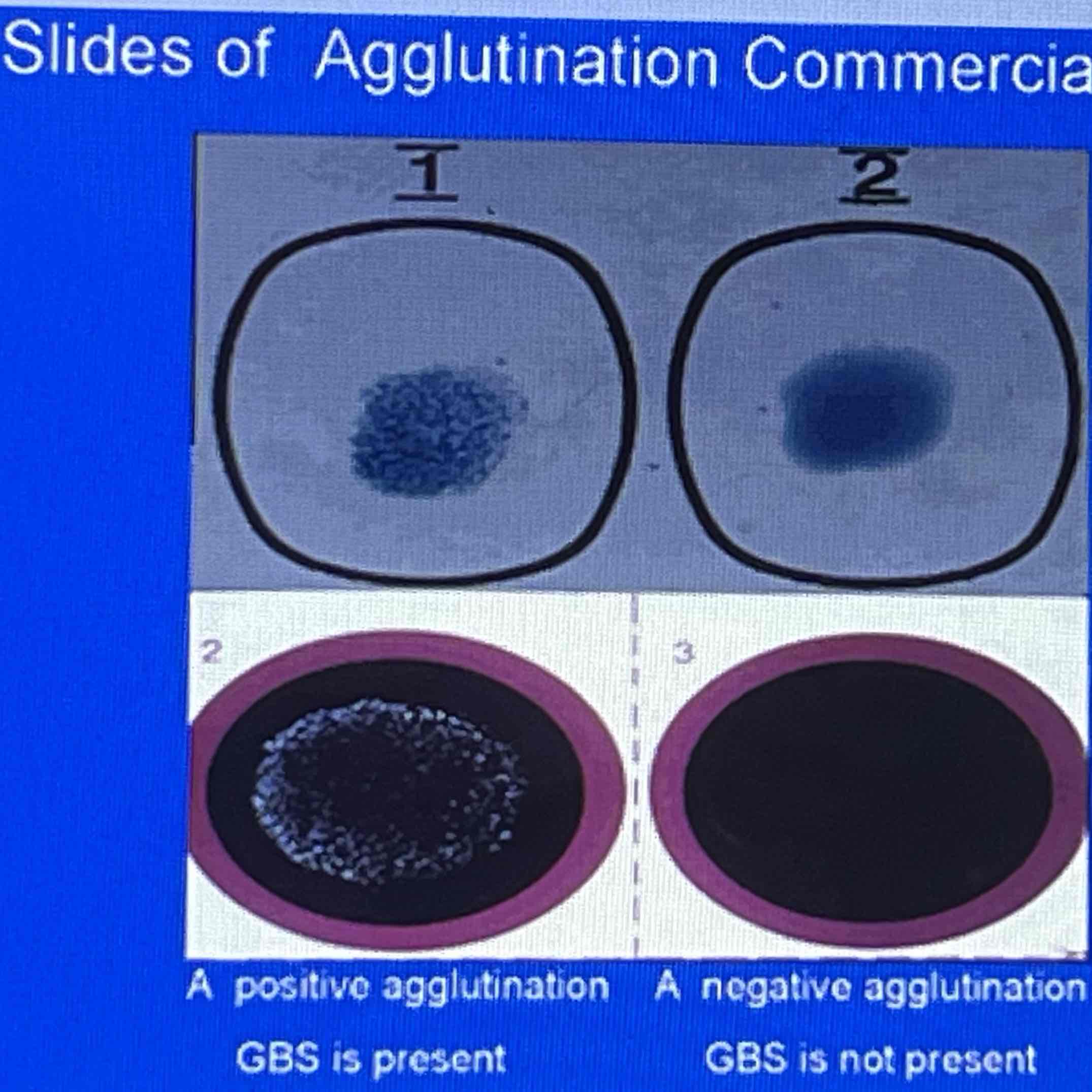 How it works: Antibodies bind to multiple antigenic particles, causing them to aggregate and form clumps that are large enough to be seen without magnification. Mainly used to groups of antigens of streptococci.
Enzymes-Linked Immunosorbent Assay
ELISA is a highly sensitive technique that uses enzyme-linked antibodies to detect the presence of specific antigens or antibodies in a sample.
How it works:
The target antigen (or antibody) is captured by a specific antibody immobilized on a surface.
A second, enzyme-linked antibody is added to bind to the target, creating a sandwich of antibodies around the antigen.
When a substrate for the enzyme is added, a color change occurs if the antigen (or antibody) is present, indicating a positive result.
Genotypic Method
Genotypic methods focus on analyzing the genetic material (DNA or RNA) of microorganisms for identification and classification. Unlike phenotypic methods that rely on observable characteristics, genotypic methods directly target the organism's genetic information. Increasingly genotypic techniques are becoming the sole means of identifying many microorganisms because of its speed and accuracy.
Media
Microbiological Media are used to grow, maintain, and study microorganisms in the lab. There are over 500 types of media, and they can be classified into three broad categories based on physical state, chemical composition, and functional type.
Physical State
Liquid Media
Liquid media are water-based solutions that do not solidify at temperatures above freezing. They are used for the growth of microorganisms in a suspension. Growth appears dispersed, particulate, or cloudy, indicating microbial proliferation throughout the medium.
Semi- Solid
Semi-solid media contain a solidifying agent (such as agar) that thickens the medium but does not create a completely firm surface. These media are used for specific applications, such as motility testing.
Characteristics:
Typically contain 0.3-0.5% agar, which allows for a viscous texture.
Can be used to determine the motility of microorganisms or to localize a reaction at a specific site.
Examples:
Sulfur Indole Motility Medium (SIM): Used to test for hydrogen sulfide production, indole formation, and motility. A stab inoculation is made in the center of the medium, and growth patterns are observed around the stab line.
Solid Media
Solid media provide a firm surface for cells to form discrete or isolated colonies during growth. They can be categorized as liquefiable (containing agar) or non-liquefiable.
Liquefiable Media: Contains agar, a complex polysaccharide that solidifies at room temperature and melts at boiling temperature (100°C). Agar remains liquid until cooled to about 42°C, allowing for inoculation at 45-50°C without harming most microorganisms.
Non-Liquefiable Media: These media do not melt and maintain their solid state regardless of temperature.
Chemical Context
Refers to the chemical component of the media. There are two types.
Defined Media (Synthetic Media)
Defined media contain known and precisely measured amounts of pure organic and inorganic compounds. The exact chemical composition is specified, allowing for reproducibility in experimental results.
Characteristics
Reproducibility: Because the components are known, defined media can produce consistent results across experiments and laboratories.
Exact Nutritional Needs: These media are designed to meet the specific nutritional requirements of microorganisms, which is particularly useful in research settings where precise conditions are necessary.
Complex Media
Complex media contain at least one ingredient that is not chemically definable, making the exact composition variable. These media are typically rich in nutrients and support the growth of a wide range of microorganisms.
Characteristics:
Nutrient-Rich: Complex media support the growth of fastidious organisms (those with complex nutritional requirements).
Variability: The composition of complex media may vary from batch to batch due to the nature of the ingredients.
Functional Type
General Media
General media are designed to support the growth of a broad spectrum of microorganisms. They typically contain nonsynthetic components and provide a basic nutrient environment. Eg TSA
Characteristics:
Nonsynthetic: These media do not have precisely defined chemical compositions. Instead, they rely on complex mixtures that provide a variety of nutrients.
Broad Spectrum: Suitable for growing many types of bacteria and fungi, making them useful for general microbiological work.
Enriched Media
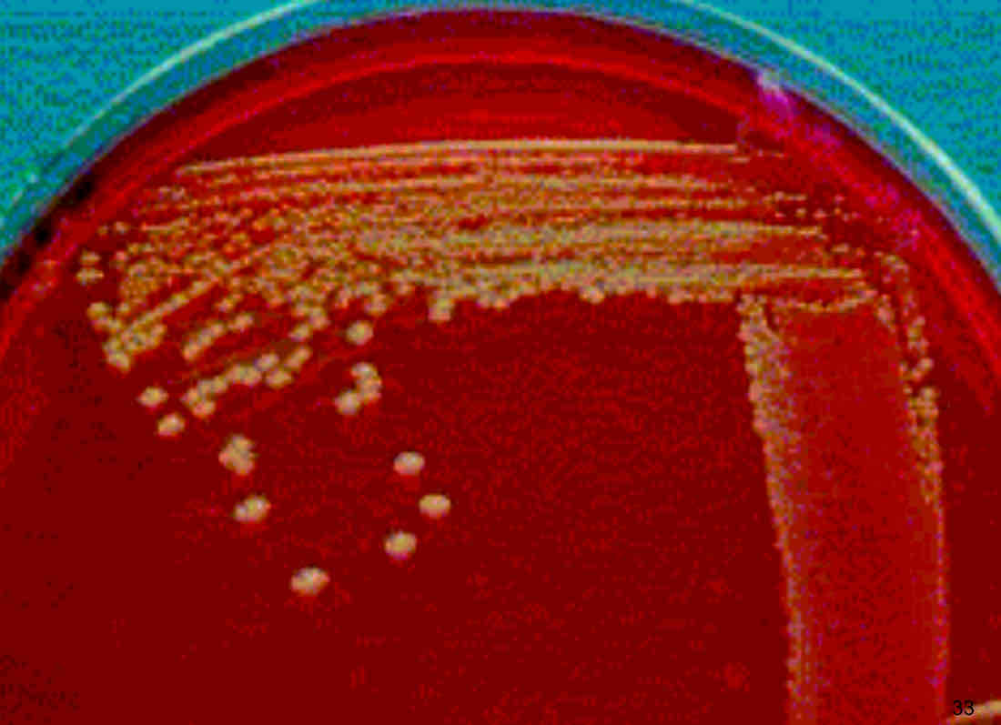
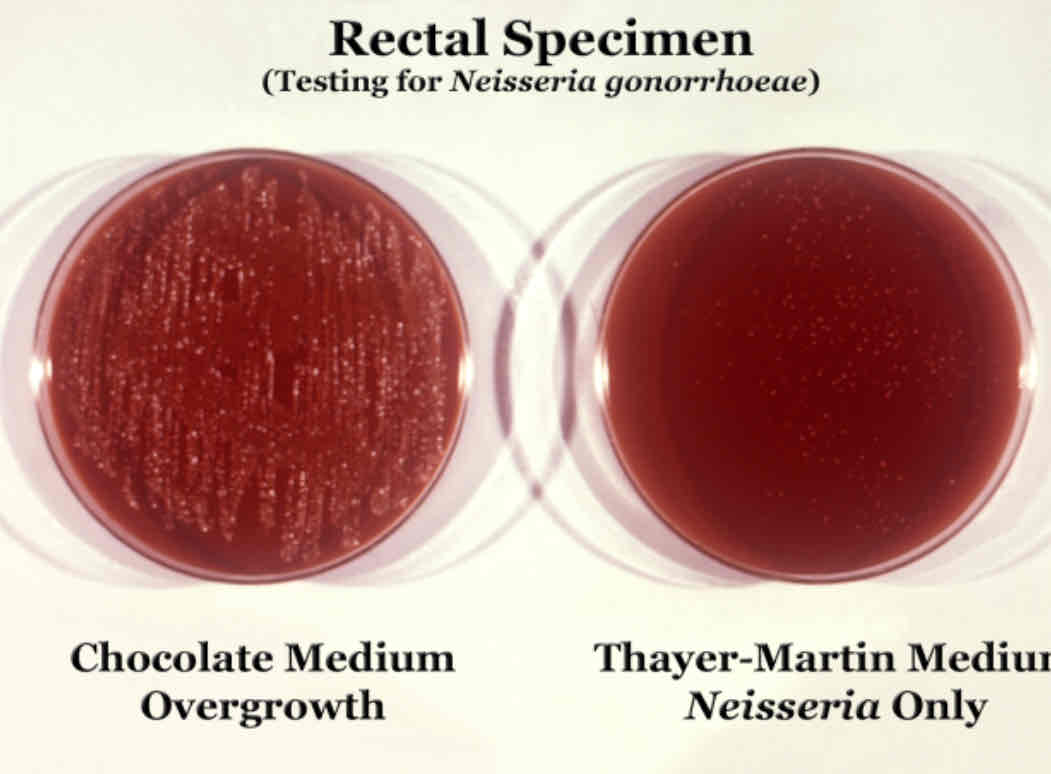
Enriched media contain complex organic substances that provide additional nutrients for microorganisms with more demanding growth requirements, often referred to as fastidious bacteria. Eg. Blood agar for fastidious streptococci, Thayer-Martin medium or chocolate agar made by heating blood agar used for testing gonorrhea.
Fastidious Bacteria: These are bacteria that have specific nutritional requirements and often require enriched media to grow.
Selective Media
Selective media are specifically designed to promote the growth of certain microorganisms while inhibiting others.
Key Features of Selective Media
Inhibitory Agents: Selective media contain one or more agents that suppress the growth of undesired microorganisms. These agents can include salts, dyes, antibiotics, or other chemicals that create a selective environment.
Primary Isolation Method: They serve as a primary method for isolating specific microbes from a sample, enhancing the efficiency and accuracy of microbiological analysis.
Examples of Selective Media
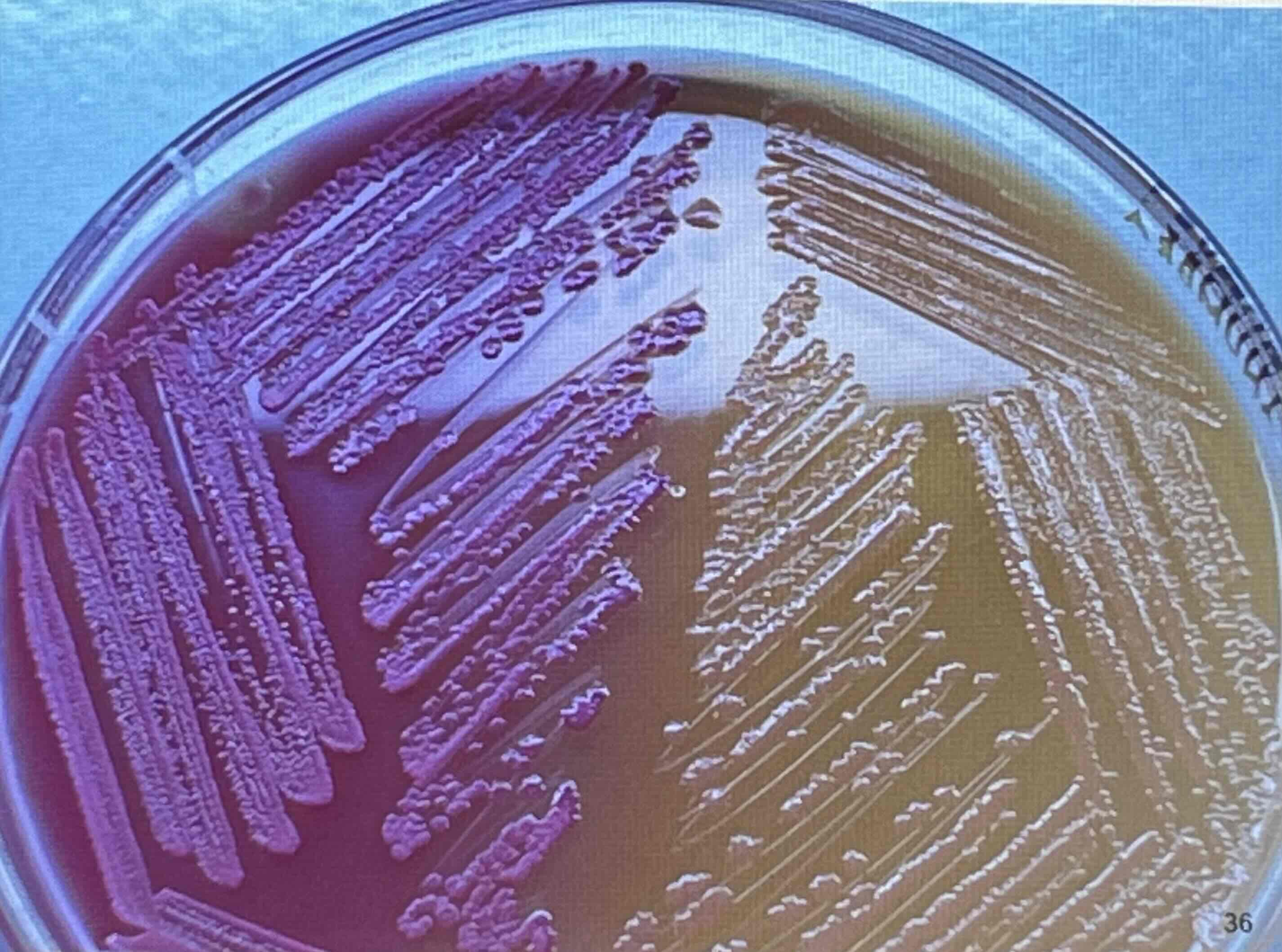
MacConkey Agar
Composition: Contains bile salts, crystal violet, lactose, and a pH indicator (neutral red).
Selectivity: Bile salts and crystal violet inhibit most Gram-positive bacteria, allowing many Gram-negative rods to grow.
Differentiation: Lactose fermenters produce acid, leading to a color change (pink colonies) due to the pH indicator, while non-fermenters remain colorless.
Differential media
Are designed to grow multiple microorganisms while providing visual indicators that distinguish between them.
Examples of Differential Media
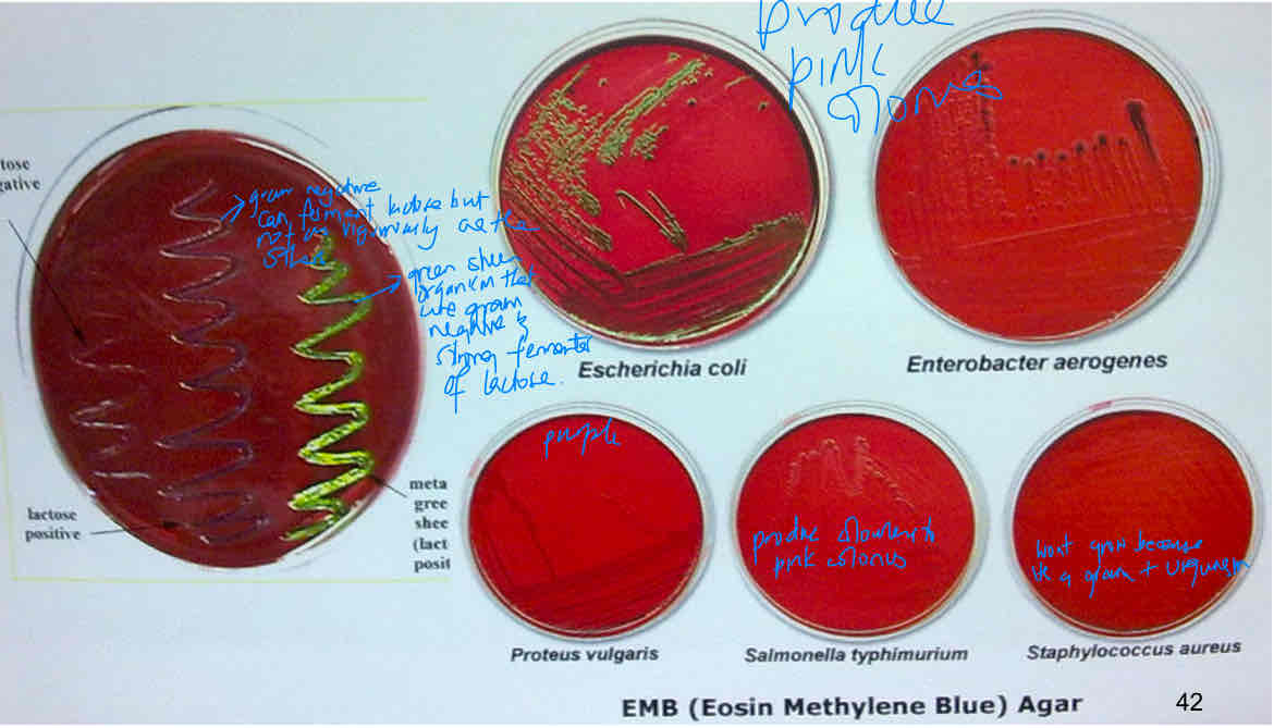
MacConkey Agar
Composition: Contains bile salts, crystal violet, lactose, and neutral red dye.
Selectivity and Differentiation: MacConkey agar is both selective (inhibiting Gram-positive bacteria) and differential (distinguishing lactose fermenters from non-fermenters).
Differentiation:
E. coli: When E. coli ferments lactose, it produces acid, lowering the pH and turning the neutral red dye pink or red, resulting in red colonies.
Salmonella: This bacterium does not ferment lactose, so it does not produce acid, and the colonies remain colorless or yellow.
Incubation, Inspection and Identification
Incubation: follows inoculation, placed in temperature controlled chamber to encourage multiplication.
Pure culture: is a container of medium that grows only a single type of microorganism.
Axenic: free of other living microbes other than the one being studied.
Subculture: making another culture from a well isolated colony.
Mixed culture
Contaminated culture
Microscope
Bright-Field
Light Transmission: The microscope directs light through the specimen, with denser and more opaque parts of the specimen absorbing more light than the surrounding areas.
Image Formation: As a result, the specimen appears darker than its surroundings on a bright background. This contrast helps highlight features of the specimen for observation.
Applications:
Live, Unstained Material: Bright field microscopes can be used to observe living organisms or cells without staining, though the natural contrast might be low.
Preserved, Stained Material: It is commonly used to view stained specimens, where dyes enhance contrast, making cellular structures or microorganisms more visible.
Dark Field Microscopy
Dark field microscopy is a technique that enhances contrast in unstained, transparent specimens. It works by blocking direct light, allowing only light that has been scattered or reflected by the specimen to reach the viewer’s eye.
Key Features
Stop on Condenser: A special stop is placed on the condenser of a bright field microscope to block direct light from entering the objective lens.
Light Reflection: Only light that is reflected or scattered by the specimen reaches the objective, making the specimen appear bright against a dark background.
Bright Specimen, Dark Background: This technique results in bright images of the specimen (such as bacteria or other small organisms) on a completely dark background.
Advantages
Live Observation: Dark field microscopy allows the observation of living, unstained cells. It is especially useful for viewing moving or swimming organisms, as it highlights their outlines and motion.
Rapid Recognition: The bright outlines make it easier to rapidly recognize the general shape and movement of cells, such as bacteria or protozoa.
Advantages
Distortion: Cells can be distorted if dried or heat-fixed, so the technique is best for fresh or live samples.
Lack of Fine Detail: While the technique offers good contrast, it does not reveal fine internal details of the cells, focusing more on the general shape and movement.
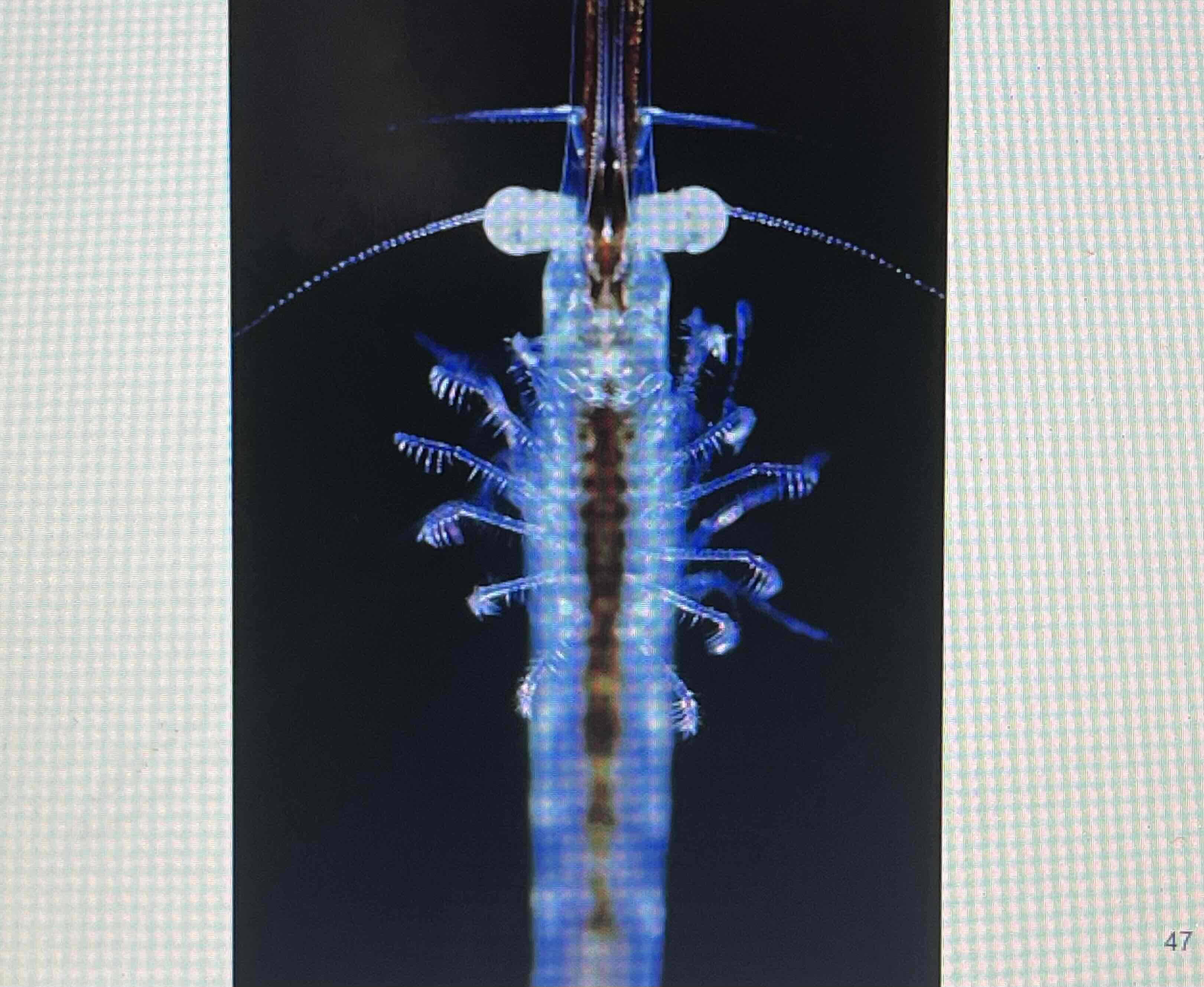
Fluorescence Microscopy
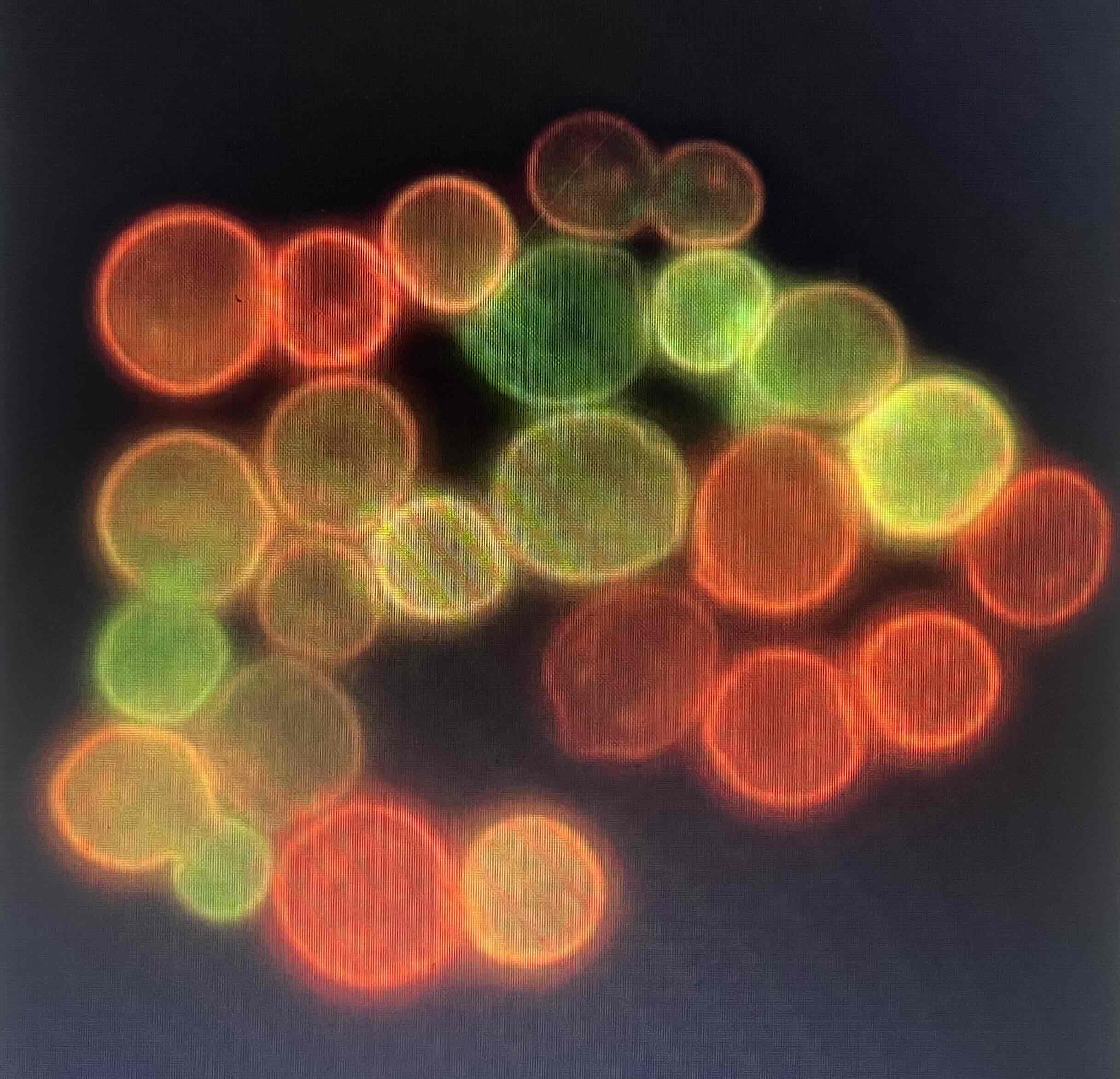
Fluorescence microscopy is a powerful technique that uses ultraviolet (UV) light to excite fluorescent dyes or natural substances within a specimen, allowing it to emit visible light, resulting in a highly detailed image.
Fluorescent Staining: Special dyes and minerals that fluoresce when exposed to UV light are used to stain specimens. These fluorescent molecules absorb the UV light and emit visible light, allowing the stained parts of the sample to glow against a dark background.
Electron Microscopy
Electron microscopy uses beams of electrons instead of light to form highly detailed images of specimens. It provides much greater magnification and resolution than light microscopy because it has a shorter wavelength than light microscope.
Magnification: Electron microscopes can magnify specimens up to 1,000,000x, far beyond the capabilities of light microscopes.
Resolution: The resolution of an electron microscope can be as fine as 0.5 nm, compared to 200 nm in a typical compound light microscope. This allows researchers to see much finer details.
Types:
Transmission Electron Microscopy
TEM directs a beam of electrons through a very thinly sliced specimen to study the internal structure of cells, organelles, and viruses at high magnification and resolution.
Scanning Electron Microscopy (SEM):
SEM scans the surface of a specimen by bombarding it with electrons. The electrons bounce off the surface and are detected to form a 3D image.
Staining
Simple Stains
Single Dye: Simple stains use only one single dye to color the cells, making them easier to see under a microscope. This technique does not differentiate between types of bacteria, but highlights basic cell morphology (shape and arrangement).
Differential Stains
Multiple Dyes: Differential stains use a combination of a primary dye and a counterstain to distinguish between different cell types or structures. These stains provide more detailed information than simple stains and are crucial in identifying bacteria and their characteristics.
Types of Differential Stains
Acid-Fast Stain
The acid-fast stain is primarily used to differentiate between acid-fast bacteria (such as Mycobacterium species) and non-acid-fast bacteria.
Results:
The cell walls of acid-fast bacteria contain mycolic acids, waxy substances that prevent ordinary dyes from penetrating. However, when treated with carbol fuchsin (a red dye) for several hours or heated briefly, the dye penetrates and binds to the cell wall. Acid-fast bacteria retain the primary dye, carbol fuchsin, and appear pink.
After staining, these bacteria resist decolorization by acid-alcohol, thus holding the dye fast, which gives them the term "acid-fast."
Non-acid-fast bacteria are decolorized by acid-alcohol and then counterstained, appearing blue.
Specialized Stains
Flagella Stain: This stain highlights bacterial flagella, structures that allow cells to move.
Endospore Stain: Detects endospores, which are highly resistant, dormant forms of certain bacteria, such as Bacillus and Clostridium species. Endospores resist most stains but can be visualized using specialized techniques.