Chapter 17 - Digestive System Disorders
Digestive System
Processes ingested food and fluids
Breaks them down into their units
Controlled by enzymes
Absorbs necessary components
Membrane transport mechanisms
Mostly in small intestine
Anatomy of the Digestive System
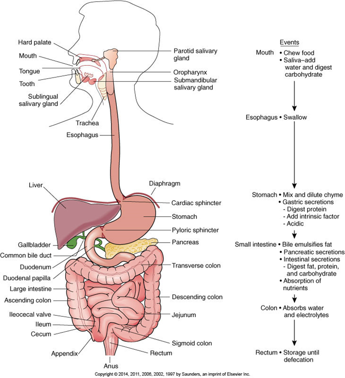
Digestive System: Introduction
Gut wall
Mucosa
Epithelium, including mucus-producing cells
Submucosa
Connective tissue—including blood vessels, nerves, lymphatics, secretory glands
Circular smooth muscle layer
Longitudinal smooth muscle layer
Serosa
Visceral peritoneum
Upper Gastrointestinal Tract
Oral cavity
Initial phase of mechanical breakdown of food
Mastication by teeth
Initial chemical digestion
Salivary amylase—starts chemical breakdown of carbohydrates
Formation of bolus
Pharynx
Swallowing (deglutition)
Esophagus
Closed except during swallowing, skeletal muscle at superior end—followed by smooth muscle
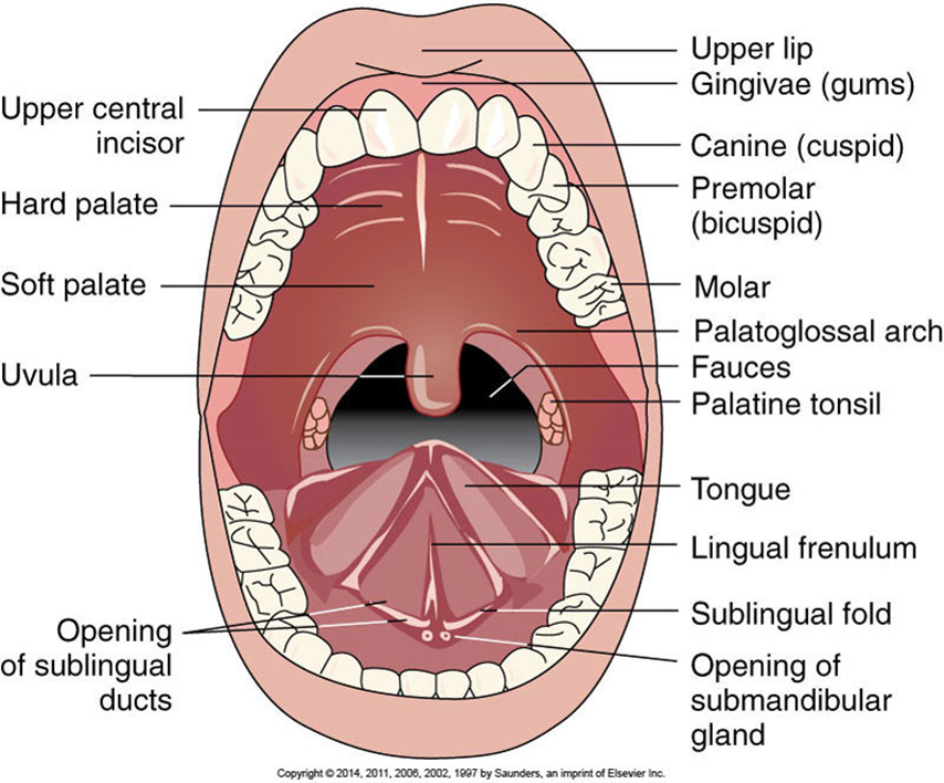
The Oral Cavity
Stomach
Expansible muscular sac—acts as reservoir for food and fluid
Three smooth muscle layers
Constant mixing and churning of food
Initial digestion of proteins
By pepsin
Formed by combination of pepsinogen and HCl
Production of intrinsic factor
Essential for absorption of vitamin B12 in the ileum
Formation of chyme
Absorption of small and lipid-soluble molecules
Liver
“Metabolic factory” of the body
Receives blood from hepatic portal vein
Transport of nutrients from intestine to liver
Hepatocytes store nutrients
Play role in carbohydrate, protein, fat metabolism
Production of plasma proteins and clotting factors
Breakdown of old and damaged erythrocytes
Bile production
Pancreas
Exocrine pancreas arranged in lobules
Secretes digestive enzymes, electrolytes
Trypsin
Chymotrypsin
Carboxypeptidase
Ribonuclease
Pancreatic amylase
Bicarbonate ions
Pancreatic duct joins bile duct to enter duodenum
Lower Gastrointestinal Tract
Small intestine
Duodenum, jejunum, ileum
Villi (folds of the mucosa) and microvilli (folds of cell membranes)
Increase surface area for absorption
Major site for absorption of nutrients
Lacteal—lymphatic vessel
Site of production of:
Mucus
Enterokinase, peptidases, nucleosidases, lipase, sucrase, maltase, lactase, cholecystokinin (hormone)
Large intestine
Peyer patches (lymphatic tissue)
Resident normal flora
Breakdown of certain food materials
Vitamin K synthesis by bacteria
Fluid and electrolyte reabsorption
Formation of solid feces
Mass movements
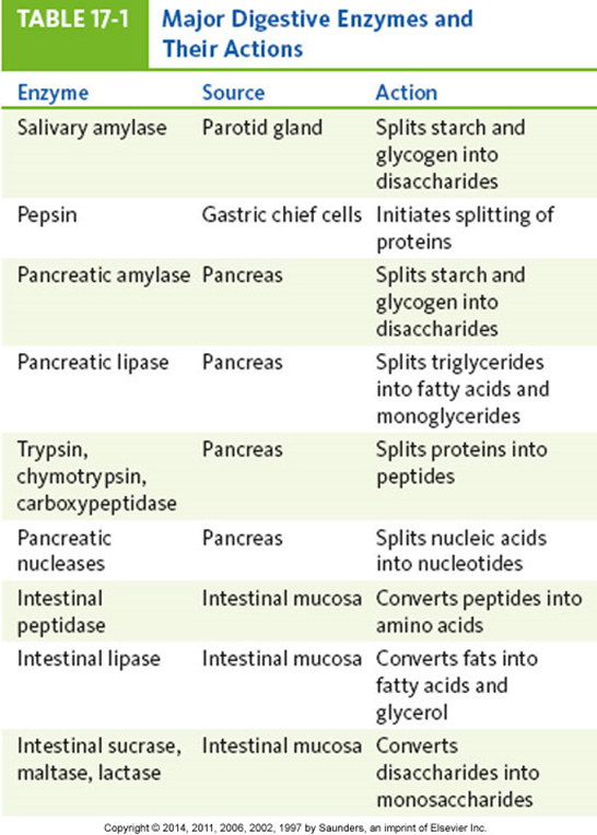
Major Digestive Enzymes and Their Actions
Neural and Hormonal Controls
Parasympathetic nervous system (PNS)
Primarily through vagus nerve (cranial nerve [CN] X)
Increased motility
Increased secretions
Sympathetic nervous system (SNS)
Stimulated by factors such as fear, anger
Inhibits gastrointestinal activity
Causes vasoconstriction
Reduced secretions and regeneration of epithelial cells
Facial (CN VII) and glossopharyngeal (CN IX) nerves
Maintain continuous flow of saliva in mouth
Distention and stretching of stomach
PNS activation
↑ Peristalsis and gastric secretions
Stomach empties within 2 to 6 hours after meal.
Food in intestine
Stimulation of intestinal activity
Enterogastric reflex
Inhibition of gastric emptying
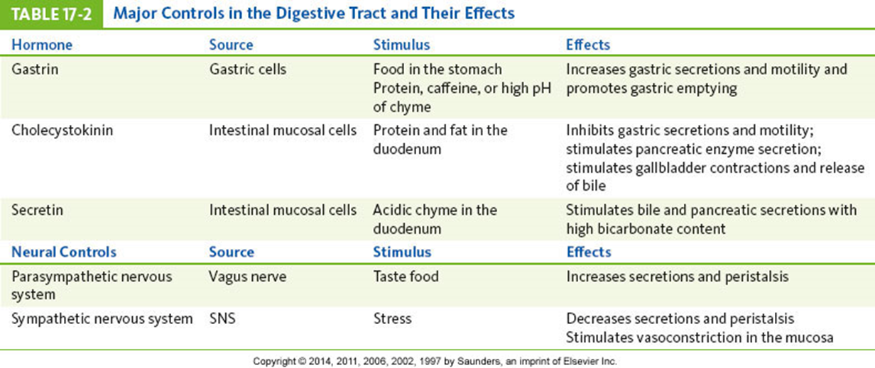
Major Controls in the Digestive Tract and Their Effects
Hormonal Controls
Gastrin
Secreted by mucosal cells (stomach) in response to distention of stomach or partially digested substances
Increases gastric motility, relaxes pyloric and ileocecal sphincters—promotes stomach emptying
Histamine
Increased secretion of hydrochloric acid
Secretin
Decreases gastric secretions
Cholecystokinin
Inhibits gastric emptying; stimulates contraction of gallbladder
Digestion and Absorption
Carbohydrates
Digestion starts in mouth
Followed by digestion in the small intestine
Proteins
Digestion starts in stomach, continues in small intestine
Lipids
Emulsified by bile prior to chemical breakdown
Action of enzymes form monoglycerides and free fatty acids
Formation of chylomicrons
Fat-soluble vitamins
Vitamins A, D, E, K
Absorbed with fats
Water-soluble vitamins
Vitamins B and C—diffuse into blood
Electrolytes
Absorbed by active transport or diffusion
Drugs are primarily absorbed in the intestine.
Various transport mechanisms
Some (e.g., aspirin) absorbed in the stomach
Water
Absorbed primarily by osmosis
About 700 mL of water is secreted into the digestive tract each day.
About 2300 mL is ingested in food and fluids
Only 50 to 200 mL leaves the body in feces.
Severe vomiting or diarrhea will interrupt this recycling mechanism.
Affects fluid and electrolyte balance of body
Anorexia, Nausea, Vomiting, and Bulimia
May be signs of digestive disorder or other condition elsewhere in the body
Systemic infection
Uremia
Emotional responses
Motion sickness
Pressure in the brain
Overindulgence of food, drugs
Pain
Anorexia and vomiting
Can cause serious complications
Dehydration, acidosis, malnutrition
Anorexia
Often precedes nausea and vomiting
Nausea
Unpleasant subjective feeling
Simulated by distention, irritation, inflammation of digestive tract
Also stimulated by smells, visual images, pain, and chemical toxins and/or drugs
Vomiting (emesis)
Vomiting center located in the medulla
Coordinates activities involved in vomiting
Protects airway during vomiting
Forceful expulsion of chyme from stomach
Sometimes includes bile from intestine
Bulimia—eating disorder
Damage to structures of the GI tract caused by recurrent vomiting
Oral mucosa
Teeth
Esophagus
Vomiting Center Activation
Distention or irritation in digestive tract
Stimuli from various parts of the brain
Response to unpleasant sights or smells, ischemia
Pain or stress
Vestibular apparatus of inner ear (motion)
Increased intracranial pressure
Sudden projectile vomiting without previous nausea
Stimulation of chemoreceptor trigger zone
By drugs, toxins, chemicals
Vomiting Reflex
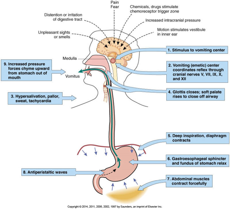
Vomiting Reflex Activities
Deep inspiration
Closing the glottis, raising the soft palate
Ceasing respiration
Minimizes risk of aspiration of vomitus into lungs
Relaxing the gastroesophageal sphincter
Contracting the abdominal muscles
Forces gastric contents upward
Reversing peristaltic waves
Promotes expulsion of stomach contents
Characteristics of Vomitus
Presence of blood—hematemesis
Coffee ground vomitus—brown granular material indicates action of HCl on hemoglobin
Hemorrhage—red blood may be in vomitus
Yellow- or green-stained vomitus
Bile from the duodenum
Deeper brown color
May indicate content from lower intestine
Recurrent vomiting of undigested food
Problem with gastric emptying or infection
Diarrhea
Excessive frequency of stools
Usually of loose or watery consistency
May be acute or chronic
Frequently with nausea and vomiting when infection or inflammation develops
May be accompanied by cramping pain
Prolonged diarrhea may lead to dehydration, electrolyte imbalance, acidosis, malnutrition
Common Types of Diarrhea
Large-volume diarrhea (secretory or osmotic)
Watery stool resulting from increased secretions into intestine from the plasma
Often related to infection
Limited reabsorption because of reversal of normal carriers for sodium and/or glucose
Small-volume diarrhea
Often caused by inflammatory bowel disease
Stool may contain blood, mucus, pus
May be accompanied by abdominal cramps and tenesmus
Steatorrhea—“fatty diarrhea”
Frequent bulky, greasy, loose stools
Foul odor
Characteristic of malabsorption syndromes
Celiac disease, cystic fibrosis
Fat usually the first dietary component affected
Presence interferes with digestion of other nutrients.
Abdomen often distended
Blood in Stool
Blood may occur in normal stools with diarrhea, constipation, tumors, or an inflammatory condition.
Frank blood
Red blood—usually from lesions in rectum or anal canal
Occult blood
Small hidden amounts, detectable with stool test
May be caused by small bleeding ulcers
Melena
Dark-colored, tarry stool
May result from significant bleeding in upper digestive tract
Gas
From swallowed air, such as drinking from a straw
Bacterial action on food
Foods or alterations in motility
Excessive gas causes:
Eructation
Borborygmus
Abdominal distention and pain
Flatus
Constipation
Less frequent bowel movements than normal
Small hard stools
Acute or chronic problem
May be caused by decreased peristalsis
Increased time for reabsorption of fluid
Periods of constipation may alter with periods of diarrhea.
Chronic constipation may cause hemorrhoids, anal fissures, or diverticulitis.
Causes of Constipation
Weakness of smooth muscle because of age or illness
Inadequate dietary fiber
Inadequate fluid intake
Failure to respond to defecation reflex
Immobility
Neurological disorders
Drugs (i.e., opiates)
Some antacids, iron medications
Obstructions caused by tumors or strictures
Fluid and Electrolyte Imbalances
Dehydration and hypovolemia are common complications of digestive tract disorders.
Electrolytes
Lost in vomiting and diarrhea
Acid-base imbalances
Metabolic alkalosis
Results from loss of hydrochloric acid with vomiting
Metabolic acidosis
Severe vomiting causes a change to metabolic acidosis because of the loss of bicarbonate of duodenal secretions.
Diarrhea causes loss of bicarbonate.
Pain: Visceral Pain
Burning sensation
Inflammation and ulceration in upper digestive tract
Dull, aching pain
Typical result of stretching of liver capsule
Cramping or diffuse pain
Inflammation, distention, stretching of intestines
Colicky, often severe pain
Recurrent sooth muscle spasms or contraction
Response to severe inflammation or obstruction
Pain: Somatic Pain
Somatic pain receptors directly linked to spinal nerves
May cause reflex spasm of overlying abdominal muscles
Steady, intense, often well-localized abdominal pain
Involvement or inflammation of parietal peritoneum
Rebound tenderness—identified over area of inflammation when pressure is released
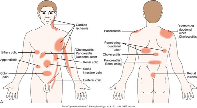
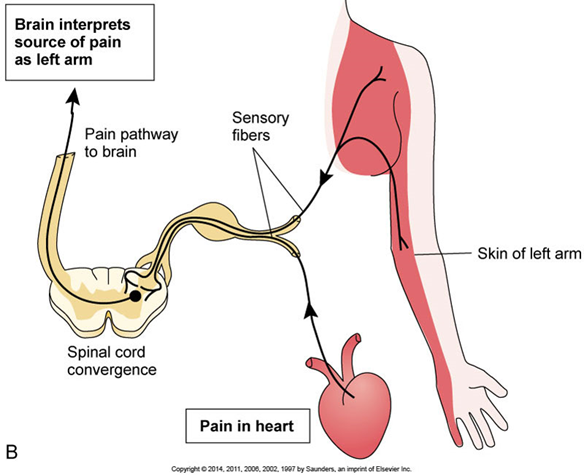
Pain: Referred Pain
Common phenomenon
Pain is perceived at a site different from origin.
Results when visceral and somatic nerves converge at one spinal cord level
Source of visceral pain is perceived as the same as that of the somatic nerve.
May assist or delay diagnosis, depending on problem
Malnutrition
May be limited to a specific nutrient or general
Causes of limited malnutrition—specific problem
Vitamin B12 deficiency
Iron deficiency
Causes of generalized malnutrition
Chronic anorexia, vomiting, diarrhea
Other systemic causes
Chronic inflammatory bowel disorders
Cancer treatments
Wasting syndrome
Lack of available nutrients
Basic Diagnostic Tests
Radiography
Contrast medium may be used.
Ultrasound
May show unusual masses
Computed tomography (CT)
Magnetic resonance imaging (MRI)
CT and MRI may use radioactive tracers.
Can be used for liver and pancreatic abnormalities
Fiberoptic endoscopy used in upper GI tract
Biopsy may be done during procedures.
Sigmoidoscopy and colonoscopy
Biopsy and removal of polyps may be done
Laboratory analysis of stool specimens
Check for infection, parasites and ova, bleeding, tumors, malabsorption
Blood tests
Liver function, pancreatic function, cancer markers
Common Therapies and Prevention
Dietary modifications
Example—gluten-free diet (celiac disease)
Reduced intake of alcohol and coffee
Increased fiber and fluid intake
Stress reduction techniques
Stress impairs immune function and tissue healing.
Drugs
Variety of medications are available.
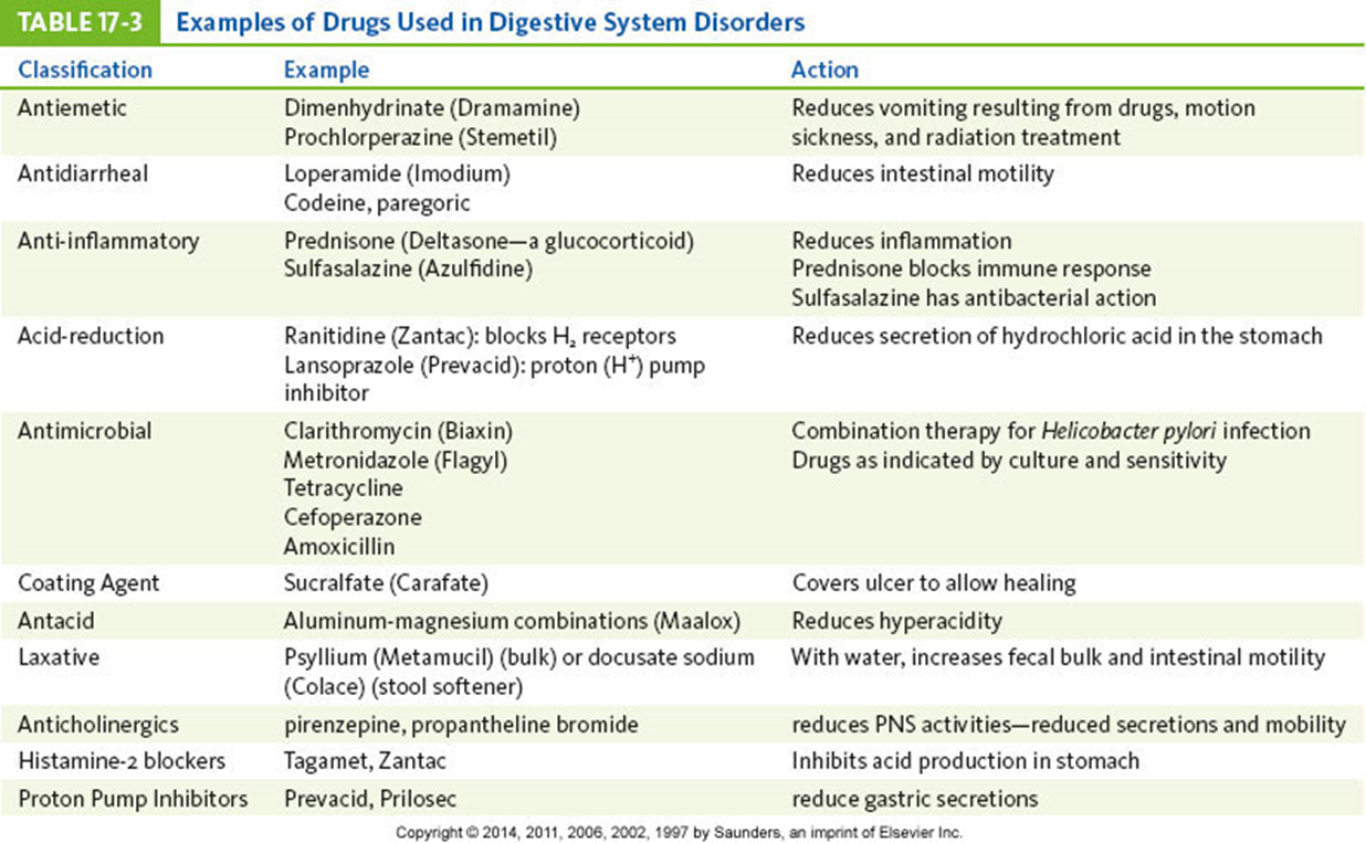
Drugs Used in Digestive System Disorders
Antacids
To relieve pyrosis
Antiemetics
To relieve vomiting
Laxatives or enemas
Treatment of acute constipation
Antidiarrheals
Reduction of peristalsis
Relieve cramps
Sulfasalazine
Anti-inflammatory and antibacterial
Used for acute episodes of inflammatory bowel disease
Clarithromycin or azithromycin
Effective against Helicobacter pylori infection
Usually combined with a proton pump inhibitor
Sucralfate
Coating agent
Enhance gastric mucosal barrier against irritants such as nonsteroidal anti-inflammatory drugs (NSAIDs)
Anticholinergic drugs
Reduce PNS activity
Reduce secretions and motility
Histamine 2 antagonists
Useful for gastric reflux
Proton pump inhibitors
Reduce gastric secretion
Disorders of the Oral Cavity
Congenital abnormalities
Cleft lip and cleft palate
Arise in sixth to seventh week of gestation
Most likely of multifactorial origin
Feeding problems of the infant
High risk of aspirating fluid into respiratory passages
Speech development impaired
Surgical repair done as soon as possible
Therapy with speech-language pathologist and orthodontist
Inflammatory lesions—aphthous ulcers
Streptococcus sanguis may be involved.
Part of the oral resident flora
Small painful lesions on:
Movable mucosa
Buccal mucosa
Floor of the mouth
Soft palate
Lateral borders of the tongue
Usually heal spontaneously
Disorders of the Oral Cavity: Infections
Candidiasis
Candida albicans—causative agent
Often part of the resident flora
Opportunistic organism
Oral candidiasis (thrush)
People receiving broad-spectrum antibiotics
During and after cancer therapy
Immunocompromised individuals or those with diabetes
May appear as red, swollen areas
May be irregular patches of a white curd-like material
Herpes simplex type 1 infection
Herpes simplex virus type 1 (HSV-1)
Transmitted by kissing or close contact
Virus remains dormant in sensory ganglion
Activated by stress, trauma, other infection
Formation of blister, ulcers, clear fluid release—contains virus; can be autoinoculated to other areas
Lesions heal spontaneously in 7 to 10 days.
Acute stage may be alleviated by antiviral medication.
May spread to eyes
Conjunctivitis and keratitis
Syphilis
Caused by Treponema pallidum
May cause oral lesions
Highly contagious during first and second stages
Primary stage
Chancre, a painless ulcer on tongue, lip, palate
Heals spontaneously (1 or 2 weeks)
Secondary stage
Red macules or papules on palate—highly infectious
Heals spontaneously
Both stages treated with long-acting penicillin
Disorders of the Oral Cavity: Dental Problems
Caries
Streptococcus mutans—initiating microbe
Lactobacillus follows in large numbers.
Bacteria break down sugars and produce large quantities of lactic acid.
Lactic acid dissolves mineral in tooth enamel
Tooth erosion and caries formation
Caries is promoted by frequent intake of sugars and acids.
Fluoride—anticaries treatment
Gingivitis
Changes in the gingivae may be a local or systemic problem.
Inflammation of the gingiva
Tissue becomes red, soft, swollen, bleeds easily
May be a result of accumulated plaque
Inadequate oral hygiene
Toothbrush trauma
Results from improper or excessive brushing
Creates extensive grooving on tooth surface
Increase plaque retention and damage to gingivae
Periodontal disease
Infection and damage to the periodontal ligament and bone
Predisposing condition is gingivitis
Caused by microorganisms as a result of poor dental hygiene
Subsequent loss of teeth possible
Several categories, depending on degree of disease
May be aggravated by systemic disease and medications that reduce salivary secretions
Periodontitis occurs when organisms enter the gingival blood vessels and travel to the connective tissues and bone of the dental arch.
Resorption of bone and loss of ligament fibers result in weakened attachment of teeth.
May result in total loss of tooth from socket
Treated by antimicrobials, local surgery of gingiva, and improved dental hygiene
Hyperkeratosis
Leukoplakia (example)
Whitish plaque or epidermal thickening of mucosa
Occurs on buccal mucosa, palate, lower lip
May be related to smoking or chronic irritation
Lesions require monitoring.
Epithelial dysplasia beneath plaque may develop into squamous cell carcinoma.
Cancer of the Oral Cavity
Squamous cell carcinoma—common type
Often develops in persons older than 40 years
Smokers, preexisting leukoplakia, alcohol abuse
Floor of the mouth, lateral borders of the tongue
Multiple lesions possible
Kaposi sarcoma in patients with AIDS
Lip cancer has a better prognosis.
Common in smokers, particularly pipe smokers
Salivary Gland Disorders
Sialadenitis
Inflammation of the salivary glands
May be infectious or noninfectious
Most commonly affected—parotid gland
Mumps—infectious parotitis
Viral infection
Vaccine available
Noninfectious parotitis
Often seen in older adults who lack adequate fluid intake and mouth care
Most malignant tumor of salivary glands is mucoepidermoid carcinoma
Dysphagia
Difficulty swallowing
Causes
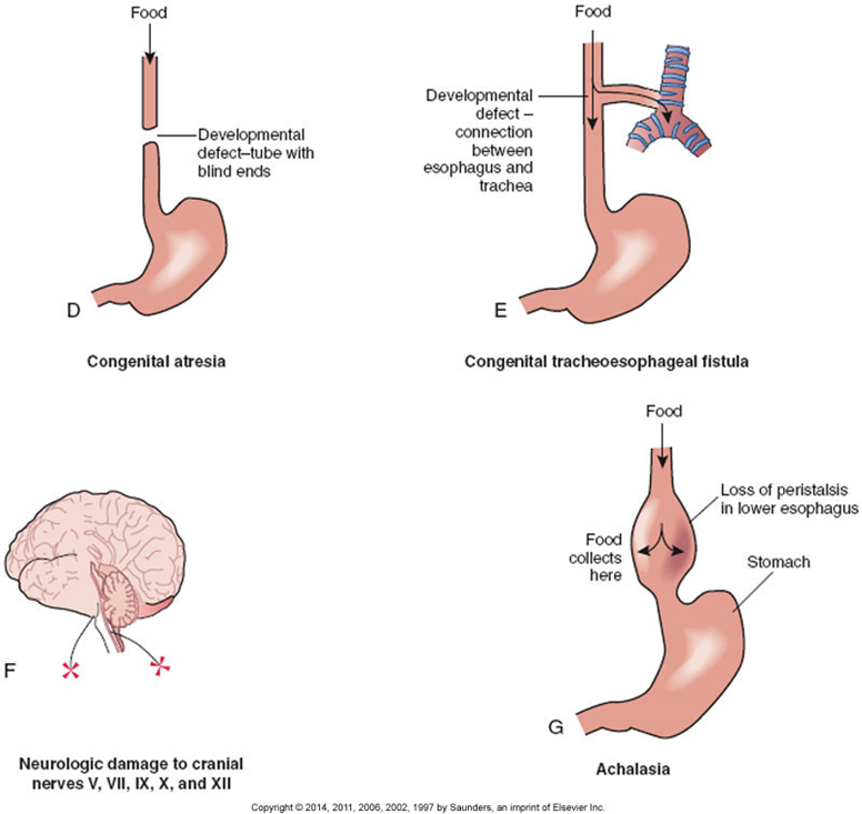
Neurological deficit
Muscular disorder
Mechanical obstruction
Results and presentation
Pain with swallowing
Inability to swallow larger pieces of solid material
Difficulty swallowing liquids
Neurological deficit
Infection
Stroke
Brain damage
Achalasia
Failure of the lower esophageal sphincter to relax because of lack of innervation
Muscular disorder
Impairment from muscular dystrophy
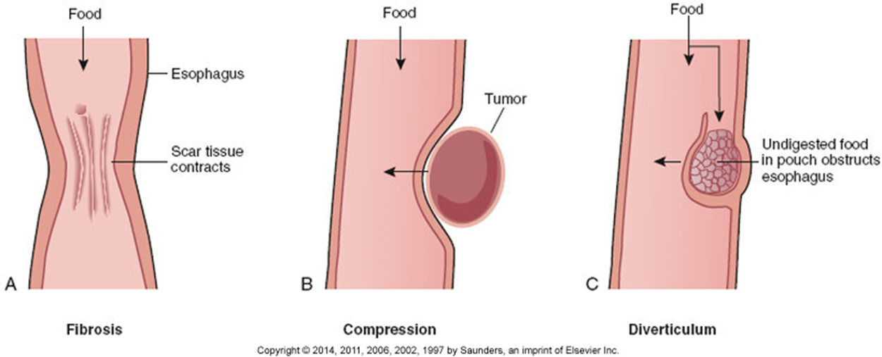
Mechanical obstruction
Congenital atresia
Developmental anomaly
Upper and lower esophageal segments are separated.
Stenosis
Narrowing of the esophagus
May be developmental or acquired
May be secondary to fibrosis, chronic inflammation, ulceration, radiation therapy
Stenosis or stricture may also result from scar tissue
May require treatment with repeated mechanical dilation
Esophageal diverticula
Outpouchings of the esophageal wall
Congenital or acquired following inflammation
Causes irritation, inflammation, scar tissue
Signs include dysphagia, foul breath, chronic cough, hoarseness
Tumors
May be internal or external
Causes of Dysphagia
Esophageal Cancer
Primarily squamous cell carcinoma
Usually in distal esophagus
Significant dysphagia in later stages
Poor prognosis because of late manifestations
Associated with chronic irritation because of:
Chronic esophagitis
Achalasia
Hiatal hernia
Alcohol abuse, smoking
Hiatal Hernia
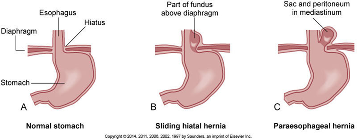
Part of the stomach protrudes into the thoracic cavity.
Sliding hernia
More common type
Portions of the stomach and gastroesophageal junction slide up above the diaphragm.
Rolling or paraesophageal hernia
Part of the fundus of the stomach moves up through an enlarged or weak hiatus in the diaphragm and may become trapped.
Food may lodge in pouch of the hernia
Causes inflammation of the mucosa
Reflux of food up the esophagus
May cause chronic esophagitis
Signs
Heartburn or pyrosis
Frequent belching
Increased discomfort when laying down
Substernal pain that may radiate to shoulder and jaw
Types of Hiatal Hernia
Gastroesophageal Reflux Disease
Periodic reflux of gastric contents into distal esophagus causes erosion and inflammation.
Often seen in conjunction with hiatal hernia
Severity depends on competence of the lower esophageal sphincter.
Delayed gastric emptying may be a factor.
Avoidance of:
Caffeine, fatty and spicy foods, alcohol, smoking, certain drugs
Use of medication may reduce reflux and inflammation
Gastritis: Acute Gastritis
Gastric mucosa is inflamed.
May be ulcerated and bleeding
May result from
Infection by microorganisms
Allergies to foods
Spicy or irritating foods
Excessive alcohol intake
Ingestion of aspirin or other NSAIDs
Ingestion of corrosive or toxic substances
Radiation or chemotherapy
Basic signs of gastrointestinal irritation
Anorexia, nausea, vomiting may develop
Hematemesis caused by bleeding
Epigastric pain, cramps or general discomfort
With infection, diarrhea may develop.
Acute gastritis is usually self-limiting.
Complete regeneration of gastric mucosa
Supportive treatment with prolonged vomiting
May require treatment with antimicrobial drugs
Gastritis: Chronic Gastritis
Characterized by atrophy of stomach mucosa
Loss of secretory glands
Reduced production of intrinsic factor
Helicobacter pylori infection is often present.
Signs may be vague.
Mild epigastric discomfort, anorexia, intolerance for certain foods
Increased risk of peptic ulcers and gastric carcinoma
Certain autoimmune disorders are associated with one type of chronic gastric atrophy.
Gastritis: Gastroenteritis
Inflammation of stomach and intestine
Usually caused by infection
May also be caused by allergic reactions to food or drugs
Microbes can be transmitted by fecally contaminated food, soil, and/or water
Most infections are self-limiting.
Serious illness may result in compromised host or virulent organisms.
May cause epidemic outbreaks in refugee or disaster settings
Safe sanitation essential for prevention
Common Infections Transmitted by Food and Water
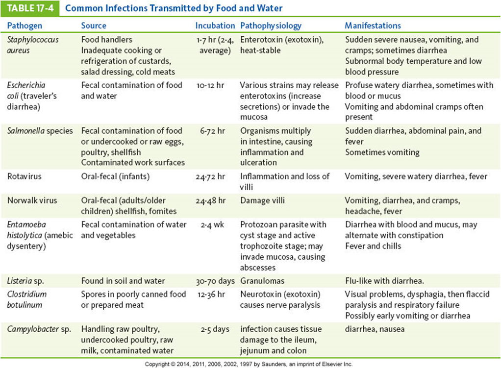
Escherichia coli Infection
Although E. coli is usually harmless as a resident in the human intestine, infective strains can cause significant problems.
Infective strains
Enterotoxigenic E. coli
Enteroinvasive E. coli
Enteropathogenic E. coli
Enteroaggregative E. coli
Enterohemorrhagic E. coli
Peptic Ulcer: Gastric and Duodenal Ulcers
Most caused by H. pylori infection
Usually occur in the proximal duodenum (duodenal ulcers)
Also found in the antrum of the stomach (gastric ulcers)
Development begins with breakdown of mucosal barrier
Decreased mucosal defense
More common in gastric ulcer development
Increased acid secretion predominant factor in duodenal ulcers
Peptic Ulcer: Common Locations
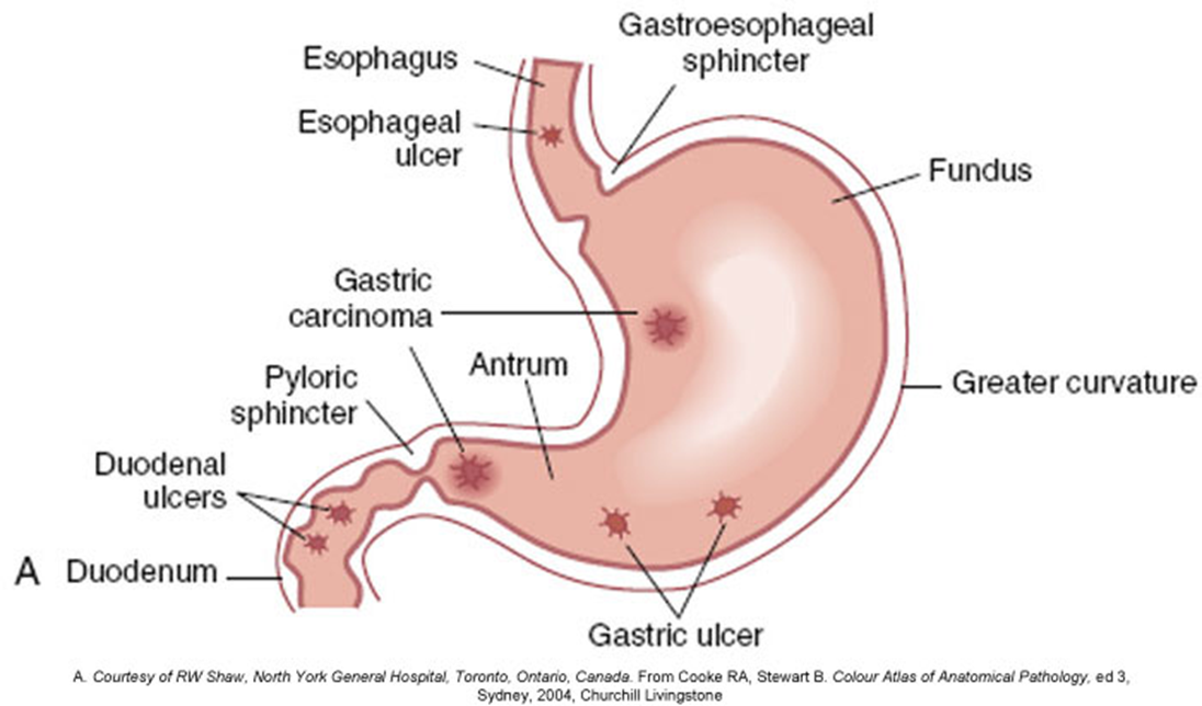
Damage to mucosal barrier predisposes to development of ulcers and is associated with:
Inadequate blood supply
Caused by vasoconstriction (e.g., by stress, smoking, shock, circulatory impairment in older adults, scar tissue, anemia)
Interferes with rapid regeneration of epithelium
Excessive glucocorticoid secretion or medication
Ulcerogenic substances break down mucous layer.
Aspirin, NSAIDs, alcohol
Atrophy of gastric mucosa
Chronic gastritis
Increased acid pepsin secretions
Increased gastrin secretion
Increased vagal stimulation
Increased sensitivity to vagal stimuli
Increased number of acid pepsin secretory cells in the stomach (genetic anomaly)
Increased stimulation of acid pepsin secretion
Alcohol, caffeine, certain foods
Interference with normal feedback mechanisms
Rapid gastric emptying
Complications of peptic ulcer
Hemorrhage
Caused by erosion of blood vessels
Common complication
May be the first sign of a peptic ulcer
Perforation
Ulcer erodes completely through the wall.
Chyme can enter the peritoneal cavity.
Results in chemical peritonitis
Obstruction
May result later because of the formation of scar tissue
Signs and symptoms
Epigastric burning or localized pain, usually following stomach emptying
Diagnostic tests
Fiberoptic endoscopy
Barium x-ray
Endoscopic biopsy
Treatment
Combination of antimicrobial and proton pump inhibitor to eliminate H. pylori
Reduction of exacerbating factors
Stress Ulcers
Associated with severe trauma or systemic problems
Burns, head injury
Hemorrhage or sepsis
Rapid onset
Multiple ulcers (usually gastric) may form within hours of precipitating event
First indicator—hemorrhage and severe pain
Gastric Cancer
Arises primarily in mucous glands
Mostly in the antrum or pyloric area
Early carcinoma
Confined to mucosa and submucosa
Later stages
Involves muscularis
Eventually invades serosa and spreads to lymph nodes
Asymptomatic in the early stages
Often, prognosis is poor on diagnosis
Diet seems to be a key factor, particularly smoked foods, nitrites, and nitrates.
Genetic influences also play a role.
Symptoms vague until cancer is advanced.
Reason for late diagnosis
Surgery together with chemotherapy and radiation may relieve symptoms.
Survival rate less than 20%
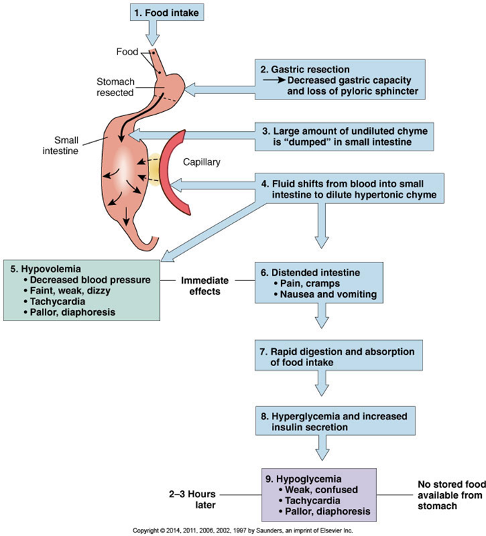
Dumping Syndrome
Control of gastric emptying is lost, and gastric contents are “dumped” into the duodenum without complete digestion.
May follow gastric resection
Hyperosmolar chyme draws fluid from vascular compartment into intestine
Intestinal distention
Increased intestinal motility
Decreased blood pressure → anxiety and syncope
Occurs during or shortly after meals
Abdominal cramps, nausea, diarrhea
Hypoglycemia 2 to 3 hours after meal
High blood glucose levels in chyme stimulate increased insulin secretion → drop in blood glucose levels
May be resolved by dietary changes
Frequent small meals—high in protein, low in simple carbohydrates
Often resolves over time
Pyloric Stenosis
Narrowing and obstruction of pyloric sphincter
May be developmental anomaly
Signs appear within several weeks after birth.
Projectile vomiting immediately after feeding
Firm mass can be palpated at pylorus.
Infant fails to gain weight, dehydration, persistent hunger
Surgery required to remove obstruction.
May be acquired later in life
Persistent feeling of fullness
Increased incidence of vomiting
Gallbladder Disorders
Cholelithiasis
Formation of gallstones
Solid material (calculi) that form in bile
Cholecystitis
Inflammation of gallbladder and cystic duct
Cholangitis
Inflammation usually related to infection of bile ducts
Choledocholithiasis
Obstruction of the biliary tract by gallstones
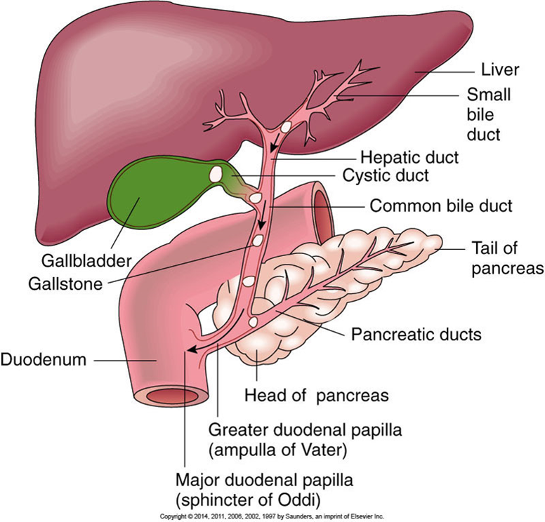
Gallstones vary in size and shape.
Form in bile ducts, gallbladder, or cystic duct
May consist of:
Cholesterol or bile pigment
Mixed content with calcium salts
Small stones
May be silent and excreted in bile
Larger stones
Obstruct flow of bile in cystic or common bile ducts; cause severe pain, which is often referred to subscapular area
Risk factors for gallstones
Women twice as likely to develop stones
High cholesterol in bile
High cholesterol intake
Obesity
Multiparity
Use of oral contraceptives or estrogen supplements
Hemolytic anemia
Alcoholic cirrhosis
Biliary tract infection
Obstruction of a duct by a large calculi
Sudden severe waves of pain
Radiating pain
Nausea and vomiting usually present
Pain continues, and jaundice develops.
Bile backs up into the liver and blood.
Risk of ruptured gallbladder if obstruction persists
Pain decreases if stone moves into duodenum
Surgical intervention may be necessary.
May be removed using laparoscopic surgery
Low-fat diet necessary following surgery
Biliary Ducts and Pancreas with Possible Locations of Gallstones
Jaundice
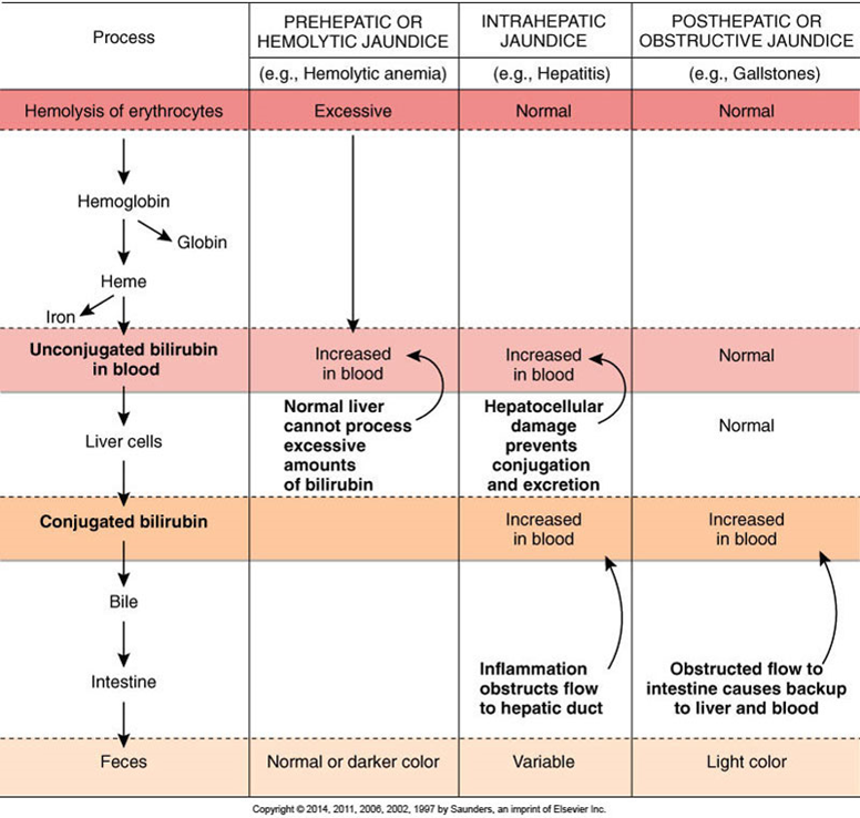
Prehepatic jaundice
Result of excessive destruction of red blood cells
Characteristic of hemolytic anemias or transfusion reactions
Intrahepatic jaundice
Occurs with disease or damage to hepatocytes
Hepatitis or cirrhosis
Posthepatic jaundice
Caused by obstruction of bile flow into gallbladder or duodenum
Tumor, cholelithiasis
Types of Jaundice
Bilirubin Measurement in Jaundice
Direct or conjugated bilirubin can be measured in the blood.
Total bilirubin is measured in blood.
Total bilirubin minus direct bilirubin = indirect or unconjugated bilirubin.
Jaundice
Type of jaundice indicated by increase in serum bilirubin level and changes in stools
Prehepatic jaundice
Unconjugated bilirubin level elevated
Intrahepatic jaundice
Both unconjugated and conjugated bilirubin levels may be elevated.
Posthepatic jaundice
Increased conjugated bilirubin level
Light-colored stool caused by absence of bile
Structure of Liver Lobule
Hepatitis
Inflammation of the liver
Alcoholic
Fatty liver
Idiopathic
Fatty liver
Viral hepatitis
Local infection
Infection elsewhere in body
Examples—infectious mononucleosis or amebiasis
Chemical or drug toxicity
Viral Hepatitis
Cell injury results in inflammation and necrosis in the liver.
Degrees of inflammation and damage vary.
Liver is edematous and tender.
Causative viruses
Hepatitis A virus (HAV)
Hepatitis B virus (HBV)
Hepatitis C virus (HCV)
Hepatitis D virus (HDV)
Hepatitis E virus (HEV)
Hepatitis A (HAV)
Small RNA virus
Infectious hepatitis
Transmitted by fecal-oral route in areas of inadequate sanitation or hygiene
Often from contaminated water or shellfish
Sexual transmission has occurred during anal intercourse.
Acute but self-limiting infection
No carrier or chronic state
Fecal shedding of virus before onset of signs
Vaccine available for travelers, food care workers, and health care workers
Hepatitis B (HBV)
Partially double-stranded DNA virus
Over 50% of HIV-positive patients are positive for HBV.
50% of patients are asymptomatic but contagious because of carrier state.
Chronic inflammation can occur.
Transmission primarily by infected blood
Sexual transmission has been noted.
Tattooing and body piercing may transmit the virus.
Vaccine available, routinely given to children
Hepatitis C (HCV)
Single-stranded RNA virus
Most common type transmitted by blood transfusion
May exist in a carrier state
About 50% of patients enter the chronic state.
Increases risk of hepatocellular carcinoma
Treated with interferon injections
Hepatitis D (HDV)
Also called delta virus
Incomplete RNA virus
Requires HBV to replicate and produce active infection
HDV infection increases severity of HBV infection
Transmitted by blood
Hepatitis E (HEV)
Single-stranded RNA virus
Transmitted by oral-fecal route
No chronic or carrier state
Viral Hepatitis: Signs and Symptoms
Preicteric stage
Fatigue and malaise
Anorexia and nausea
General muscle aching
Icteric stage
Onset of jaundice
Stools light in color, urine becomes darker
Liver tender and enlarged, mild aching pain
Posticteric stage—recovery stage
Reductions in signs
Weakness persists for weeks
Course of Hepatitis B Infection
Viral Hepatitis (Cont.)
Only body defense is formation of antibodies via vaccination
Supportive measures
Rest, diet high in protein, carbohydrate, and vitamins
Chronic hepatitis can be treated with interferon.
Decreases viral replication
Effective in only 30% to 40% of individuals
Drug combination (slow-acting interferon plus antiviral drug) more effective
Toxic or Nonviral Hepatitis
Variety of hepatotoxins can cause inflammation and necrosis of the liver.
Drugs include:
Acetaminophen, halothane, phenothiazines, tetracycline
Chemicals include:
Carbon tetrachloride (not used currently), toluene, ethanol
Direct effect of toxins
May result from sudden exposure to large amounts or from lower dose and long-term exposure
Cirrhosis
Progressive destruction of the liver
Causes
Alcoholic liver disease
Biliary cirrhosis
Associated with immune disorders
Postnecrotic cirrhosis
Linked with chronic hepatitis or long-term exposure to toxic materials
Metabolic
Usually caused by genetic metabolic storage disorders
Extensive diffuse fibrosis
Interferes with blood supply
Bile may back up.
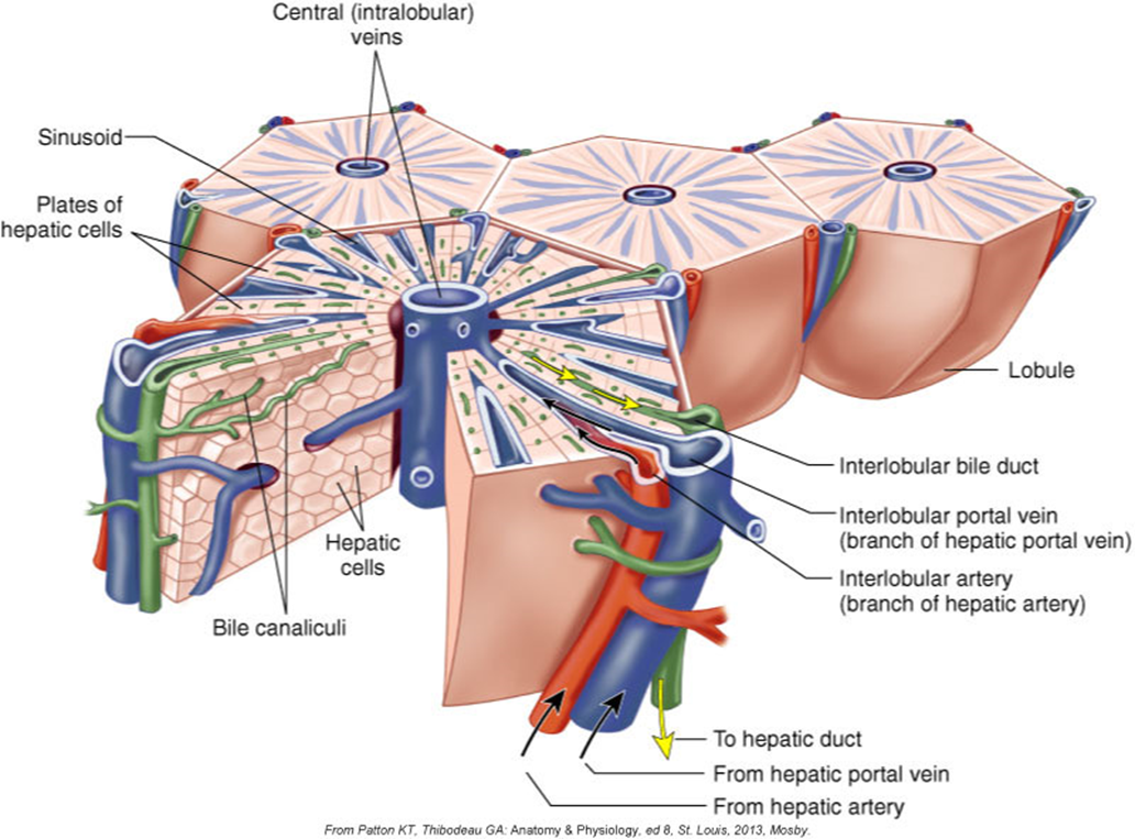
Loss of lobular organization
Degenerative changes may be asymptomatic until disease is well advanced.
Liver biopsy and serologic test to determine cause and extent of damage
Cirrhosis: Alcoholic Liver Disease
Initial stage—fatty liver
Enlargement of the liver
Asymptomatic and reversible with reduced alcohol intake
Second stage—alcoholic hepatitis
Inflammation and cell necrosis
Fibrous tissue formation—irreversible change
Third stage—end-stage cirrhosis
Fibrotic tissue replaces normal tissue.
Little normal function remains.
Functional Losses with Cirrhosis
Decreased removal and conjugation of bilirubin
Decreased production of bile
Impaired digestion and absorption of nutrients
Decreased production of blood-clotting factors
Impaired glucose and glycogen metabolism
Impaired conversion of ammonia to urea
Decreased inactivation of hormones and drugs
Drug dosages must be carefully monitored to avoid toxicity.
Decreased removal of toxic substances
Reduction of bile entering the intestine
Impairs digestion and absorption
Backup of bile in the liver
Leads to obstructive jaundice
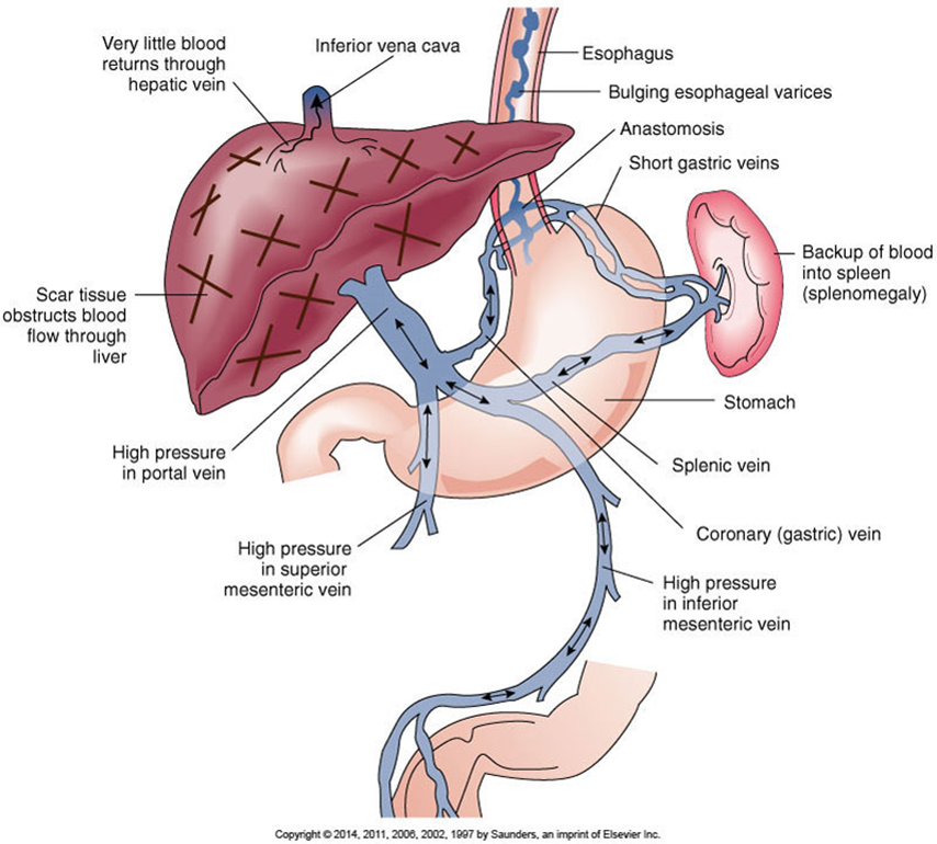
Blockage of blood flow through the liver
Leads to portal hypertension
Congestion in the spleen
Increases hemolysis
Inadequate storage of iron and vitamin B12
Congestion in intestinal walls and stomach
Impairing digestion and absorption
Development of esophageal varices
Hemorrhage
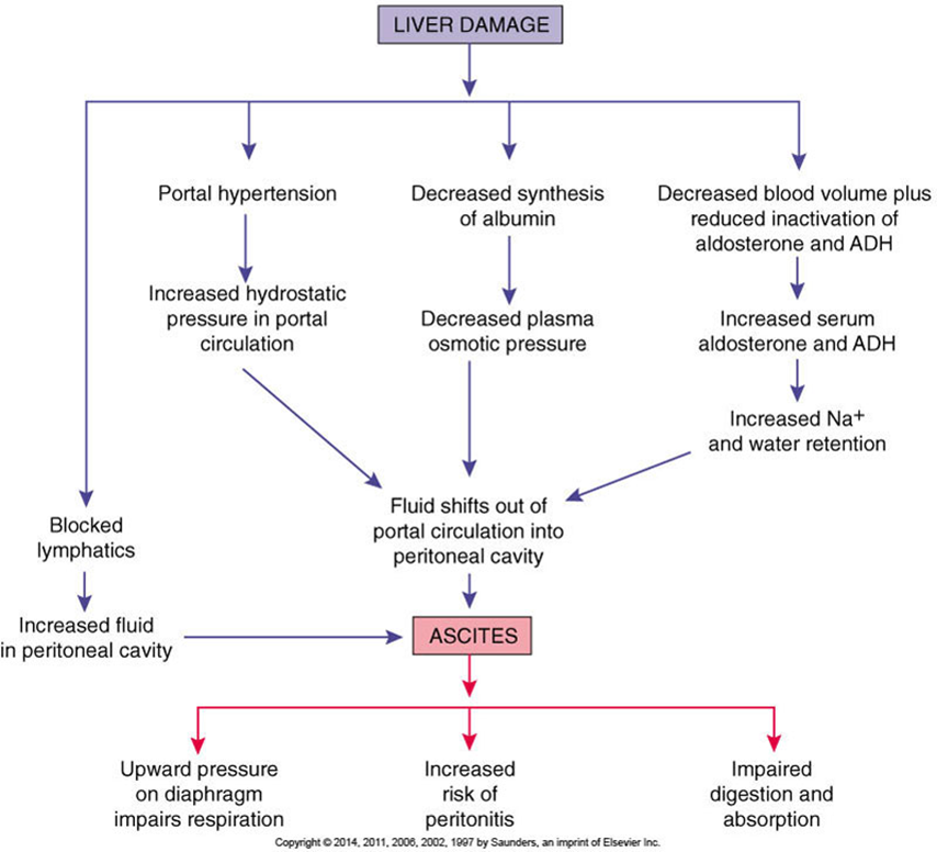
Development of ascites, an accumulation of fluid in the peritoneal cavity
Causes abdominal distention and pressure
Development of Esophageal Varices
Development of Ascites with Cirrhosis
Cirrhosis
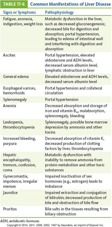
Initial manifestations often mild and vague
Fatigue, anorexia, weight loss, anemia, diarrhea
Dull aching pain may be present in upper right abdominal quadrant.
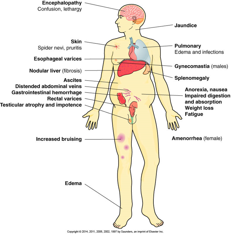
Advanced cirrhosis
Ascites and peripheral edema
Increased bruising
Esophageal varices
May rupture, leading to hemorrhage, circulatory shock
Jaundice, encephalopathy
Effects of Advanced Cirrhosis
Cirrhosis: Treatment
Avoidance of alcohol or specific cause
Supportive or symptomatic treatment
Dietary restrictions
Balancing serum electrolytes
Paracentesis
Antibiotics to reduce intestinal flora
Emergency treatment if esophageal varices rupture
Liver transplantation
Common Manifestations of Liver Disease
Liver Cancer
Hepatocellular carcinoma
Most common primary tumor of liver
More common in cirrhotic livers
Secondary or metastatic cancer
Arises from areas served by the hepatic vein or spread along the peritoneal membranes
Initial signs are mild and general.
Diagnosis usually occurs with advanced stages
Chemotherapy, possible lobectomy or radiofrequency ablation (RFA) procedure
Acute Pancreatitis
Inflammation of the pancreas
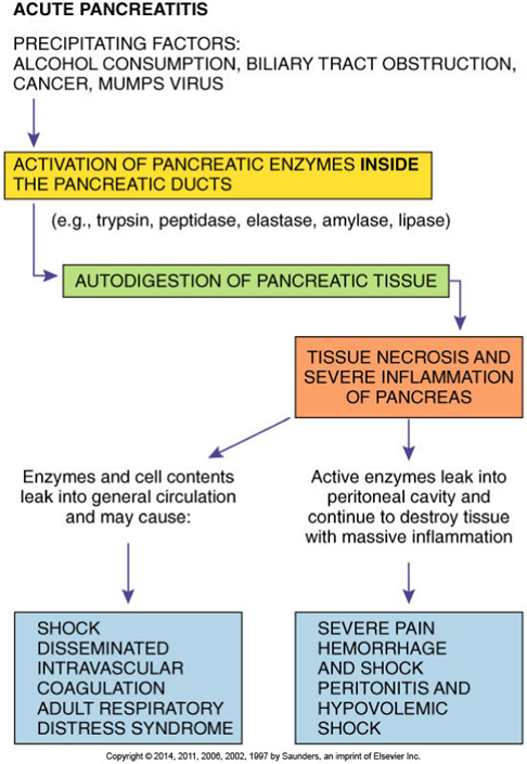
Results in autodigestion of the tissue
May be acute or chronic
Acute form considered a medical emergency
Pancreas lacks a fibrous capsule
Destruction may progress into tissue surrounding the pancreas
Substances released by necrotic tissue lead to widespread inflammation
Hypovolemia and circulatory collapse may follow.
Chemical peritonitis results in bacterial peritonitis.
Septicemia may result.
Adult respiratory distress syndrome and acute renal failure are possible complications.
Causes
Gallstones
Alcohol abuse
Sudden onset may follow intake of large meal or large amount of alcohol
Acute Pancreatitis: Signs and Symptoms
Severe epigastric or abdominal pain radiating to the back—primary symptoms
Signs of shock
Caused by o hypovolemia
Low-grade fever until infection develops
Body temperature may then rise significantly.
Abdominal distention and decreased bowel sounds
Decreased peristalsis and paralytic ileus
Diagnostic tests
Serum amylase levels—first rise, then fall after 48 hours
Serum lipid levels are elevated.
Hypocalcemia
Leukocytosis
Treatment
Oral intake is stopped.
Treatment of shock and electrolyte imbalances
Analgesics for pain relief
Pathophysiology of Acute Pancreatitis
Pancreatic Cancer
Risk factors
Smoking
Pancreatitis and dietary factors
Adenocarcinoma—most common form
Arises from the epithelial cells in the ducts
Weight loss and jaundice early manifestations
Frequently asymptomatic until well advanced
Metastases occur early.
Mortality is close to 95%.
Celiac Disease
Malabsorption syndrome
Primarily a childhood disorder
May occur in adults in middle age
Appears to have genetic link
Defect in intestinal enzyme
Prevents further digestion of gliadin (breakdown product of gluten)
Toxic effect on intestinal villi—atrophy of villi
Malabsorption and malnutrition result.
First signs appear when cereals are added.
At about 4 to 6 months of age
Manifestation
Steatorrhea, muscle wasting, failure to gain weight
Irritability and malaise common
Diagnosed by a series of blood tests
Gluten-free diet for treatment
Intestinal mucosa returns to normal after a few weeks without gluten intake.
Chronic Inflammatory Bowel Disease
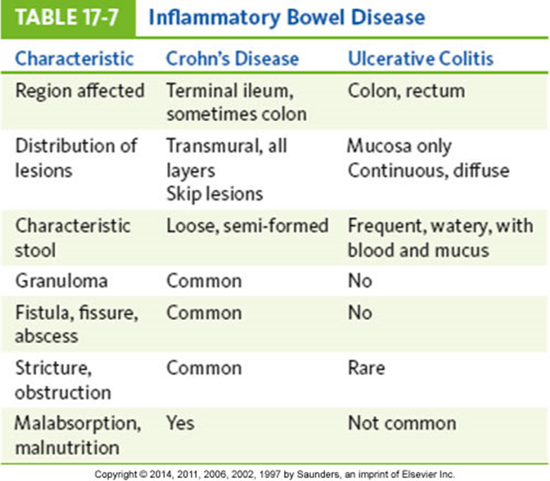
Crohn’s disease and ulcerative colitis are chronic inflammatory bowel diseases (IBDs).
Causes unknown
Genetic factor appears to be involved.
Crohn’s disease—often during adolescence
Ulcerative colitis—second or third decade
Many similarities between Crohn’s disease and ulcerative colitis
Inflammatory Bowel Disease
Crohn’s Disease
May affect any area of the digestive tract
Usually small intestine affected
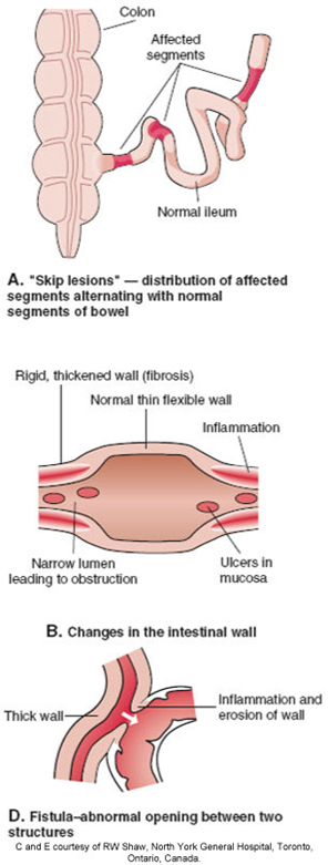
Inflammation occurs in characteristic distribution
“Skip lesions”—affected areas separated by areas of normal tissue
Progressive inflammation and fibrosis may cause obstructed areas.
Damaged walls impair processing and absorption of food.
Inflammation stimulates intestinal motility.
Interference with digestion and absorption
Hypoproteinemia, avitaminosis, malnutrition, possibly steatorrhea
Other complications
Adhesions between loops may form and fistulas may develop.
Children
Delayed growth and sexual maturation
Glucocorticoid used in treatment
Ulcerative Colitis
Inflammation starts in the rectum
Progresses through the colon
Mucosa and submucosa are inflamed.
Tissue destruction interferes with absorption of fluid and electrolytes in the colon.
Severe acute episodes—toxic megacolon may develop.
Marked diarrhea, with up to 12 stools per day
Contains blood and mucus
Accompanied by cramping pain
Treatment of IBD
Team approach
Anti-inflammatory medications
Sulfasalazine
Glucocorticoids
Antimotility agents
Nutritional supplements
Antimicrobials
Immunotherapeutic agents
Surgical resection
Usually ileostomy or colostomy
Irritable Bowel Syndrome
Types
Abnormal gastrointestinal mobility and secretion
Visceral hypersensitivity
Postinfectious IBS
Overgrowth of flora
Food allergy or intolerance
Psychosocial factors
Manifestations
Lower abdominal pain
Diarrhea
Constipation, alternating with diarrhea
Bloating, nausea
Diagnosis
Based on signs and symptoms
Testing for food allergies
Testing for bacterial or parasitic infections
No single cure for IBS
Appendicitis: Development
Obstruction of the appendiceal lumen
By a fecalith, gallstone, or foreign material
Fluid builds up inside the appendix.
Microorganisms proliferate
Appendiceal wall becomes inflamed.
Purulent exudate forms
Appendix is swollen.
Ischemia and necrosis of the wall
Results in increased permeability
Bacteria and toxins escape into surroundings.
Leads to abscess formation or localized bacterial peritonitis
Abscess may develop when inflamed area is walled off.
Inflammation and pain may temporarily subside.
Localized infection or peritonitis develops around the appendix.
May spread along the peritoneal membranes
Increased necrosis and gangrene in the wall
Caused by increasing pressure in the appendix
Appendix ruptures or perforates
Release of contents into peritoneal cavity
Generalized peritonitis
May be life-threatening
Treatment
Surgical removal of appendix and antimicrobial drugs
Signs and Symptoms
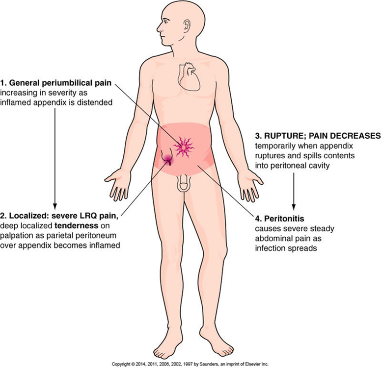
General periumbilical pain
Related to the inflammation
Nausea and vomiting common
Pain becomes severe and localized in lower right quadrant (LRQ).
LRQ rebound tenderness develops.
Involvement of parietal peritoneum over appendix
After rupture
Pain subsides temporarily.
Pain recurs—severe, generalized abdominal pain and guarding
Low-grade fever and leukocytosis
Development of inflammation
Boardlike abdomen, tachycardia, hypotension
As peritonitis develops, abdominal wall muscles spasm.
Toxins lead to reduced blood pressure.
Typical Progression of Pain in Acute Appendicitis
Diverticular Disease
Development of diverticula
Diverticulum
Outpouching (herniation) of the mucosa through the muscular layer of the colon
Diverticulosis
Asymptomatic diverticular disease
Diverticulitis
Inflammation of the diverticula
Form at gaps between muscle layers
Congenital weakness of wall may be a factor
Weaker areas bulge when pressure increases.
Many cases are asymptomatic.
Diverticulitis stasis of material in diverticula leads to inflammation and infection.
Cramping, tenderness, nausea, vomiting
Slight fever and elevated white blood cell count
Treatment of diverticulitis
Antimicrobial drugs
Dietary modifications to prevent stasis
Colorectal Cancer
Most malignancies develop from adenomatous polyps.
Early diagnosis is essential.
Cancer occurs primarily in persons older than 50 years.
Risk factors
Familial multiple polyposis
Long-term ulcerative colitis
Genetic factors
Environmental factors
Diet low in fiber
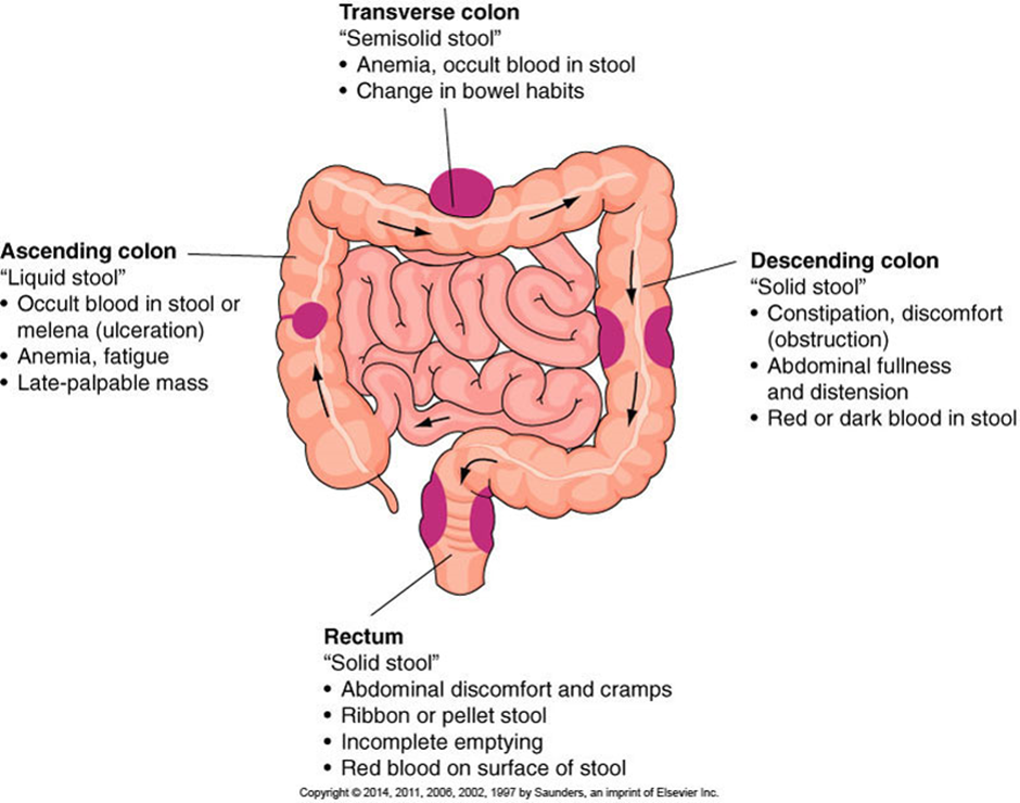
Initial signs depend largely on the location of the growth.
General signs
Change in bowel habits
Alternating diarrhea and constipation
Bleeding
Fatigue, weight loss, anemia
Treatment
Surgical removal with radiation and/or chemotherapy
Common Signs and Symptoms of Colorectal Cancer
Intestinal Obstruction
Lack of movement of intestinal contents through the intestine
More common in small intestine
Mechanical obstructions
Result from tumors, adhesions, hernias, other tangible obstructions
Functional or adynamic obstructions
Result from impairment of peristalsis
Spinal cord injury
Paralytic ileus caused by toxins or electrolyte imbalance
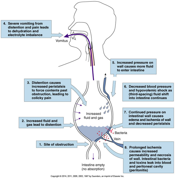
Gases and fluids accumulate proximal to the blockage, distending the intestine.
Increasingly strong contractions of proximal intestine
Effort to move contents along
Pressure increases in lumen.
More secretions enter the intestine.
Compression of veins in wall
Intestinal wall becomes edematous
Prevention of absorption
Intestinal distention leads to persistent vomiting.
Additional loss of fluid and electrolytes
Hypovolemia can result.
Intestinal wall becomes ischemic and necrotic.
If obstruction is not removed, gangrene ensues.
Ischemia and necrosis → decreased innervation and cessation of peristalsis
Paralytic ileus occurs if it is not a cause to begin with
Obstruction promotes rapid reproduction of intestinal bacteria.
Some produce endotoxins.
Affected wall becomes necrotic and more permeable
Bacteria and toxins leak into peritoneal cavity (peritonitis) or into blood (bacteremia and septicemia).
Perforation of the necrotic segment may occur.
Generalized peritonitis and septic shock
Functional obstructions or paralytic ileus from:
Abdominal surgery (follows surgery)
Spinal shock following spinal cord injuries
Inflammation related to severe ischemia
Pancreatitis, peritonitis, infection in the abdominal cavity
Hypokalemia
Mesenteric thrombosis
Toxemia
Mechanical obstruction from:
Adhesions that twist or constrict intestine
Hernias
Strictures caused by scar tissue
Masses—tumors or foreign bodies
Intussusception
Volvulus
Hirschsprung’s disease
Gradual obstruction from chronic inflammatory conditions
Mechanical obstruction of small intestine
Severe colicky abdominal pain
Intermittent bowel sounds can be heard.
Paralytic ileus
Pain is steady.
Bowel sounds decrease or are absent.
Vomiting and abdominal distention
Occurs quickly with obstruction of small intestine
Vomiting is recurrent, eventually with bile-stained content
Obstruction of the small intestine is a medical emergency!
Obstruction of large intestine
Develops slowly, with mild signs
Constipation
Mild abdominal pain, followed by abdominal distention
Anorexia, vomiting, more severe pain
Treatment
Treatment of underlying cause
Fluid and electrolyte replacement
Surgery and antimicrobial therapy
Effects of Intestinal Obstruction
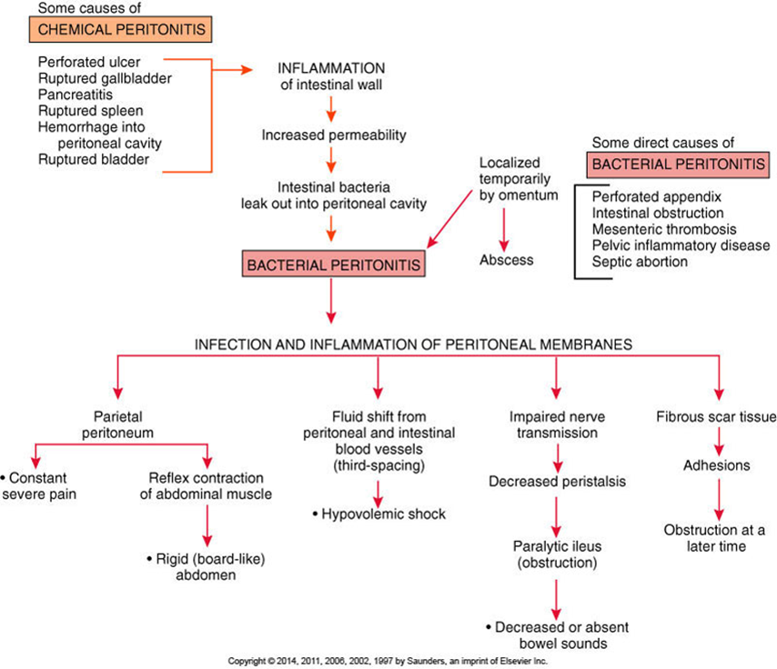
Peritonitis
Inflammation of the peritoneal membranes
Chemical peritonitis may result from:
Enzymes released with pancreatitis
Urine leaking form a ruptured bladder
Chyme spilled from a perforated ulcer
Bile escaping from the ruptured gallbladder
Blood
Any other foreign material in the cavity
Bacterial peritonitis caused by:
Direct trauma affecting the intestine
Ruptured appendix
Intestinal obstruction and gangrene
Any abdominal surgery
If foreign material is left or infection develops
Pelvic inflammatory disease in women
When infection reaches the cavity through fallopian tubes
Signs and symptoms
Sudden, severe, generalized abdominal pain
Localized tenderness at site of underlying problem
Vomiting common, abdominal distention
Dehydration, hypovolemia, low blood pressure
Decreased blood pressure, tachycardia, fever, leukocytosis
Treatment
Depends on primary cause
Surgery might be required.
Massive antimicrobial drugs—specific to causative organism
Development of Peritonitis
Development of Peritonitis