Ion Channels and Transporters
Vocabulary:
Electrical signaling: Proteins utilizing ions to maintain potentials, ions utilized are not in electrochemical equilibrium across the semipermeable plasma membrane. With neural signaling, ions travel down the electrochemical gradient to generate current, with this being permitted by channels and kinetic energy only
Active transporter: A protein within a membrane that allows for the passage of molecules, utilizing energy. Used to reestablish concentration gradients and move ions against the electrochemical gradient, requiring ATP directly or indirectly. Have multiple subtypes. Slower than channel-mediated transport, can be electrogenic
Primary: ATP will phosphorylate it to open/close, transports things against their gradient using energy
Secondary: Energy stored in concentration gradients is used to transport one molecule along its gradient and another molecule against it. Relies on primary subtype to establish concentration gradients. Conformation change only occurs when both molecules are bound
Symport: Co-transport, both molecules move in the same direction (often Na+ into a cell)
Antiport: One molecule moving into a cell, another moving out, opposite directions
Electrogenic: Describes proteins that create uneven exchanges between sides of the membrane to generate charge
Ion channel: An integral protein within a membrane that can open/close to regulate ions flowing down their electrochemical gradients. Regulated by diverse mechanisms, exhibit some degree of specificity (can let in single or multiple ions). Multiple channels may exist for one ion. More than 220 genes make these. Most are gated, though some are non-gated leak channels. Often have multiple helical transmembrane segments, water-filled pores, charges in pores to attract ions, and pore loops to form a selectivity filter. Can only accommodate a single type (cation/anion) of partially hydrated ion, multiple ions at a time enhances ion flux. Permit the electrochemical gradient to generate current
Leak channel: A non-gated ion channel
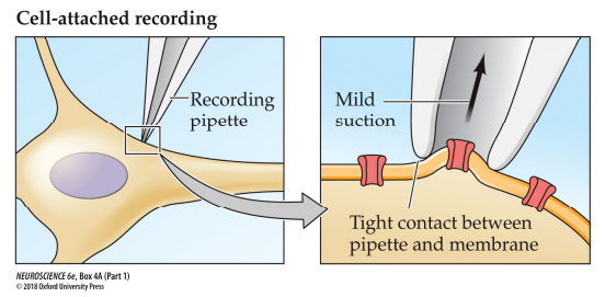
Patch clamp: A technique used to study current flow through ion channels, uses refinement of voltage clamps. Uses a glass micropipette to form a tight seal with a small area of the cell membrane, ideally isolating a single channel. This pipette serves as the recording electrode and the current injecting electrode Current flowing through the channel is measured by the pipette, has the ability to clamp voltage
Cell-attached recording: The cell is intact, it can measure current through channels under physiological conditions, with intracellular signaling paths intact. It measures spontaneous activity, ideally just one channel
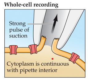
Whole-cell recording: The pipette suction disrupts membrane/cytoplasm, so that the cytoplasm is continuous with the micropipette electrolyte solution, allowing it to measure whole cell currents or membrane potentials in response to extracellular signals or signals introduced by the micropipette. Will alter the cytoplasmic contents after several minutes of exposure
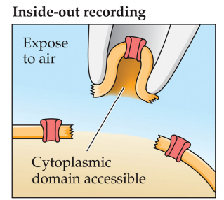
Inside-out recording: The micropipette extracts part of the whole cell and detaches it before retracting, allowing manipulation of the internal surface and studying of intracellular signaling molecules and their roles (like cyclic nucleotides). Best for detecting impacts of intracellular signals on a single channel
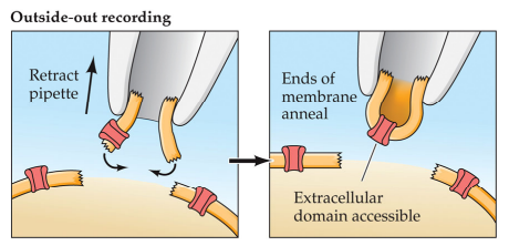
Outside-out recording: Most difficult subtype as there are many points of potential failure, requires strong suction to generate free ends when breaking the membrane in two (like with inside out recording, but split), free ends join together, so that the membrane is shaped like an upright U inside the micropipette, used to examine effects of multiple external conditions (like with a dose-response curve)
Ion movement: Hodgkin and Huxley hypothesized that this created APs, fast rates create a large ion current. Caused by the electrochemical gradient across the plasma membrane. Can be individual to an ion, K+ isn’t the same as Na+. Can “sense” voltage, may utilize voltage-sensitive ion channels. Found that depolarization increased the probability of channel opening
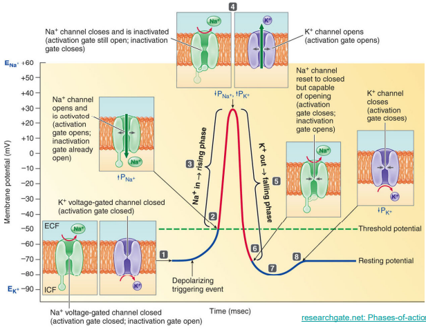
Voltage-gated Na+ channel: Contains 2 gates what move in response to voltage changes. Make by 10 SCN genes. Generate APs, regulate AP threshold and repetitive firing. Contains alpha and beta subunits. During depolarization, activation opens rapidly , at peak inactivation and closed and activation are open, during repolarization the activation gates close and inactivation gates open. Absolute refractory period is due to opening of inactivation gates, relative refractory period where APs are hard to generate
Activation gate: Closed below a voltage threshold, rapidly opens above the threshold, closes during repolarization
Inactivation gate: Open below a voltage threshold, slowly closes above the threshold, reopens during repolarization. While closed, APs cannot be generated (makes the absolute refractory period)
Alpha subunit: 6 transmembrane regions repeated 4 times, with voltage sensors located on the 4th helix of each group. The pore loop (selectivity filter) is between the 5th and 6th, forming a selectivity filter
Beta subunit: Has 2 subunits, with 4 possibilities
Voltage-gated K+ channel: Has one gate, closed below a threshold and opens slowly above threshold. Closes slowly to cause hyperpolarization. Opening coincidentally occurs when Na+ inactivation channels close. Created by 78 KCN genes. help repolarize the membrane after APs, specialized for transport in one direction
Passive K+ channels: Always open, for “leaks”
Passive Na+ channels: Always open
Inward rectifying K+ channels: Help maintain the RMP, blocked by intracellular molecules like Mg2+ when the membrane potential is less negative than RMP, allows potassium to flow in one direction rather than the other
Voltage-gated Ca2+ channels: Made by 10 CACNA genes, responsible for AP generation and intracellular signaling
Voltage-gated Cl- channels: Make by CLCN genes, present in every neuron. Control excitability and contribute to RMP, with ECl- being more negative that threshold potential, help regulate cell volume by contributing to intracellular osmolarity. Has two different transmembrane subunits, each of which forms a pore
Ligand-gated channels: Can be permeable to multiple ions, open/close due to present of another chemical. Used for rapid and brief signaling. Extracellularly, less specific than most ion channels, allow more than one ion to pass through and can be activated by multiple molecules/ligands. Intracellularly, secondary messengers bind to the channel to open them (like with CNG)
Ionotropic: Controlled by neurotransmitters
Cyclic nucleotide-gated (CNG): Controlled by cyclic nucleotides binding to the internal domains of the channel. Has 4 subunits and 6 transmembrane sections with pore loops, permeable to various cations
Acid-sensing (ASICs): Open in response to proton presence
Transient receptor protein (TRP): Open to extracellular ligands and some intracellular ligands. Can also respond to physical pressure and temperature, diverse types
Transient receptor protein channels (TRP): Respond to multiple modes of stimuli like temperature (thermoreceptors), stretch, some tastes, and pain (nociception). Can be multimodal integrators of sensory stimuli, permeable to cations
Thermosensitive channels: Part of the TRP superfamily, activate with temperature. Temperature change displaces membrane lipids, permeable to various cations. Temperature changes lipid structure to pull open
Mechanosensitive channels: Open/close with mechanical force
Piezo channels: Nonselective to cations. Has 3 subunits with 38 transmembrane sections. Some form ‘blades’
Some TRP channels
Acid-sensing channels (ASIC)
Bacterial K+ channel (KcsA): A pH-gated K+ channel, first to be atomically visualized via X-ray crystallography. Has 4 subunits with 2 helical transmembrane sections (alpha helices). Looks like an inverted tee-pee, or a downward cone. Connected by a poor loop inserted into the membrane, pores formed by these and transmembrane helices
Pore loop: Forms the selectivity filter, determines which ions can pass through
Pore helix: Negatively charged, attracts cations
Narrow portion: Only allows partially hydrated K+, smaller ions can’t be stabilized, charge repulsion promotes ion flux
Voltage-gated mammalian K+ channel: Similar in structure to KscA, has 4 subunits with 2 transmembrane segments and a pore loop that for the pore. Has beta subunits that are intracellularly regulatory and attached by the T1 domain. Each subunit has 4 additional transmembrane subunits acting as voltage sensors, containing strings of positively charged amino acids, attached to the pore by helical linkers. The voltage sensor, when hyperpolarized, has a linker that is pulled down towards the intracellular side to close the pore. When depolarized, the linker is pulled towards the extracellular side to open the pore. Doesn’t use ATP
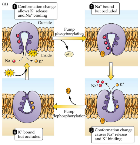
Na+/K+ ATPase: A primary active transporter, in every cell. Has at least 2 subunits encoded by separate genes. Key structures include a nucleotide binding site, a phosphorylation site, ion binding sites, and a phosphatase actuator domain. With 1 ATP, moves 2 K+ into the cell and 3 Na+ out of the cell, uneven exchange makes it electrogenic