Comprehensive Biology: Cell Biology, Membrane Transport, Bioenergetics, and Cytoskeletal Dynamics
Cellular Organization: Prokaryotic and Eukaryotic Architecture
Foundational Principles of Cell Biology:
The cell represents the fundamental structural and functional unit of all living organisms.
Organisms are categorized into two major cellular archetypes based on internal compartmentalization: prokaryotes and eukaryotes.
Prokaryotic Cellular Characteristics:
Organismal Nature: Exclusively unicellular organisms encompassing Bacteria and Archaea.
Dimensions: Typically range in diameter from .
Internal Compartmentalization: Entirely devoid of membrane-bound organelles or a membrane-enclosed nucleus.
Ribosomes: Contain small ribosomes (composed of and subunits) dispersed throughout the cytoplasm.
Genetic Material Organization: Genetic material is localized in a poorly delineated, non-membrane-bound cytoplasmic region termed the nucleoid. The genome consists predominantly of a single circular double-stranded DNA chromosome, often supplemented by small, autonomous circular extrachromosomal DNA molecules known as plasmids.
Reproduction: Reproduce asexually, primarily via binary fission.
Surface Structures: Outer cellular architecture includes a rigid peptidoglycan cell wall, an outer capsule or slime layer, surface pili (for adhesion and conjugation), and flagella for motility.

Eukaryotic Cellular Characteristics:
Organismal Nature: Predominantly found in multicellular organisms (animals, plants, fungi) but also encompass complex unicellular lineages (protists and protozoa).
Dimensions: Typically range in diameter from .
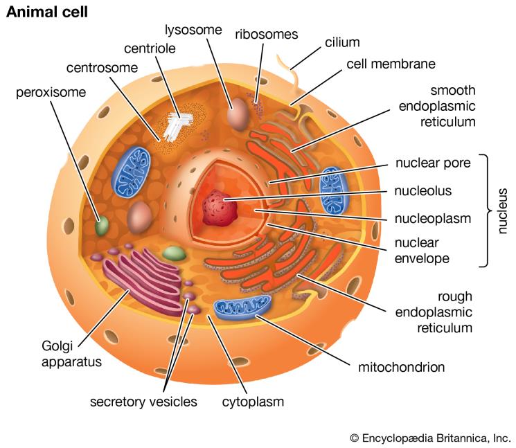
Compartmentalization: Feature extensive internal compartmentalization defined by biological membranes, establishing distinct organelles such as the nucleus, endoplasmic reticulum, Golgi apparatus, mitochondria, lysosomes, and peroxisomes.
Evolutionary Advantage of Compartmentalization: Establishing membrane-enclosed microenvironments allows a single cell to simultaneously run incompatible metabolic processes. Distinct compartments maintain specialized local environments (e.g., maintaining an acidic of in lysosomes while the surrounding cytosol remains at ) suited to specific enzymatic reactions, preventing hydrolytic destruction of cytosolic machinery.
Ribosomes: Possess large ribosomes (composed of and subunits) located free in the cytosol or bound to the endoplasmic reticulum membrane.
Genetic Material Organization: Linear double-stranded DNA molecules complexed with basic histone proteins to form chromatin, organized into discrete chromosomes and sealed within a double-membrane nuclear envelope.
Cell Differentiation: During embryonic development, unspecialized eukaryotic stem cells undergo differentiation, acquiring distinct structural and biochemical specializations tailored to precise physiological roles (e.g., neurons, hepatocytes, myocytes).
Virology: Virion Architecture, Taxonomy, and Infection Lifecycles
Biological Nature of Viruses:
Viruses are non-cellular, sub-microscopic macromolecular assemblies lacking metabolic machinery, autonomous energy production, and the ability to reproduce outside of a permissive host cell.
They function as obligate intracellular parasites, completely reliant on host cellular enzymes, ribosomes, transfer RNAs, and metabolic building blocks to replicate.
Virion: The extracellular, inanimate, infectious physical state of a virus.
Viral Dimensions: Viruses exhibit extreme miniaturization, with diameters spanning from approximately up to giant viruses:
Polio virus: in diameter ().
Zika virus: in diameter ().
Adenovirus: in diameter ().
Human Immunodeficiency Virus 1 (HIV-1): in diameter ().
Mimivirus: in diameter ().
Unit conversion rule: ; therefore, a virus equals .
Taxonomic Diversity: While every cellular microbe and higher organism is thought to host at least one specific virus (suggesting billions of viral species exist globally), approximately virus species have been formally classified.
Virion Structural Components:
Viral Genome: Core of genetic material consisting of double-stranded DNA (dsDNA), single-stranded DNA (ssDNA), double-stranded RNA (dsRNA), or single-stranded RNA (ssRNA; positive-sense or negative-sense), encoding from several to several hundred genes.
Capsid: A protective outer protein shell assembled from protein subunits called capsomers, arranged in helical, icosahedral, or complex geometries.
Lipid Envelope: Present in enveloped viruses; a lipid bilayer derived directly from host cell membranes (via budding) embedded with virus-encoded glycoproteins.
Bacteriophage Structural Prototype (T4-like Complex Phage):
Icosahedral Capsid Head: Encloses the viral dsDNA genome.
Collar and Sheath: Contractile helical protein cylinder through which DNA is injected.
Baseplate and Spikes: Anchor the phage onto the bacterial cell envelope.
Tail Fibers: Mediate specific recognition and binding to outer bacterial surface receptors.
Influenza Virus Structural Prototype (Enveloped Orthomyxovirus):
Lipid Envelope: Host-derived lipid bilayer containing viral membrane channels (e.g., M2 ion channel).
Haemagglutinin (HA): Surface spike glycoprotein responsible for binding sialic acid receptors on host respiratory epithelial cells to mediate viral entry.
Neuraminidase (NA / Sialidase): Surface spike enzyme that cleaves terminal sialic acid residues to release newly budded virions from the host cell.
Ribonucleoprotein (RNP) Core: Contains segmented negative-sense RNA genomes complexed with nucleoprotein and viral RNA-dependent RNA polymerase.

Host Range and Tissue Tropism:
Viral entry is dictated by high-affinity, specific molecular binding between viral attachment proteins (spikes or capsid domains) and cognate surface receptors on host cells.
Narrow Host Range: Specific to a single species or specialized cell type within that species. For example, human rhinoviruses and human influenza viruses strictly infect epithelial cells lining the human respiratory tract.
Wide Host Range: Ability to bind evolutionary conserved surface receptors across multiple taxonomic classes. For example, the rabies virus infects central nervous system tissues across canines (dogs), foxes, bats, raccoons, and humans.
Mechanisms of Viral Infection and Lifecycles:
Lytic Infection Cycle:
The virus binds to and enters the permissive host cell.
The viral genome immediately redirects the transcriptional and translational machinery of the host toward viral nucleic acid replication and viral protein synthesis.
Viral capsids assemble and package genomes.
Lysis of the host cell occurs (often via virus-encoded endolysins), rupturing the plasma membrane, destroying the host cell, and liberating progeny virions into the surroundings (e.g., T4 bacteriophage in Escherichia coli, lytic influenza replication).
Non-Lytic (Lysogenic / Integrative) Infection Cycle:
The virus injects or releases its nucleic acid into the host.
Rather than immediately destroying the cell, the viral genome integrates directly into the host chromosomal DNA as a stable, dormant genetic element termed a provirus (in animal cells) or prophage (in bacteria).
The host cell survives and divides, replicating the integrated viral genome alongside its own chromosomal DNA. Function may become impaired, or latency may persist until an environmental stressor reactivates the lytic cycle (e.g., Chickenpox / Varicella-zoster virus establishing latency in sensory ganglia; HIV establishing persistent infection in CD4+ T helper cells).
Positive-Sense RNA Viral Infection (e.g., Zika Virus):
Zika virus possesses a positive-sense single-stranded RNA () genome.
Because the genomic RNA is identical in polarity to host messenger RNA, upon uncoating in the cytoplasm, the genomic RNA immediately serves as a translation template for host cell ribosomes without requiring a transcription intermediate.
Retroviral Integration Mechanics (e.g., HIV-1):
Docking: Viral envelope glycoprotein gp120 binds the CD4 receptor and chemokine coreceptors (CCR5 or CXCR4) on helper T cells.
Fusion: Direct fusion of the viral lipid envelope with the host plasma membrane, injecting the capsid and viral enzyme core into the cytosol.
Reverse Transcription: Viral reverse transcriptase synthesizes a complementary DNA (cDNA) copy from the viral RNA template, yielding double-stranded viral DNA.
Integration: Viral integrase shuttles the dsDNA into the nucleus and catalyzes permanent integration into host chromosomal DNA, creating a provirus.


Biological Membranes: Composition, Bilayer Dynamics, and Fluidity Regulation
General Functions of Biological Membranes:
Define external cellular boundaries (plasma membrane), insulating the living cytoplasm from the non-living extracellular environment.
Enclose internal compartments (organelles) to enable metabolic compartmentalization.
Regulate and control the selective movement of solutes, ions, nutrients, and macromolecules into and out of the cell.
Facilitate detection of, and response to, external environmental stimuli through cell-surface receptors.
Mediate intercellular interactions, tissue adhesion, and chemical communication.
Provide a physical, semi-fluid two-dimensional scaffold for organizing biochemical activities and enzymatic cascades.
Ultrastructure and Chemical Composition of the Bilayer:
Trilaminar Morphology: Under transmission electron microscopy (TEM), biological membranes appear as a distinctive three-layered sandwich: two electron-dense outer lines separated by an electron-lucent central layer, representing a continuous phospholipid bilayer.
Thickness: Uniformly measures approximately across diverse eukaryotic and prokaryotic lineages.
Amphipathic Nature: Phospholipids possess structurally distinct polar (hydrophilic) and nonpolar (hydrophobic) domains:
Polar Head Group: Consists of a charged moiety (such as choline, ethanolamine, or serine) attached via an ester linkage to a negatively charged phosphate residue.
Glycerol Backbone: A 3-carbon glycerol molecule linking the phosphate group to fatty acyl chains.
Hydrophobic Fatty Acyl Tails: Two nonpolar long-chain fatty acids extending into the core.
Spontaneous Bilayer Formation: When placed in an aqueous environment, amphipathic phospholipids spontaneously assemble to minimize unfavorable thermodynamic interactions between water and hydrophobic hydrocarbon chains. Nonpolar tails bury inward via the hydrophobic effect, while polar phosphate heads form electrostatic and hydrogen bonds with surrounding water molecules.
Liposomes: Artificially synthesized spherical lipid bilayers enclosing a central aqueous lumen. Phospholipids align with polar heads facing the external water and the internal aqueous core, while the nonpolar tails face the hydrophobic interior of the bilayer.


The Fluid Mosaic Model and Membrane Asymmetry:
Fluidity Concept: The lipid bilayer exists as a dynamic, two-dimensional liquid crystal wherein individual lipid molecules move continuously within their respective leaflet.
Mosaic Concept: Diverse functional components, including integral membrane proteins, peripheral proteins, lipid-anchored proteins, carbohydrates, and sterols, penetrate or associate with the bilayer.
Molecular Kinetics of Lipids:
Flexion of Fatty Acyl Tails: Rapid intramolecular bending occurring on a timescale of .
Lateral Shift Diffusion: Individual lipids rapidly exchange places with adjacent lipids within the same monolayer leaflet on a timescale of .
Transverse Diffusion ("Flip-Flop"): Migration of a phospholipid from one leaflet across the hydrophobic core to the opposite leaflet is thermodynamically unfavorable and slow (, equivalent to several days), requiring active enzymatic catalysis by flippases, floppases, or scramblases in vivo.
Membrane Asymmetry: Leaflets exhibit non-identical biochemical compositions. The outer (extracellular) leaflet of animal plasma membranes is enriched in phosphatidylcholine, sphingomyelin, glycolipids, and glycoproteins. Carbohydrate chains attached to lipids (glycolipids) and proteins (glycoproteins) project exclusively into the extracellular space, forming the glycocalyx.
Protein Mobility and Constraints: Membrane proteins diffuse laterally through the lipid sea, but their mobility is significantly restricted relative to lipids. Rapid movement is spatially bounded by membrane microdomains (lipid rafts), tethering to the underlying intracellular cortical cytoskeleton (actin/spectrin network), or interactions with large extracellular matrix complexes.

Biophysical Regulation of Membrane Fluidity:
Transition Temperature (): The specific characteristic temperature at which a biological membrane undergoes a reversible phase transition from an ordered, rigid crystalline gel to a disordered, flexible liquid crystal.
Warming above : Increases kinetic thermal motion, yielding a fluid, liquid-crystalline state.
Cooling below : Restricts hydrocarbon tail movement, packing lipids into a frozen, crystalline gel.
Fatty Acid Saturation Dynamics:
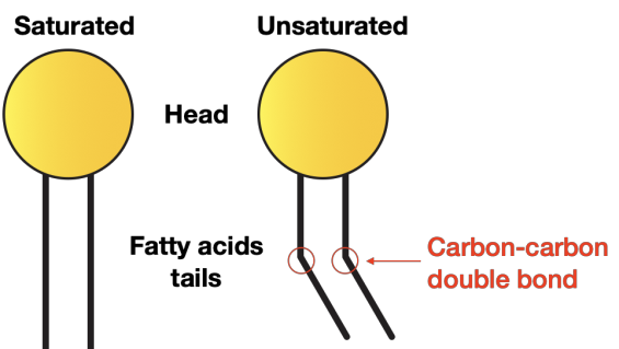
Saturated Fatty Acids: Lack carbon-carbon double bonds ( single bonds only), allowing straight hydrocarbon chains to pack together via van der Waals interactions, reducing fluidity and increasing .
Unsaturated Fatty Acids: Contain one or more cis carbon-carbon double bonds (), introducing rigid kinks in the acyl chains. These kinks prevent tight intermolecular packing, increasing membrane fluidity and lowering .
Cholesterol as a Fluidity Buffer:
Cholesterol is an amphipathic sterol featuring a tiny polar hydroxyl group (), a rigid planar four-ring steroid core, and a short nonpolar hydrocarbon tail. It wedges between phospholipids, with its hydroxyl aligned near the phosphate heads.
At Elevated Temperatures (Liquid Crystal Phase): The rigid steroid ring system interferes with acyl chain movement, reducing membrane permeability and decreasing membrane fluidity.
At Reduced Temperatures (Crystalline Gel Phase): Cholesterol prevents saturated hydrocarbon tails from packing into a rigid lattice, preserving fluidity and preventing membrane freezing.


Classification of Membrane Proteins:
Integral Membrane Proteins (Transmembrane Proteins): Permanently embedded within the lipid bilayer, spanning across both leaflets. Transmembrane segments typically form amphipathic -helices consisting of approximately 20 hydrophobic amino acids whose nonpolar side chains interact with the fatty acyl interior. Examples include nutrient transporters, ion channels, G-protein coupled receptors (GPCRs), and integrins.
Peripheral Membrane Proteins: Do not penetrate the hydrophobic core of the bilayer and lack covalently attached lipids. They associate with membrane surfaces (cytosolic or extracellular) through non-covalent interactions (electrostatic bonds, hydrogen bonds) with polar lipid head groups or with the hydrophilic domains of integral membrane proteins.
Lipid-Anchored Membrane Proteins: Localized outside the lipid bilayer on either surface, but covalently tethered to a lipid molecule embedded within the membrane (e.g., GPI anchors on the extracellular face, or myristoyl/farnesyl anchors on the cytosolic face).

Membrane Transport Mechanisms: Passive Diffusion and Active Transport Systems
Membrane Permeability Hierarchy:
High Permeability: Small, nonpolar, uncharged molecules (, , , ) dissolve directly into the hydrophobic core and pass freely.
Moderate to Low Permeability: Small, uncharged polar molecules (, urea, glycerol) cross at slow, regulated basal rates.
Impermeable: Large, uncharged polar molecules (glucose, sucrose) and all charged inorganic ions (, , , , ), regardless of size, are excluded by the hydrophobic core.
The Four Major Transport Modalities:

1. Simple Diffusion (Non-mediated Passive Transport):
Driven exclusively by random thermal motion down an electrochemical or concentration gradient (downhill flow).
Requires no metabolic energy input ( independent) and involves no binding interaction with transport proteins.
Restricted to small nonpolar solutes such as molecular oxygen () and carbon dioxide ().
2. Diffusion Through a Channel (Non-mediated Protein-Assisted Passive Transport):
Carried out by integral membrane protein complexes composed of multiple subunits spanning the bilayer to form a central, water-filled aqueous pore.
Mediates rapid passage of inorganic ions (, , , ) down their electrochemical gradients.
Selectivity Filters: Channels contain narrow pore regions lined with specific amino acid residues that coordinate ions of a precise atomic radius and hydration shell, excluding other ions.
Gating Mechanisms: Channels alternate between non-conducting (closed) and conducting (open) conformations in response to physiological signals:
Voltage-Gated Channels: Modulate opening/closing based on changes in electrical potential across the plasma membrane (e.g., voltage-gated and channels propagating action potentials in excitable cells).
Ligand-Gated Channels: Open or close in response to non-covalent binding of a specific chemical molecule (ligand) to an extracellular or intracellular receptor domain (e.g., nicotinic acetylcholine receptors at the neuromuscular junction).

3. Facilitated Diffusion (Transporter- / Carrier-Mediated Passive Transport):
Transfers polar molecules (e.g., D-glucose, amino acids) down their concentration gradients without the expenditure of cellular metabolic energy.
Mechanism: Solute binds specifically to an exposed active binding pocket on a facilitative transporter. Binding induces a reversible conformational shift in the carrier protein, exposing the solute to the opposite side of the bilayer where reduced binding affinity promotes dissociation.
Carrier Functional Types:
Uniporter: Translocates a single specific solute across the membrane in one direction (e.g., GLUT family glucose transporters).
Symporter (Cotransporter): Obligatory coupled transport of two distinct solutes in the same direction across the membrane (e.g., symporter utilizing the inward gradient to pull glucose into intestinal epithelial cells).
Antiporter (Exchanger): Obligatory coupled transport of two distinct solutes in opposite directions across the membrane (e.g., antiporter, which couples the downhill entry of into the cell with the active extrusion of into the extracellular fluid to regulate intracellular ).


4. Active Transport (Transporter-Mediated Uphill Transport):
Moves solutes against their steep thermodynamic electrochemical or concentration gradients ("uphill" movement).
Requires direct coupling to metabolic energy input, typically via adenosine triphosphate () hydrolysis.
The Sodium-Potassium ATPase ( Pump):
A foundational primary active transporter present in animal plasma membranes.
Stoichiometry and Catalytic Cycle: For every single molecule of hydrolyzed to and inorganic phosphate (), the pump exports three sodium ions () out of the cell and imports two potassium ions () into the cell.
Physiological Significance: Generates and sustains the resting membrane potential (electrogenic pump), maintains osmotic balance preventing cell swelling, and establishes the steep transmembrane gradient that fuels secondary active transport across tissues.

Extracellular Matrix (ECM), Cell Walls, and Signal Transduction Pathways
Molecular Architecture of the Extracellular Matrix (ECM):
The ECM is an intricate, non-cellular three-dimensional meshwork of self-assembling macromolecules secreted locally by cells into surrounding intercellular spaces.
Functions: Establishes tissue shape, provides physical volume and tensile strength, anchors cells, guides cell migration during morphogenesis, and transmits extracellular cues.
Major Biochemical Components:
Collagens: Fibrous glycoproteins with high tensile strength that resist mechanical stretching forces.
Proteoglycans: Core proteins covalently decorated with glycosaminoglycan (GAG) chains carrying dense negative charges, which attract water to form a hydrated gel that resists compressive forces.
Fibronectin and Laminin: Multi-domain adhesive glycoproteins that crosslink collagens and proteoglycans with cell-surface receptors.

Integrin Anchor Receptors:
Integrins are heterodimeric (composed of non-covalently associated and subunits) transmembrane anchor glycoproteins.
Extracellular Domain: Binds specific sequences (e.g., RGD peptide motif) present on ECM components like fibronectin, laminin, and collagen.
Intracellular Cytoplasmic Domain: Links to the internal cortical actin cytoskeleton via adapter proteins (talin, vinculin, -actinin).
Signaling Role: Integrins function as bi-directional mechanotransducers. Ligand binding at the cell surface triggers intracellular signaling cascades altering gene expression, cell survival, cell motility, and cytoskeletal remodeling.

Cell Walls of Non-Animal Lineages:
Plants, fungi, and bacteria assemble rigid extracellular cell walls that perform protective and load-bearing functions analogous to animal ECM.
Plant Cell Wall Organization:
Cellulose: Linear polymers of -linked D-glucose bundled into rigid microfibrils that provide tensile strength.
Matrix Glycans (Hemicellulose): Branched polysaccharides that crosslink individual cellulose microfibrils into a coherent network.
Pectins: Negatively charged, branched polysaccharides that form a hydrated gel phase filling the interstitial spaces.
Lignin: A complex, hydrophobic polyphenolic polymer deposited in the secondary cell walls of woody tissues to provide rigidity, compressive strength, and waterproof seals.
Middle Lamella: The pectin-rich outermost layer gluing adjoining plant cells together.
Turgor Pressure: The rigid cell wall counteracts hydrostatic osmotic pressure generated by the central vacuole. Loss of vacuolar water decreases turgor pressure, causing soft plant tissues to wilt.

The Principles and Stages of Signal Transduction:
Signal transduction is the biochemical process by which an extracellular signaling molecule (ligand) binds to a cell-surface receptor, converting an external chemical cue into a specific intracellular physiological response without the ligand entering the cytoplasm.
Stage 1: Reception: High-affinity binding of an extracellular ligand (e.g., peptide hormone, growth factor) to the extracellular binding domain of an integral membrane receptor, inducing a conformational change in the receptor's structure.
Stage 2: Transduction: The activated receptor engages intracellular transducers, generating short-lived, small, non-protein intracellular signaling molecules termed second messengers (e.g., cyclic adenosine monophosphate [], calcium ions [], inositol 1,4,5-trisphosphate []) or activating heterotrimeric G-proteins. This initiates an enzymatic phosphorylation cascade.
Stage 3: Cellular Response: Downstream target effector proteins undergo functional activation or repression. Biological endpoints include transcriptional activation, cell division, directional cell motility, cytoskeletal restructuring, altered metabolic pathways, and wound healing.
Model Cascade: Epinephrine-Induced Glycogenolysis:
Epinephrine binds to cell-surface -adrenergic GPCRs in hepatocytes.
The coupled heterotrimeric G-protein () binds , dissociates, and activates the membrane-bound effector enzyme adenylate cyclase.
Activated adenylate cyclase converts into the second messenger .
binds and activates Protein Kinase A (PKA) by releasing its catalytic subunits.
Active PKA utilizes to phosphorylate and activate Phosphorylase Kinase (.
Active Phosphorylase Kinase phosphorylates and activates Glycogen Phosphorylase (.
Active Glycogen Phosphorylase degrades glycogen into glucose-1-phosphate, which is converted to glucose and released into the bloodstream.
If adenylate cyclase is chemically inhibited, synthesis from stops, terminating downstream phosphorylation and halting glucose mobilization.

Cellular Bioenergetics: Respiration, Photosynthesis, and ATP Generation
Energy Transduction and Thermodynamics:
Living cells operate as open non-equilibrium thermodynamic systems requiring continuous energy flux to counteract entropy, synthesize macromolecules, transport solutes uphill, and power mechanical movement.
Animal cells derive chemical free energy exclusively via the oxidation of organic nutrients in mitochondria (cellular respiration).
Plant cells combine mitochondrial aerobic respiration with solar phototrophic energy harvesting via chloroplasts.
Chloroplast Architecture and Photosynthetic Pathways:
Photosynthesis builds high-energy carbohydrates from low-energy inorganic compounds using solar photon energy:
Light-Dependent Reactions: Occur across thylakoid membranes; absorbed photons excite chlorophyll reaction centers, driving photolysis of water () and electron transport to generate a proton gradient powering ATP synthesis, reducing to .
Light-Independent Reactions (Calvin-Benson Cycle): Localized in the aqueous chloroplast stroma. Consumes the and produced by the light reactions to fix carbon dioxide () via the carboxylating enzyme RuBisCO, synthesizing triose phosphate carbohydrates.

Mitochondrial Ultrastructure and Compartmentalization:
Mitochondria typically measure in length, bounded by two distinct membranes that establish two internal fluid spaces:
Outer Mitochondrial Membrane (OMM): Highly permeable to small solutes () due to abundant transmembrane beta-barrel channel proteins called porins.
Intermembrane Space: Chemically contiguous with the cytosol regarding small ions, but sequesters proteins like cytochrome c.
Inner Mitochondrial Membrane (IMM): Highly impermeable barrier enriched in the unique dimeric phospholipid cardiolipin. Folded into invaginations termed cristae to expand surface area for the complexes of oxidative phosphorylation, the electron transport chain (ETC), and synthase.
Mitochondrial Matrix: Dense, gel-like internal compartment containing circular mitochondrial DNA (mtDNA), mitochondrial ribosomes, enzymes for lipid -oxidation, and the complete enzymatic machinery of the tricarboxylic acid (TCA / Krebs) cycle.

The Catabolic Stages of Cellular Respiration:
Total chemical conversion:
1. Glycolysis: Operates in the cytosol. One 6-carbon glucose molecule is broken down into two 3-carbon pyruvate molecules, generating a net yield of (via substrate-level phosphorylation) and . In the absence of oxygen, cells enter anaerobic fermentation, reducing pyruvate to lactate to regenerate .
2. Pyruvate Decarboxylation and the TCA Cycle: In the presence of , pyruvate is transported into the mitochondrial matrix, oxidatively decarboxylated to Acetyl-CoA (), and channeled into the cyclic TCA pathway, yielding , , , and .
ATP Generation Mechanisms:
Substrate-Level Phosphorylation: Direct chemical transfer of a high-energy phosphate group from a phosphorylated metabolic intermediate to . Accounts for only of total cellular .
Oxidative Phosphorylation: Electron carriers (, ) donate electrons to the ETC, generating an electrochemical proton gradient across the IMM that powers synthase. Generates of total cellular .

Oxidative Phosphorylation Mechanism (The Electron Transport Chain):
Complex I (NADH Dehydrogenase): Accepts high-energy electrons from matrix , passes them through iron-sulfur clusters to lipid-soluble Coenzyme Q (Ubiquinone / CoQ), and translocates protons () from the matrix into the intermembrane space.
Complex II (Succinate Dehydrogenase): Catalyzes succinate oxidation in the TCA cycle, transferring electrons from onto CoQ without translocating protons.
Complex III (Cytochrome Complex): Receives electrons from reduced CoQ, passes them onto the mobile peripheral membrane protein Cytochrome c, and pumps protons across the IMM.
Complex IV (Cytochrome c Oxidase): Collects electrons from Cytochrome c and catalyzes the final reduction of molecular oxygen: Molecular oxygen () acts as the obligate terminal electron acceptor. Protons are simultaneously translocated into the intermembrane space.
ATP Synthase ( Complex): Transmembrane proton translocation back down the electrochemical gradient through the channel drives physical rotation of the catalytic headpiece, synthesizing from and .

Programmed Cell Death: The Intrinsic Apoptotic Pathway
Cellular and Morphological Hallmarks of Apoptosis:
Apoptosis is an evolutionarily conserved, genetically encoded mechanism of programmed cell death used to remove redundant, damaged, or infected cells without provoking an inflammatory response.
Distinct from necrotic lysis; features cell shrinkage and cytoplasmic condensation.
Dynamic blebbing and protrusion of the plasma membrane.
Systematic nuclear chromatin condensation and internucleosomal cleavage of nuclear DNA into fragments.
Loss of adhesive contacts with surrounding extracellular matrix and neighbor cells.
Fragmentation of the dying cell into membrane-bound apoptotic bodies display "eat-me" signals (e.g., phosphatidylserine flipped to the outer leaflet), triggering rapid phagocytosis by macrophages or neighboring cells.
Developmental Role: Critical for tissue morphogenesis, such as interdigital cell death sculpting digits during vertebrate limb development. Inhibiting apoptosis during embryogenesis causes interdigital soft tissue to persist, resulting in webbed digits.
The Intrinsic (Mitochondrial) Apoptotic Pathway:
Triggering Stimuli: Activated by internal cellular damage, severe DNA lesions, oxidative stress, hypoxia, or viral infection.
Pro-apoptotic Activation: Intracellular sensors activate pro-apoptotic BH3-only proteins, which neutralize anti-apoptotic regulators and activate the pro-apoptotic pore-forming effector proteins BAX and BAK.
Mitochondrial Outer Membrane Permeabilization (MOMP): Activated BAX and BAK oligomerize in the outer mitochondrial membrane, forming microscopic pores that compromise outer membrane integrity.
Cytochrome c Release: The hemoprotein Cytochrome c, normally sequestered in the intermembrane space where it participates in the respiratory chain, leaks through BAX/BAK pores into the cytosol.
Apoptosome Assembly: Cytosolic Cytochrome c binds the adapter protein Apaf-1 (Apoptotic Protease Activating Factor 1) in the presence of , triggering Apaf-1 heptamerization into a wheel-like complex called the apoptosome.
Initiator Caspase Recruitment: The apoptosome binds and activates the initiator procaspase, procaspase-9, cleaving it into active caspase-9.
Executioner Caspase Cascade: Caspase-9 proteolytically activates downstream executioner caspases (such as caspase-3 and caspase-7), which cleave vital structural proteins, nucleases, and cytoskeletal filaments to dismantle the cell.

The Endomembrane System and Protein Targeting Pathways
Architecture of the Endomembrane System:
A coordinated dynamic continuum of internal membrane-bound compartments that collaborate to translate, modify, sort, package, and transport lipids and proteins.
Includes the nuclear envelope, rough and smooth endoplasmic reticulum, Golgi complex, endosomes, lysosomes, transport vesicles, and the plasma membrane.

Endoplasmic Reticulum Functional Divergence:
Rough Endoplasmic Reticulum (RER): Characterized by flattened membranous cisternae studded on their cytosolic face with ribosomes. Dedicated to:
Cotranslational translocation of secretory, resident endomembrane, and transmembrane proteins.
Synthesis of membrane phospholipids.
N-linked core glycosylation of nascent polypeptides.
Chaperone-mediated protein folding and quality control (utilizing luminal chaperones like BiP and protein disulfide isomerase). Misfolded proteins that fail quality control are retained and targeted to the proteasome for degradation.
Smooth Endoplasmic Reticulum (SER): Irregular tubular networks devoid of ribosomes. Dedicated to:
De novo lipid and fatty acid synthesis.
Biosynthesis of steroid hormones from cholesterol (prominent in endocrine tissues like the adrenal cortex and gonads).
Metabolic detoxification of lipophilic drugs and organic toxins (via cytochrome P450 monooxygenases in hepatocytes).
Sequestration and regulated release of calcium ions (, abundant in the sarcoplasmic reticulum of myocytes).

Global Overview of Protein Synthesis and Sorting:
Transcription and Splicing: Pre-mRNA is transcribed from genomic DNA in the nucleus, processed with a 7-methylguanosine cap, spliced by spliceosomes to remove introns and ligate exons, and terminated with a poly(A) tail.
Nuclear Export: Mature mRNA exits the nucleus through nuclear pore complexes into the cytoplasm.
Translational Initiation: Translation of all nuclear-encoded proteins begins on free cytosolic ribosomes.
Bifurcation of Fate: Translation continues to completion on free ribosomes unless the nascent peptide displays a specific ER-targeting signal sequence directing it to the RER membrane.

Proteins Translated Completely on Free Ribosomes (Post-Translational Targeting):
Cytosolic Proteins: Lack targeting sequences; remain in the cytosol upon release (e.g., glycolytic enzymes, tubulin, actin).
Peripheral Membrane Proteins: Associate with the cytosolic face of cellular membranes post-translationally.
Nuclear Proteins: Possess a Nuclear Localization Signal (NLS) and are imported through nuclear pores in a fully folded state.
Mitochondrial and Chloroplast Proteins: Possess basic amino-terminal targeting presequences; imported post-translationally in an unfolded conformation mediated by cytosolic chaperones (Hsp70) and outer-membrane translocases (e.g., the mitochondrial TOM complex [Translocase of the Outer Membrane], composed of receptor units Tom20, Tom22 and channel core Tom40, working with Tom5, Tom6, and Tom7).
Peroxisomal Proteins: Synthesized on free ribosomes and imported post-translationally via carboxy-terminal or amino-terminal peroxisomal targeting signals (PTS).


Cotranslational Translocation into the Rough ER:
The Signal Sequence: Located at the amino-terminus () of the nascent polypeptide. Encompasses a central stretch of 6 to 15 hydrophobic amino acids () flanked by a positively charged amino-terminal region () and a polar carboxy-terminal region () containing a specific recognition sequence for cleavage by signal peptidase (typically at positions and relative to the cleavage site).
Step-by-Step Translocation Mechanism:
SRP Binding: As the hydrophobic signal sequence emerges from the ribosome exit tunnel, it is bound by the Signal Recognition Particle (SRP), a cytosolic ribonucleoprotein complex.
Translational Arrest: SRP binding halts further translation, preventing premature folding of the nascent chain in the cytosol.
Receptor Docking: The ribosome-SRP-nascent chain complex diffuses to the RER membrane, where SRP binds to the membrane-bound SRP receptor in a GTP-dependent interaction.
Translocon Engagement: SRP dissociates (hydrolyzing GTP), handing the ribosome over to an aqueous protein translocase pore termed the Sec61 translocon. The ribosome docks onto the translocon, displacing an internal plug domain, and translation resumes.
Pore Insertion and Signal Cleavage: The nascent polypeptide loop threads through the translocon channel into the ER lumen. A membrane-bound signal peptidase cleaves off the amino-terminal signal peptide.
Folding in Lumen: The luminal chaperone BiP binds the entering chain, preventing aggregation and driving directional entry into the ER lumen where it is folded and modified.


Insertion of Transmembrane Integral Proteins:
Transmembrane proteins contain hydrophobic signal-anchor sequences or stop-transfer sequences (segments of hydrophobic amino acids).
When a stop-transfer sequence enters the translocon, translocation ceases; the translocon opens laterally, discharging the hydrophobic segment into the fatty acyl core of the bilayer where it is anchored as an -helix.
Single-Pass Proteins: Contain a single stop-transfer or signal-anchor segment, leaving one terminal domain in the ER lumen and the other in the cytosol.
Multi-Pass Proteins: Possess alternating start-transfer and stop-transfer sequences that weave back and forth across the lipid bilayer (e.g., seven-transmembrane GPCRs, multi-subunit ion channels).

Vesicular Trafficking: Budding, Coat Systems, and Membrane Fusion
Vectorial Transport and Directionality:
Transport vesicles ( diameter) bud from a donor compartment membrane and fuse with a specific recipient acceptor compartment.
Anterograde Transport (Outward / Forward): Directional movement flowing from the ER to the cis-Golgi network, across the Golgi cisternae to the trans-Golgi network (TGN), and out to the plasma membrane or lysosomes.
Retrograde Transport (Inward / Retrieval): Backward flow from the trans-Golgi to medial/cis-cisternae, or from the cis-Golgi network back to the ER to retrieve escaped resident proteins (e.g., BiP carrying a carboxy-terminal KDEL retrieval sequence).
Golgi Architecture and Cisternal Maturation:
The Golgi apparatus comprises polarized flattened, membrane-bound cisternae arranged proximal to distal:
cis-Golgi Network (CGN): Entry sorting station that receives vesicular tubular clusters from the ER; sorts whether proteins progress forward or recycle back to the ER.
cis-, medial-, and trans-Cisternae: Successive functional sub-compartments containing unique resident glycosyltransferases and modifying enzymes, catalyzing stepwise modification of N-linked glycans and assembly of O-linked oligosaccharides.
trans-Golgi Network (TGN): Exit sorting station that segregates cargo into distinct transport vesicles targeted for constitutive secretion, regulated secretion, or endosomes/lysosomes.
Cisternal Maturation Model: Golgi cisternae are dynamic structures that physically mature over time—new cis-cisternae form via fusion of ER-derived vesicles, progress physically forward to become medial- and trans-cisternae, and disperse at the TGN. Resident Golgi enzymes are continuously retrieved from later to earlier cisternae via retrograde COPI vesicles.

Coated Vesicle Systems:
Vesicle budding is initiated by cytoplasmic protein coats that perform two functions: mechanically curving the donor membrane into an emerging bud and selectively capturing cargo molecules.
COPII Coated Vesicles:
Pathway: Mediates anterograde transport from the ER to the cis-Golgi network.
Assembly: Initiated by the small GTPase Sar1. Sar1-GDP exchanges GDP for GTP via a membrane GEF, extending an amphipathic helix into the cytosolic leaflet of the ER membrane. Activated Sar1 recruits the inner coat heterodimer Sec23/Sec24 (Sec24 directly binds sorting signals on cargo receptors). The inner coat then recruits the outer structural cage heterodimer Sec13/Sec31, driving membrane curvature and bud scission.
COPI Coated Vesicles:
Pathway: Mediates retrograde transport from the cis-Golgi back to the rough ER, and retrograde recycling between Golgi cisternae.
Clathrin-Coated Vesicles:
Pathway: Transports cargo from the trans-Golgi network outward to late endosomes/lysosomes, and from the plasma membrane inward during receptor-mediated endocytosis.
Structure: Composed of clathrin triskelions (three heavy chains and three light chains) assembling into a polyhedral cage, linked to cargo receptors via adaptor protein complexes (e.g., AP-1, AP-2).

Tethering, Docking, and SNARE-Mediated Fusion:
Upon scission from the donor membrane, vesicles shed their protein coats, exposing integral fusion proteins.
v-SNAREs (Vesicle SNAREs): Specifically incorporated into the budding vesicle membrane (e.g., synaptobrevin / VAMP).
t-SNAREs (Target SNAREs): Localized in the cytosolic leaflet of the target membrane, typically consisting of two or three proteins (e.g., syntaxin and SNAP-25).
Fusion Mechanics: The cytosolic helical domains of a v-SNARE and cognate t-SNAREs wind around one another to form an extremely stable, parallel four-helix bundle termed the SNARE complex. The mechanical force generated by this trans-SNARE pairing pulls the opposing lipid bilayers into close proximity, displacing intervening water and driving hemifusion and complete bilayer fusion.

Exocytic and Endocytic Pathways:
Constitutive Secretory Pathway: Continuous, unregulated default pathway operating in all eukaryotic cells. Vesicles continuously bud from the TGN and fuse with the plasma membrane, replenishing membrane lipids/proteins and secreting soluble ECM components (e.g., collagen).
Regulated Secretory Pathway: Restricted to specialized secretory cells (endocrine, exocrine, neurons). Cargo is concentrated and stored in dense-core secretory granules/vesicles in the cytosol. Exocytosis is triggered by a specific extracellular stimulus (e.g., hormone, neurotransmitter) that causes a transient spike in cytosolic free calcium ().
Endocytosis: Inward budding of the plasma membrane to internalize extracellular fluid and cell-surface receptors:
Receptor-Mediated Endocytosis: Internalization of specific ligands via clathrin-coated pits into early endosomes, which mature into late endosomes and fuse with lysosomes.
Phagocytosis: Specialized internalization mechanism employed by phagocytes (macrophages, neutrophils). Dynamic actin polymerizations project pseudopods around large foreign particles or microbes, enclosing them in a large vesicle called a phagosome.
Lysosomal Degradation, Autophagy, and Vacuolar Compartmentalization
Lysosomal Ultrastructure and Physiology:
Lysosomes are membrane-enclosed digestive organelles ranging from to in diameter.
Acid Hydrolase Suite: Contain approximately 50 distinct hydrolytic enzymes (proteases, nucleases, glycosidases, lipases, phosphatases, sulfatases) that degrade biological macromolecules into monomeric subunits (amino acids, nucleotides, monosaccharides, free fatty acids) for metabolic recycling.
Acidic pH Requirement: Lysosomal acid hydrolases operate at an optimal acidic of . At neutral cytosolic (), these enzymes display negligible activity, protecting the host cell from catastrophic self-digestion in the event of accidental lysosomal leakage.
The Vacuolar -ATPase (Proton Pump): An electrogenic ATP-driven pump embedded within the lysosomal membrane that hydrolyzes cytosolic to pump protons () against their concentration gradient into the lysosomal lumen, maintaining its low internal .
Consequence of -ATPase Inhibition: If the proton pump is chemically inhibited, luminal concentration decreases ( rises), and acid hydrolases become less efficient or completely inactivated, halting macromolecular degradation.
Membrane Glycosylation Shield: The internal luminal face of the lysosomal membrane is lined with integral proteins bearing dense, branched oligosaccharide chains that form a protective barrier against internal acid hydrolases.

Three Major Degradation Pathways Converging on the Lysosome:
1. The Endocytic Pathway: Extracellular macromolecules and activated cell-surface receptors are internalized via endocytic vesicles, delivered to early endosomes, sorted through multivesicular bodies to late endosomes, and delivered to lysosomes via vesicle fusion.
2. The Autophagic Pathway (Macroautophagy / Organelle Turnover):
Constitutes an evolutionarily conserved recycling mechanism for degrading obsolete, damaged, or excess intracellular organelles (e.g., depolarized mitochondria) and cytosolic aggregates.
Phase 1 (Initiation): Formation of an isolation membrane (phagophore) derived from the endoplasmic reticulum.
Phase 2 (Vesicle Elongation): The phagophore membrane curves around the targeted organelle or cytosolic cargo.
Phase 3 (Maturation): Membrane ends fuse to form a closed, double-membrane vesicle called an autophagosome.
Phase 4 (Docking and Fusion): The outer membrane of the autophagosome fuses with a lysosome, forming an autolysosome.
Phase 5 (Breakdown and Degradation): Lysosomal acid hydrolases degrade the inner membrane and the engulfed cargo. The resulting degradation products are exported into the cytosol via membrane transporters for reuse.
3. The Phagocytic Pathway (Pathogen Clearance):
Phagocytes extend pseudopods to engulf antibody-opsonized microbes, sealing them in an intracellular phagosome.
The phagosome fuses with primary lysosomes to generate a phagolysosome.
Acid hydrolases kill and enzymatically digest the pathogen. Non-digestible microbial debris is expelled via exocytosis.


The Plant Central Vacuole:
A large, fluid-filled, single-membrane-bound organelle that occupies up to of the total volume of mature plant cells.
Tonoplast: The vacuolar bounding membrane containing active transport systems and aquaporins.
Enzymatic Degradation: Functions as the plant equivalent of the animal lysosome, maintaining an acidic internal () and housing acid hydrolases.
Turgor Pressure Generation: Maintains high internal solute concentrations (organic acids, sugars, inorganic ions), driving osmotic water influx that presses the cytoplasm against the cell wall, generating hydrostatic turgor pressure that provides mechanical support to non-woody plant structures.
Storage and Sequestration: Stores metabolic intermediates (malate for sequestration), amino acids, hydrolytic enzymes, anthocyanin pigments, and toxic secondary metabolites for herbivore defense.
The Cytoskeleton: Microtubules, Microfilaments, and Intermediate Filaments
Comparative Cytoskeletal Architecture:
The eukaryotic cytoskeleton is a dynamic network of interconnected protein polymers responsible for cell shape, structural integrity, cytoplasmic organization, chromosome segregation, and cell motility.
Divided into three primary filament systems based on subunit composition, diameter, and mechanical properties:


1. Microtubules (MTs):
Dimensions: The thickest cytoskeletal elements, forming hollow, unbranched cylinders with an outer diameter of approximately .
Subunit Composition: Assembled from stable heterodimers of -tubulin and -tubulin non-covalently linked together. Both subunits bind GTP.
Protofilament Lattice: Typically composed of 13 parallel linear protofilaments arranged in a cylinder surrounding a central hollow core (lumen).
Structural Polarity: Protofilaments align with the same orientation, creating a polar filament:
Plus (+) End: Terminates with exposed -tubulin subunits; fast-growing, highly dynamic.
Minus (-) End: Terminates with exposed -tubulin subunits; slow-growing, typically capped and stabilized at the MTOC.
Dynamic Instability: Individual microtubules switch stochastically between phases of sustained polymerization (growth) and rapid depolymerization (catastrophe), governed by whether the rate of GTP-tubulin addition at the plus end exceeds the rate of GTP hydrolysis to GDP.
Microtubule-Organizing Centers (MTOCs):
The central nucleation site for microtubule assembly in vivo.
Centrosome: The primary MTOC in non-mitotic animal cells, positioned adjacent to the nucleus. Contains a pair of orthogonal centrioles surrounded by a pericentriolar material (PCM) rich in -tubulin ring complexes (-TuRCs) that nucleate microtubules with their minus ends anchored at the centrosome and plus ends projecting toward the cell periphery.
Basal Bodies: MTOCs positioned at the cell cortex that nucleate the axoneme of cilia and flagella.
Spindle Poles: Formed during mitosis by duplicate centrosomes to generate the bipolar mitotic spindle.
Axonemal vs. Cytoplasmic Microtubules:
Cytoplasmic MTs: Dynamic, transient arrays involved in directional vesicle trafficking, organelle positioning, and mitotic chromosome movement.
Axonemal MTs: Highly stable, permanent, organized structures found inside cilia and eukaryotic flagella, arranged in an evolutionary conserved axonemal architecture (nine doublet microtubules surrounding a central central singlet pair) linked by nexin and outer/inner dynein arms.
Microtubule-Associated Proteins (MAPs): Structural proteins that bind microtubules to regulate stability and spacing. For example, Tau is a specialized non-motor MAP abundant in neurons that bundles and stabilizes parallel axonal microtubules; hyperphosphorylation of Tau disrupts the microtubule network and is implicated in neurodegenerative diseases.

2. Microfilaments (Actin Filaments):
Dimensions: The thinnest cytoskeletal elements, forming flexible, two-stranded helical polymers with a diameter of approximately .
Subunit Assembly: Monomeric globular actin (G-actin) binds and polymerizes in a head-to-tail orientation to form filamentous actin (F-actin).
Structural Polarity: Features a fast-growing plus (+) end (barbed end) and a slower-growing minus (-) end (pointed end).
Treadmilling Kinetics: G-actin with bound adds to the plus end. Following incorporation, actin hydrolyzes bound to and inorganic phosphate. -actin subunits at the minus end have lower affinity for neighbors and depolymerize. At steady state, the rate of subunit addition at the plus end matches the rate of subunit loss at the minus end, producing a steady flux of subunits along the filament known as treadmilling.
Capping Proteins: Specialized proteins that bind to filament ends. Binding of a capping protein to the plus end stops subunit addition and loss at that end, halting treadmilling and promoting filament stability.
Functions: Driving cell locomotion (via lamellipodia and filopodia), establishing cell surface shape (microvilli), generating contractile forces (muscle contraction, cytokinesis contractile rings), and serving as tracks for short-distance vesicle transport.
3. Intermediate Filaments (IFs):
Dimensions: Tough, rope-like fibers with an intermediate average diameter of approximately .
Subunit Architecture: Built from elongated fibrous proteins rather than globular enzymes. A monomer features a central -helical rod domain flanked by non-helical globular head and tail domains.
Assembly Hierarchy:
Two monomers align in parallel to form a coiled-coil dimer.
Two dimers associate in an antiparallel, staggered orientation to form a non-polar tetramer.
Eight tetramers associate laterally to form a bundle, which twists into a rope-like filament ( thick).
Lack of Structural Polarity and Absence of Motors: Because tetramers assemble from antiparallel dimers, the opposite ends of intermediate filaments are structurally identical. Consequently, intermediate filaments lack polarity and cannot support motor proteins.
Mechanical Integrity and Types: Provide high tensile strength, dissipate mechanical shear stresses, and anchor internal organelles:
Keratins: In epithelial cells; anchor desmosomes and hemidesmosomes. Mutations weaken keratin networks, leading to skin blistering under mechanical stress.
Vimentin and Desmin: In mesenchymal and muscle tissues.
Neurofilaments: In axons, establishing and maintaining axonal caliber.
Nuclear Lamins (Lamins A, B, C): Form a supporting meshwork on the inner nuclear membrane.
Molecular Motor Proteins and Intracellular Motility
Mechanochemical Motor Energetics:
Cytoskeletal motor proteins convert chemical free energy from hydrolysis into mechanical work along cytoskeletal tracks.
Motors move processively by coupling ATP binding, hydrolysis, and product release to cycles of conformational changes in their catalytic motor head domains.
Microtubule-Based Motors: Kinesins and Dyneins:
Kinesins:
Directionality: Primarily plus-end directed motors mediating anterograde transport (transporting cargo outward from the MTOC near the cell center toward the plasma membrane).
Structure: Heterotetramers comprising two heavy chains (containing catalytic motor heads with ATP- and tubulin-binding sites, joined by a coiled-coil stalk) and two light chains (mediating cargo binding).
Autoinhibition: In the absence of cargo, kinesin folds into a compact, inactive conformation. Binding of cargo to the tail relieves this autoinhibition, allowing the motor to bind microtubules and processively walk.
Cytoplasmic Dynein:
Directionality: Exclusively a minus-end directed motor mediating retrograde transport (transporting cargo inward from the cell periphery toward the MTOC/nucleus).
Cargo Specialization: Transports endocytic vesicles, late endosomes, lysosomes, misfolded protein aggregates, and worn-out organelles toward the cell center.
Mitotic Function: Generates sliding forces between antiparallel microtubules to drive chromosome segregation during mitosis.
Cooperative Organelle Transport in Melanophores:
Zebrafish pigment cells (melanophores) use cytoskeletal motors to adapt coloration: Dynein pulls pigment granules (melanosomes) inward toward minus ends at the centrosome (causing skin lightening), whereas Kinesin transports melanosomes outward toward plus ends at the cell periphery (causing skin darkening).
Cooperation with Actin: Kinesin transports melanosomes along microtubules outward to the cell cortex, then hands the cargo over to the actin-based motor Myosin Va for short-range distribution along cortical actin filaments.


Actin-Based Motors: The Myosin Superfamily:
Motors that walk along actin microfilaments, with most moving toward the plus (barbed) end.
Conventional Myosins (Myosin II): Bipolar multi-subunit complexes with two heavy chains and four light chains. Assemble into thick bipolar filaments in muscle and non-muscle cells, sliding actin filaments to drive muscle contraction, cytokinesis, and cell motility.
Unconventional Myosins (Myosins I, III through XVIII): Primarily monomeric or dimeric motors involved in short-range vesicle trafficking, endocytosis, and organelle transport along actin tracks.
The Nucleus, Nuclear Pore Complexes, and Ribosome Biogenesis
The Nuclear Envelope:
The nucleus houses the eukaryotic genome and compartmentalizes gene transcription away from cytoplasmic translation.
Dual-Membrane System: Composed of two parallel phospholipid bilayers separated by a perinuclear lumen ():
Outer Nuclear Membrane (ONM): Continuous with the rough endoplasmic reticulum membrane and studded with active ribosomes on its cytosolic surface.
Inner Nuclear Membrane (INM): Encloses the nucleoplasm, containing integral membrane proteins that anchor the underlying nuclear lamina and chromatin.
The Nuclear Lamina: A fibrous meshwork ( thick) lining the inner surface of the INM. Composed of intermediate filament proteins (lamins A, B, and C). Provides structural support to the nuclear envelope, maintains nuclear shape, and acts as an anchoring scaffold for heterochromatin.

The Nuclear Pore Complex (NPC):
Large supramolecular channels (molecular mass in vertebrates) embedded at sites where the inner and outer nuclear membranes fuse.
Mammalian nuclei contain between individual NPCs across the envelope.
Architectural Features:
Displays an octagonal rotational symmetry.
Cytoplasmic Filaments: Eight protein filaments projecting into the cytosol to capture incoming cargo.
Scaffold Nucleoporins (Y-Complex): Structural framework anchoring the complex into the curved membrane pore.
Nuclear Basket: Eight flexible protein struts extending into the nucleoplasm that converge on a terminal ring.
Central FG-Nucleoporins: Channel interior lined with unstructured nucleoporin domains containing repetitive hydrophobic Phenylalanine-Glycine () peptide motifs. These motifs form an entropic hydrogel mesh that limits diffusion.
Nucleocytoplasmic Transport Kinetics:
Passive Diffusion: Small molecules, metabolites, and proteins with a molecular mass of or less diffuse passively through the hydrogel meshwork rapidly (rates up to .
Facilitated Active Transport: Macromolecules exceeding the cutoff cannot freely enter and require active, receptor-mediated transport (rates of .
Mechanisms of Regulated Nuclear Import:
Nuclear cargo proteins carry a Nuclear Localization Signal (NLS), which consists of one or two short clusters of basic, positively charged amino acids (typically Lysine and Arginine; e.g., .
The NLS is bound by soluble nuclear transport receptors called importins in the cytoplasm.
The importin-cargo complex interacts with the hydrophobic -repeats, transiently melting through the central channel into the nucleoplasm.
In the nucleoplasm, the small GTPase Ran bound to binds importin, triggering cargo dissociation.

The Nucleolus and Ribosome Biogenesis:
The nucleolus is a dense, non-membrane-bound subnuclear compartment assembled around the Nucleolar Organizer Regions (NORs) of chromosomes encoding ribosomal RNA () genes.
Primary site of ribosomal subunit assembly in eukaryotic cells.
Four Successive Steps of Ribosome Biogenesis:
Transcription of Pre-rRNA: RNA Polymerase I transcribes long tandem arrays of genes into a single large primary pre-rRNA transcript ( in mammals).
rRNA Processing: The pre-rRNA transcript is chemically modified (pseudouridylation, 2'-O-methylation) by small nucleolar ribonucleoproteins (snoRNPs) and endonucleolytically cleaved into mature , , and rRNAs. (The rRNA is transcribed independently by RNA Polymerase III outside the nucleolus).
Subunit Assembly: Ribosomal proteins synthesized in the cytoplasm are imported through NPCs into the nucleolus. These proteins complex with the processed rRNAs to form nascent (small) and (large) pre-ribosomal subunits.
Subunit Export and Functional Maturation: The assembled and ribosomal subunits are exported through nuclear pore complexes into the cytoplasm. Once in the cytoplasm, they remain separate until translation initiates, coming together to form a functional ribosome on an mRNA molecule.