Brain Organization, Functional Dissociation, and Stroke
Phrenology (1808)
focused on measurements of the human skull
based on the concept that the brain is the organ of the mind
certain brain areas have localized, specific functions or modules
brain composed of 21 personality organs with a specific mental function, each found at a specific location in the cerebral convolutions of the brain
conclusions based on “extremes of society” such as criminals or clergyman
Wilder Penfield (1951/54)
treated epilepsy patients by destroying nerve cells in the brain where the seizures originated
stimulated the brain with electric probes while patients were conscious and observed their responses
accurately targeted areas of the brain responsible, reducing side effects of the surgery
technique allowed for creation of sensory and motor homunculus
Brain Anatomy (Cortex)
Frontal Lobe
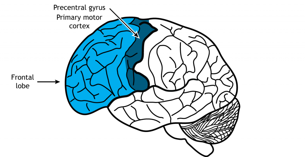
located in the front of the brain, the most rostral
posterior border is the central sulcus
largest of the four lobes
responsible for higher level executive functions
attention, critical thinking, impulse control
influences personality
last brain region to fully develop
location of the primary motor cortex
responsible for planning and executing movement
specifically located in the precentral gyrus
Broca’s Area
sections 44 and 45
speech production
articulation, language comprehension, grammar, sound sequencing, semantics
Parietal Lobe
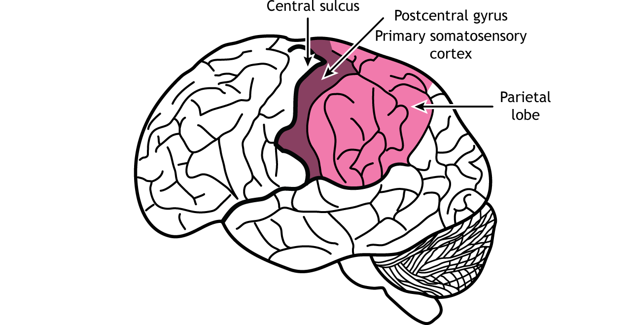
divided from frontal lobe by central sulcus
sense different tactile properties of things in the world around us with our body
proprioception: the ability to identify where parts of your body are located
higher level visual processing
sensory cortex
interprets sensry input like touch, temperature, pain, vision, sound, taste, and smell
primary somatosensory cortex
in postcentral gyrus
sections 1, 2, 3
responsible for the perception of touch and pain
represented by sensory homunculus
neural spatial representation of sensors for body parts, disproportionate
Temporal Lobes
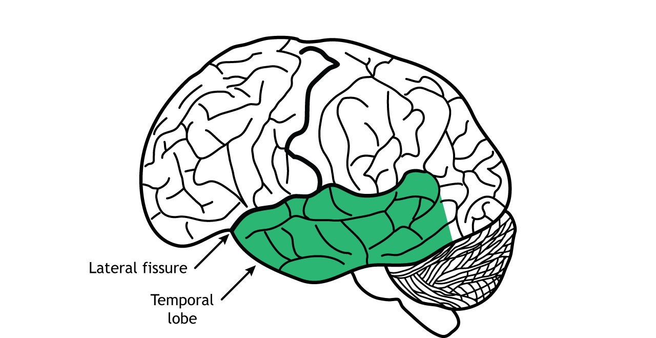
side of the brain, separated from frontal and parietal by lateral fissue
sensory processing (hearing, smell, taste and higher level visual processing)
hippocampus
buried medially and ventrally in temporal lobe
ability to remember important facts
memory-related processes
primary auditory cortex
section 41: initial processing of sound, detecting frequencies
section 42: basic processing of auditory information (pitch rhythm and volume
Wernicke’s Area
comprehension and interpretation of speech and complex sounds
Occipital Lobes
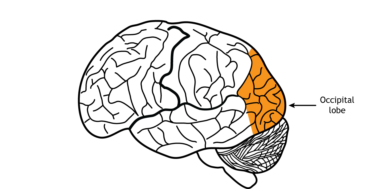
back of the brain
processing of visual stimuli (light, objects in motion, object orientation, color)
primary visual cortex
section 17
receives raw visual data from the retina
initial processing of visual input
visual association cortex
section 18
interpretation and recognition of visual images
complex processing
Anatomical Planes
frontal/coronal plane
vertical plane
runs parallel to eyes or ears
divides into front and back regions
sagittal plane
vertical plane
runs perpendicular to the eyes or ears
divides into left and right regions
horizontal plane
runs parallel to the ground
divide into top and bottom regions
Nervous System
divided into CNS and PNS
CNS includes brain and spinal cord
PNS includes cranial and spinal nerves
info travelling towards the brain is afferent
info travelling from the brain is efferent
multiple sclerosis affects afferent and efferent
Sensory Afferent Pathways
sensory info can be external or internal (skin, gut, heart, etc.)
dorsal column lemniscal pathways
touch, pressure
spinothalamic pathways
pain, temperature
Motor Efferent Pathways
carries from CNS (primary motor cortex) down into brainstem then to muscles/glands
Vascular Innervation
brain requires oxygen and nutrients to function
accomplished by blood pumped around ithe body using a network of blood vessels (circulatory system)
brain has a high demand for oxygen and nutrients
2% of total body weight but receives 15% of total cardiac output
Blood Supply to Brain
from two major arteries
common carotid arteries
major blood vessels in the neck that supply oxygenated blood to the head, neck, and brain
branches into internal and external carotid arteries
anterior cerebral artery (ACA)
supplies the medial surface of the frontal and parietal lobes, the frontobasal cortex, most of the corpus callosum
middle cerebral artery (MCA)
supplies the largest area, including the lateral surfaces of the frontal, parietal, and temporal lobes, as well as the insular cortex
supplies blood to broca’s area, when blocked can cause broca’s aphasia (difficulty saying words)
supplies to wernicke’s area, when blocked can cause wernicke’s aphasia (difficulty understanding language)
deep branches supply the basal ganglia and internal capsule
lenticulostriate arteries supply basal ganglia (caudate nucleus, etc. ) and internal capsule
common site for small strokes from bloackage, cause motor deficits
anterior choroidal artery
supplies parts of the hippocampus, amygdala, posterior limb of the internal capsule and the thalamus
ophthalmic artery
supplies the structures of the eye/orbit
vertebral arteries
provide 20-30% of the blood supply to the brain
supply the brainstem, cerebellum, and posterior cerebral hemispheres
posterior inferior cerebellar artery
supplies the inferior cerebellum and parts of the brainstem
basilar artery
formed by the joining of vertebral arteries, supplies the pons and cerebellum
superior cerebellar artery
supplies the superior cerebellum and parts of the midbrain
posterior cerebral arteries
supply the occipital lobes (visual cortex) and the inferior part of the temporal lobes
particular blood flow to specific regions
blockage can cause specific deficits
Stroke
the 3d leading cause of death in canada (after cancer and heart disease)
caused by disruption of blood flow to part of the brain
deprives neurons and other brain cells of glucose and oxygen leading to cell death
results in impaired or lost function
hemorrhagic stroke = hemorrhage/blood leaks into brain tissue
20% of strokes
caused by uncontrolled bleeding in the brain
weakened/diseased blood vessels rupture
subarachnoid hemorrhage = uncontrolled on the surface of the brain in the area between the brain and the skull (direct impact to head)
intracerebral hemorrhage = when an artery deep within the brain ruptures
aneurysm = a weakened area in the blood vessel wall of the blood vessel that fills with blood and bulges
high blood pressure or trauma can cause the bulge to rupture, resulting in uncontrolled bleeding into the brain
ischemic stroke
80% of strokes
interruption of blood flow due to a blood clot
plaque (fatty materials, calcium, and scar tissue) contributes to ost ischemic strokes by narrowing the arteries that supply blood to the brain (atherosclerosis sclerosis)
thrombotic strokes = blood clot forms in an artery directly leading to the brain
embolic strokes = clot develops somewhere else in the body and travels to the brain
core is where there are dying neurons (necrosis) and quick cell death
cells have no energy, loss of oxygen and glucose
blood flow below 10-25%
penumbra is surrounding core
blood flow is ~25-50%
cells may be viable for hours, reversible damage
can eventually die by aptosis - recognize low oxygen and glucose levels and start cell destruction pathways
can be brought back with the return of blood flow
transient ischemic attack = mini stroke
caused by temporary interruption of blood flow to the brain
many people can have a TIA without even knowing it
can put a person at an increased risk of a full blown stroke, vascular dementia or neurodegeneration
clot breaks off and travels to smaller arteries
watershed areas = areas highly susceptible to stroke due to low perfusion pressure
stroke kills neurons, neurons are responsible for behaviour
decreased blood flow causes metabolic dysfunction (no oxygen, glucose, ATP), disrupts neural activity in area
Neuropathology of a Stroke
reduced cerebral blood flow (can be ischemic (thrombotic or embolic) or hemorrhagic) causes ATP depletion (lack of oxygen and glucose)
ATP depletion leads to:
neurochemical injury
lactic acidosis
edema
oxidative stress
neuroinflammation injury
reactive astrocytes
activated microglia
neutrophil infiltration
neurochemical injury and neuroinflammation injury lead to cell damage and neuronal death
Pathophysiology of Stroke
“neural shock”
areas distant from the damage are functionally depressed (penumbra)
both pathogenic and self-protective mechanisms occur in penumbra
ischemic penumbra is functionally compromised but potentially “salvageable”
areas related to the damaged region suffer sudden withdrawal of excitation or inhibition (core)
irreversible bran damage in ischemic core
may be followed by changes in the metabolism of the injured hemisphere, glucose utilization, or both, which may persist for days
Ischemic Cascade
neurons and glial cells (astrocytes, oligodendrocytes) lose their ability to produce energy in the absence of glucose due to interrupted blood flow
astrocytes bring glucose to neurons
when astrocytes react, blood brain barrier breaks down
in ischemic stroke: reduced blood flow to brain —> reduced glucose to astrocytes —> glucose cannot go to neurons —> cannot make ATP —> cell death