Study Notes on the Appendicular Skeleton (Chapter 8)
INTRODUCTION
Chapter 8: The Appendicular Skeleton
General Overview
The skeleton provides an internal support structure for the body, protects vital organs, and serves as a framework for muscle attachment and movement.
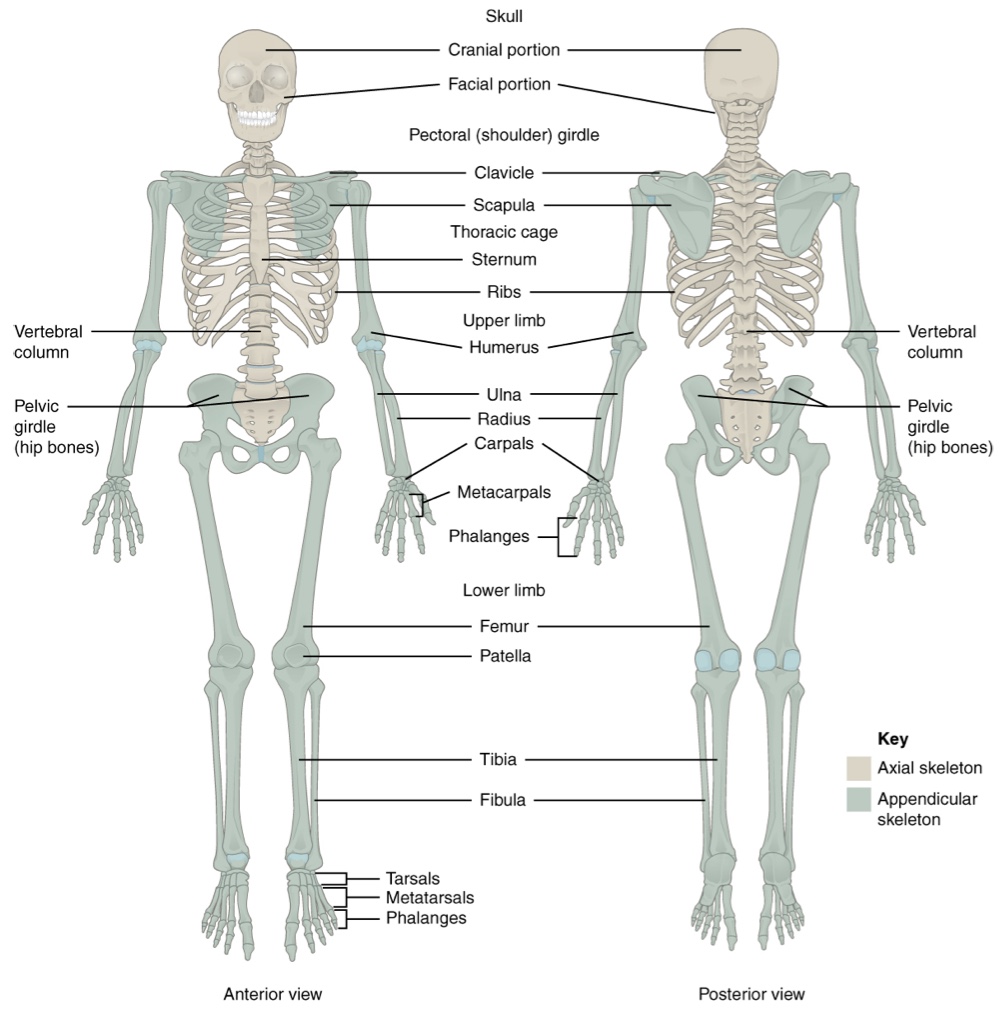
The adult axial skeleton has 80 bones forming the central axis of the body, including the head (skull), neck (vertebrae), and trunk (rib cage and vertebral column).
The appendicular skeleton contains 126 bones, representing the limbs and the girdles that attach them to the axial skeleton. These bones are divided into two main groups:
Bones within the upper and lower limbs themselves.
Girdle bones that attach these limbs to the axial skeleton.
The shoulder region forms the pectoral girdle, which anchors the upper limb to the thoracic cage, providing extensive mobility.
The lower limb is connected to the vertebral column by the strong, weight-bearing pelvic girdle.
Functional Adaptations
Upper limbs are highly mobile, allowing diverse activities such such as reaching, grasping, manipulation of objects, and complex fine motor skills.
Lower limbs are adapted for weight-bearing, stability, and efficient locomotion (walking, running), providing strong support for the body.
8.1 THE PECTORAL GIRDLE
Appendicular Skeleton Breakdown
The appendicular skeleton includes:
The bones of the limbs (humerus, radius, ulna, femur, tibia, fibula).
The bones of the hands (carpals, metacarpals, phalanges) and feet (tarsals, metatarsals, phalanges).
The girdle bones which connect the limbs to the axial skeleton: the pectoral girdle (clavicle and scapula) and the pelvic girdle (hip bones).
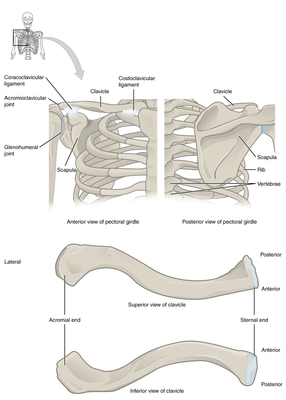
Components of the Pectoral Girdle
Formed by:
The scapula (shoulder blade), a large, flat, triangular bone located posteriorly on the thorax.
Supported by the clavicle and articulates with the humerus to form the shoulder joint.
The clavicle (collarbone)—an S-shaped bone that provides the only bony attachment of the upper limb to the axial skeleton.
Clavicle Details: The medial (sternal) end articulates with the manubrium of the sternum at the sternoclavicular joint, which is a synovial saddle joint that allows significant movement. The lateral (acromial) end articulates with the acromion of the scapula, forming the acromioclavicular joint, located just above the shoulder joint.
Functions of the clavicle:
Serves as a strut or brace, extending laterally to support the scapula and keep the shoulder joint away from the trunk, allowing for a wide range of arm motion.
Transmits physical forces from the upper limb to the sternum and axial skeleton.
Protects the underlying nerves and blood vessels (brachial plexus and subclavian vessels) that pass to the upper limb.
Clavicle Structure
Regions of Clavicle:
Sternal end (medial end): triangular in shape; articulates with the manubrium of the sternum via articular facet for sternum.
This forms the sternoclavicular joint, which is the only bony articulation between the pectoral girdle of the upper limb and the axial skeleton.
This joint allows considerable mobility, enabling movement in upward/downward and anterior/posterior directions during shoulder movements.
It is indirectly supported by the costoclavicular ligament.
Acromial end: flatter; articulates with the acromion of the scapula via articular facet for acromion. It features the conoid tubercle inferiorly for attachment of the conoid ligament (part of coracoclavicular ligament).
Shaft: the long, curved portion between the two ends. It has a rough inferior surface with the subclavian groove and impression for the costoclavicular ligament.
Sex Differences in Clavicle:
Females: typically shorter, thinner, and less curved. The muscle attachment surfaces are generally less prominent.
Males: typically heavier, longer, exhibit greater curvature, and have rougher muscle attachment surfaces due to stronger muscle development, features that are more pronounced in manual workers.
Fascinating Facts about the Clavicle
Most commonly fractured bone in the body, accounting for about 5% of all adult fractures.
Fractures are most commonly caused by direct falls onto the shoulder, a direct impact on its lateral aspect, or falls onto outstretched arms. Because the sternoclavicular joint is strong and rarely dislocated, excessive force typically results in a break, usually occurring in the middle third of the shaft (or between the middle and lateral portions).
Symptoms often include sagging shoulders, severe pain, and swelling. Patients typically support the injured limb with the uninjured hand. In a complete fracture, the lateral clavicle fragment and shoulder will drop due to the upper limb's weight, and muscles acting across the shoulder can pull fragments anteriorly and medially, leading to overriding. Fortunately, despite overlying important blood vessels and nerves, these structures are rarely affected, often due to the anterior displacement of the broken clavicle.
Scapula Overview:
Located posteriorly on the posterior side of the shoulder, overlying ribs 2-7.
It is part of the pectoral girdle, playing an important role in anchoring the upper limb to the body.
Surrounded by muscles on both anterior (deep) and posterior (superficial) sides, it does not articulate with the ribs of the thoracic cage.
Provides a broad surface for muscle attachment and serves as a mobile base for the humerus.
Functions with the clavicle to form the pectoral girdle, enabling complex movements of the shoulder joint.
Scapula Anatomy
Borders of Scapula: It has three distinct margins or borders, named for their positions within the body:
Superior border: the shortest and sharpest border, featuring the scapular notch (suprascapular notch) located lateral to its midpoint for the suprascapular nerve.
Medial (vertebral) border: runs parallel to the vertebral column.
Lateral (axillary) border: thicker, pointing towards the armpit.
Angles: The triangular scapula has three angles:
Superior angle: formed by the junction of the superior and medial borders.
Inferior angle: formed by the junction of the medial and lateral borders, serving as the most inferior portion and an important landmark for muscle attachment (for powerful muscles involved in shoulder and upper limb movements) and movement assessment.
Lateral angle: composed of the glenoid cavity, where the humerus articulates, located between the superior and lateral borders.
Glenoid cavity (fossa): A shallow, pear-shaped depression located at the lateral angle. It articulates with the head of the humerus to form the glenohumeral (shoulder) joint.
The shallow nature of the glenoid cavity allows for great mobility but sacrifices stability.
Immediately above and below the glenoid cavity are the small bony bumps: the supraglenoid tubercle and the infraglenoid tubercle, respectively, which provide attachments for muscles of the arm.
Prominent Projections of Scapula
Coracoid Process: Shaped like a crow's beak (coracoid = "shaped like a crow's beak"); this prominent, hook-like anterior projection extends superolaterally from the lateral angle.
This process projects anteriorly and curves laterally, and is located inferior to the lateral end of the clavicle.
It serves as an attachment site for several muscles (e.g., pectoralis minor, coracobrachialis, muscles of the anterior chest and arm) and is anchored to the clavicle by a strong ligament, and other ligaments (e.g., coracoclavicular, coracoacromial).
Spine of Scapula: A long and prominent bony ridge that projects posteriorly from the dorsal surface of the scapula across its upper portion.
It divides the posterior surface into the supraspinous and infraspinous fossae.
Acromion or Acromial Process: This flattened, expanded lateral end of the scapular spine arches anteriorly over the shoulder joint.
It forms the bony tip of the superior shoulder region and articulates with the clavicle, forming the acromioclavicular joint, and serves as an attachment point for the deltoid and trapezius muscles.
Together, the clavicle, acromion, and spine of the scapula form a V-shaped bony line that provides for the attachment of neck and back muscles that act on the shoulder, as well as muscles that pass across the shoulder joint to act on the arm.
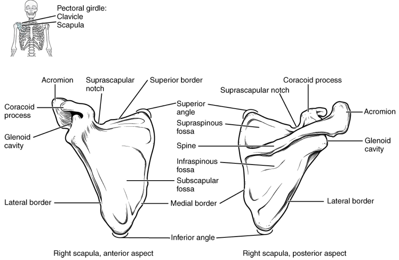
Important Depressions and Fossae of Scapula
Fossae: These depressions provide extensive surface area for muscle attachment for muscles that cross the shoulder joint to act on the humerus:
Supraspinous fossa: the narrow depression located superior to the scapular spine, accommodating the supraspinatus muscle.
Infraspinous fossa: the broad depression located inferior to the scapular spine, accommodating the infraspinatus muscle.
Subscapular fossa: the broad, shallow depression on the anterior (costal) surface of the scapula, accommodating the subscapularis muscle.
Acromioclavicular Joint
This joint is crucial for transmitting forces from the upper limb to the clavicle and axial skeleton.
The ligaments that directly surround this joint are relatively weak. A hard fall onto the elbow or an outstretched hand can stretch or tear the acromioclavicular ligaments, resulting in a moderate injury to the joint.
However, the primary support for the acromioclavicular joint comes from a very strong ligament called the coracoclavicular ligament. This connective tissue band anchors the coracoid process of the scapula to the inferior surface of the acromial end of the clavicle and thus provides important indirect support for the acromioclavicular joint.
Following a strong blow to the lateral shoulder (e.g., common in contact sports like hockey, football, or martial arts), a complete dislocation of the acromioclavicular joint can result. In this case, the acromion is thrust under the acromial end of the clavicle, leading to ruptures of both the acromioclavicular and coracoclavicular ligaments.
This dislocation injury is known as a "shoulder separation," where the scapula separates from the clavicle, and the weight of the upper limb pulls the shoulder downward.
8.2 Bones of the Upper Limb
The upper limb is divided into three regions:
Arm: located between the shoulder and elbow joints.
Forearm: located between the elbow and wrist joints; contains the ulna (medially) and the radius (laterally).
Hand: located distal to the wrist; contains eight carpal bones, five metacarpal bones (forming the palm), and 14 phalanges (finger and thumb bones).
There are 30 bones in each upper limb.
Humerus
The humerus is the single bone of the upper arm region.
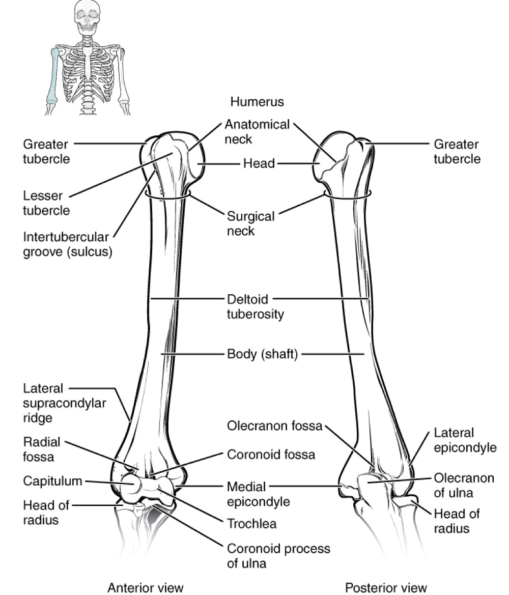
Proximal End features:
Head of the humerus: large, round, smooth region facing medially, articulates with the glenoid cavity of the scapula to form the glenohumeral (shoulder) joint.
Anatomical neck: the margin of the smooth area of the head.
Greater tubercle: an expanded bony area on the lateral side of the proximal humerus.
Lesser tubercle: smaller tubercle found on the anterior aspect of the humerus.
Both tubercles serve as attachment sites for muscles that act across the shoulder joint.
Intertubercular groove (sulcus) (also known as the bicipital groove): a narrow groove between the greater and lesser tubercles; provides passage for a tendon of the biceps brachii muscle.
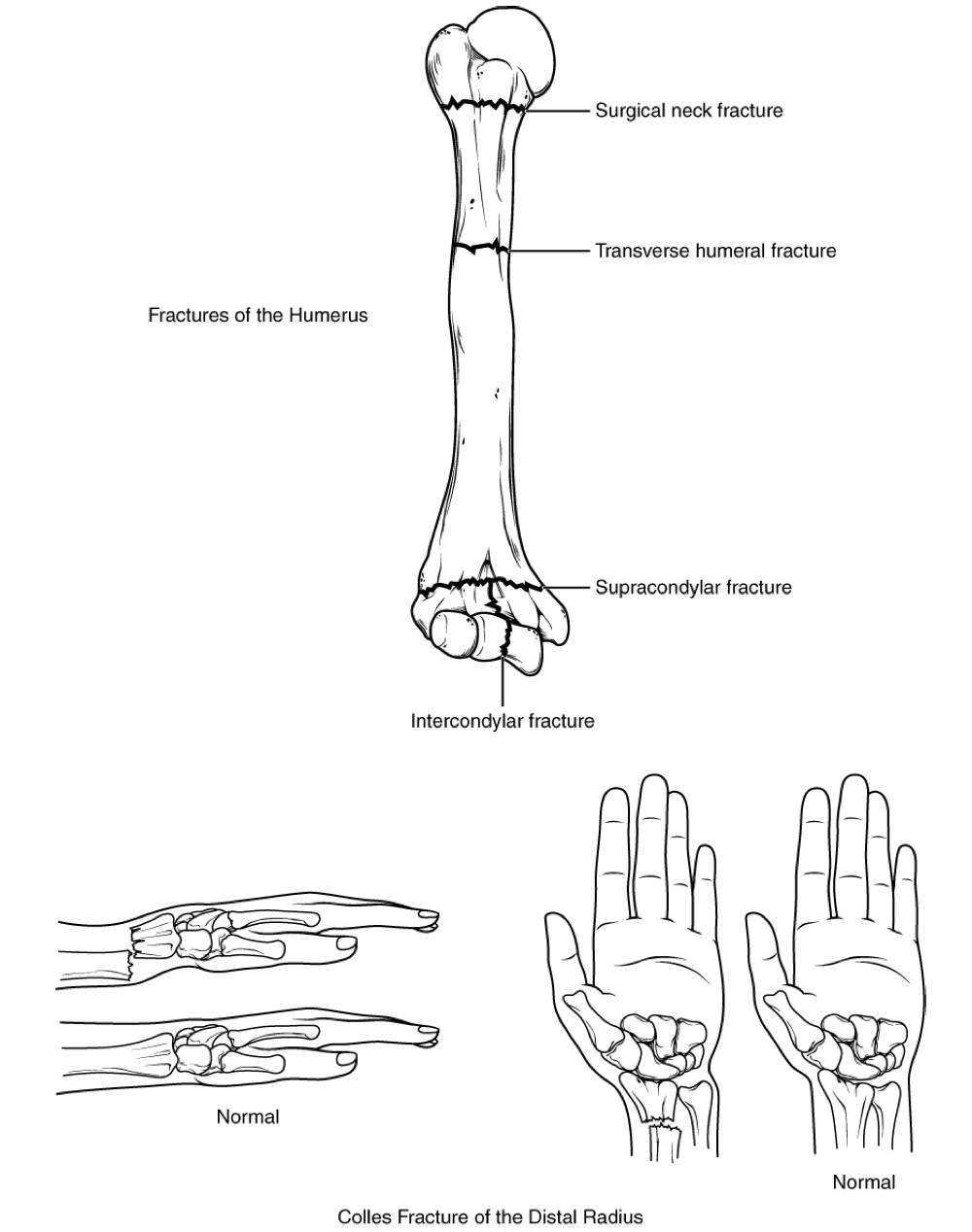
Surgical neck: located at the base of the expanded, proximal end of the humerus, where it joins the narrow shaft of the humerus; a common site of arm fractures.
Deltoid tuberosity: a roughened, V-shaped region on the lateral side in the middle of the humerus shaft; site of attachment for the deltoid muscle.
Distal End features (flattened):
Medial epicondyle: prominent bony projection on the medial side.
Lateral epicondyle: smaller projection on the lateral side.
Lateral supracondylar ridge: roughened ridge of bone above the lateral epicondyle.
These areas serve as attachment points for muscles that act on the forearm, wrist, and hand
The distal end has two articulation areas that join the ulna and radius to form the elbow joint:
Trochlea: a spindle- or pulley-shaped region (trochlea = "pulley"), more medial; articulates with the ulna.
Capitulum: a knob-like structure (capitulum = "small head") located immediately lateral to the trochlea on the anterior surface; articulates with the radius.
Small depressions above these bony areas accommodate forearm bones when the elbow is fully bent (flexed):
Coronoid fossa: superior to the trochlea, receives the coronoid process of the ulna.
Radial fossa: superior to the capitulum, receives the head of the radius.
Olecranon fossa: a larger depression on the posterior humerus, receives the olecranon process of the ulna when the forearm is fully extended.
Ulna
The ulna is the medial bone of the forearm, running parallel to the radius (lateral bone).
Proximal end of the ulna features:
Trochlear notch: a large, C-shaped notch resembling a crescent wrench, articulates with the trochlea of the humerus as part of the elbow joint.
Coronoid process of the ulna: a prominent lip of bone forming the inferior margin of the trochlear notch.
Ulnar tuberosity: a roughened area on the anterior ulna, just below the coronoid process.
Radial notch of the ulna: a small, smooth area located lateral and slightly inferior to the trochlear notch, serving as the articulation site between the proximal radius and the ulna, forming the proximal radioulnar joint.
Olecranon process: the posterior and superior portions of the proximal ulna, forming the bony tip of the elbow.
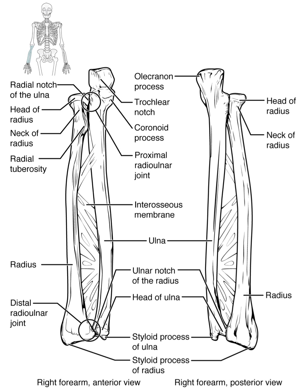
Distal end of the ulna features:
Line of attachment for the interosseous membrane of the forearm, a sheet of dense connective tissue that unites the ulna and radius bones.
Small, rounded area forms the head of the ulna.
Projecting from the posterior side of the ulnar head is the styloid process of the ulna, a short bony projection which serves as an attachment point for a connective tissue structure that unites the distal ends of the ulna and radius.
In the anatomical position, with the elbow fully extended and the palms facing forward, the arm and forearm do not form a straight line. Instead, the forearm deviates laterally by 5−155−15 degrees from the line of the arm. This deviation is called the carrying angle. It allows the forearm and hand to swing freely or to carry an object without hitting the hip. The carrying angle is larger in females to accommodate their wider pelvis.
Radius
The radius runs parallel to the ulna, on the lateral (thumb) side of the forearm.
Proximal end of the radius features:
Head of the radius: a disc-shaped structure that forms the proximal end. The small depression on its surface articulates with the capitulum of the humerus as part of the elbow joint, whereas the smooth, outer margin articulates with the radial notch of the ulna at the proximal radioulnar joint.
Neck of the radius: the narrowed region immediately below the expanded head.
Radial tuberosity: an oval-shaped, bony protuberance on the medial side, serving as a muscle attachment point.
Shaft of the radius:
Slightly curved and has a small ridge along its medial side.
This ridge forms the interosseous border of the radius, which, like the similar border of the ulna, is the line of attachment for the interosseous membrane that unites the two forearm bones.
Distal end of the radius features:
Smooth surface for articulation with two carpal bones to form the radiocarpal joint or wrist joint.
On the medial side of the distal radius is the ulnar notch of the radius. This shallow depression articulates with the head of the ulna, which together form the distal radioulnar joint.
The lateral end of the radius has a pointed projection called the styloid process of the radius. This provides attachment for ligaments that support the lateral side of the wrist joint. Compared to the styloid process of the ulna, the styloid process of the radius projects more distally, thereby limiting the range of movement for lateral deviations of the hand at the wrist joint.
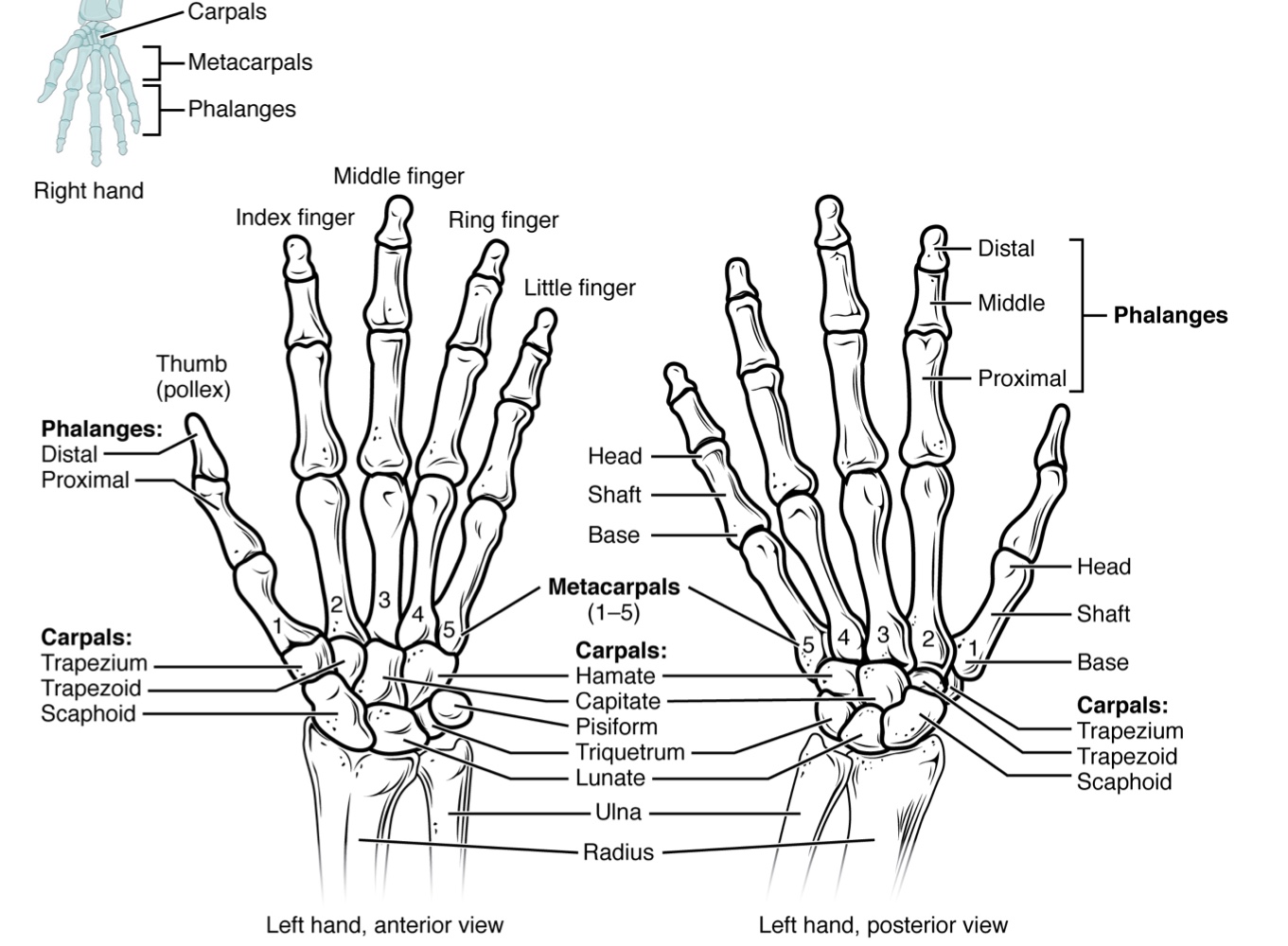
Carpal Bones
The wrist and base of the hand are formed by a series of eight small carpal bones arranged in two rows: a proximal row of four carpal bones and a distal row of four carpal bones.
The bones in the proximal row, running from the lateral (thumb) side to the medial side, are:
scaphoid ("boat-shaped")
lunate ("moon-shaped")
triquetrum ("three-cornered")
pisiform ("pea-shaped") bones.
The small, rounded pisiform bone articulates with the anterior surface of the triquetrum bone. The pisiform thus projects anteriorly, where it forms the bony bump that can be felt at the medial base of your hand.
The distal bones (lateral to medial) are the trapezium ("table"), trapezoid ("resembles a table"), capitate ("head-shaped"), and hamate ("hooked bone") bones.
The hamate bone is characterized by a prominent bony extension on its anterior side called the hook of the hamate bone.
A helpful mnemonic for remembering the arrangement of the carpal bones is "So Long To Pinky, Here Comes The Thumb."
This mnemonic starts on the lateral side and names the proximal bones from lateral to medial (scaphoid, lunate, triquetrum, pisiform),
then makes a U-turn to name the distal bones from medial to lateral (hamate, capitate, trapezoid, trapezium). Thus, it starts and finishes on the lateral side.
The carpal bones form the base of the hand. The four proximal carpal bones are united to each other by ligaments to form a unit.
Only three of these, the scaphoid, lunate, and triquetrum, contribute to the radiocarpal joint.
The scaphoid and lunate bones articulate directly with the distal end of the radius.
The triquetrum bone articulates with a fibrocartilaginous pad that spans the radius and styloid process of the ulna.
The distal end of the ulna thus does not directly articulate with any of the carpal bones.
The four distal carpal bones are also held together as a group by ligaments.
The proximal and distal rows of carpal bones articulate with each other to form the midcarpal joint.
Together, the radiocarpal and midcarpal joints are responsible for all movements of the hand at the wrist.
The distal carpal bones also articulate with the metacarpal bones of the hand.
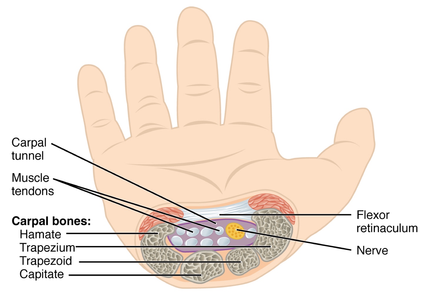
In the articulated hand, the carpal bones form a U-shaped grouping.
A strong ligament called the flexor retinaculum spans the top of this U-shaped area to maintain this grouping of the carpal bones.
Flexor retinaculum details:
Attached laterally to the trapezium and scaphoid bones.
Attached medially to the hamate and pisiform bones.
Together, the carpal bones (forming walls and floor) and the flexor retinaculum (forming the roof) create a passageway called the carpal tunnel.
The carpal tunnel is a narrow tunnel through which tendons of nine muscles of the anterior forearm and an important nerve pass to enter the hand.
Overuse of muscle tendons or wrist injury can cause inflammation and swelling within this space.
This compression of the nerve results in carpal tunnel syndrome, characterized by pain or numbness, and muscle weakness in the areas of the hand supplied by this nerve.
Metacarpal Bones
The palm of the hand contains five elongated metacarpal bones.
These bones lie between the carpal bones of the wrist and the bones of the fingers and thumb.
Each metacarpal bone has a proximal end articulating with one of the distal carpal bones, forming a carpometacarpal joint.
The expanded distal end of each metacarpal bone articulates at the metacarpophalangeal joint with the proximalphalanx bone of the thumb or one of the fingers.
The distal end also forms the knuckles of the hand, at the base of the fingers. The metacarpal bones are numbered 1−51−5, beginning at the thumb.
The first metacarpal bone, at the base of the thumb, is separated from the other metacarpal bones. This allows it a freedom of motion that is independent of the other metacarpal bones, which is very important for thumb mobility.
The remaining metacarpal bones are united together to form the palm of the hand. The second and third metacarpal bones are firmly anchored in place and are immobile. However, the fourth and fifth metacarpal bones have limited anterior-posterior mobility, a motion that is greater for the fifth bone. This mobility is important during power gripping with the hand.
Phalanx Bones
The fingers and thumb contain 14 bones, each of which is called a phalanx bone (plural = phalanges), named after the ancient Greek phalanx (a rectangular block of soldiers).
The thumb (pollex) is digit number 11 and has two phalanges, a proximal phalanx, and a distal phalanx bone.
Digits 22 (index finger) through 55 (little finger) have three phalanges each, called the proximal, middle, and distal phalanx bones.
An interphalangeal joint is one of the articulations between adjacent phalanges of the digits
8.3 The Pelvic Girdle & Pelvis
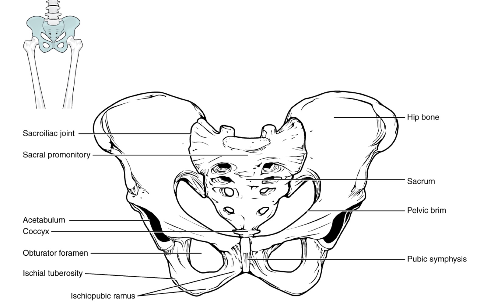
The pelvic girdle (hip girdle) is formed by a single bone, the hip bone or coxal bone (coxal = "hip"), which serves as the attachment point for each lower limb.
Each hip bone is firmly joined to the axial skeleton via its attachment to the sacrum of the vertebral column.
The right and left hip bones also converge anteriorly to attach to each other.
The bony pelvis is the entire structure formed by:
The two hip bones.
The sacrum.
The coccyx (attached inferiorly to the sacrum).
Unlike the pectoral girdle, the pelvis is a largely immobile, weight-bearing structure.
This stability is crucial for transferring body weight laterally from the vertebral column, through the pelvic girdle and hip joints, and into the lower limbs when one limb is not bearing weight.
The immobility of the pelvis provides a strong foundation for the upper body, resting atop the mobile lower limbs.
Hip Bone
The hip bone, or coxal bone, forms the pelvic girdle portion of the pelvis.
The paired hip bones are large, curved bones that form the lateral and anterior aspects of the pelvis.
Each adult hip bone is formed by 3 separate bones that fuse together during the late teenage years.
These bony components are the ilium, ischium, and pubis
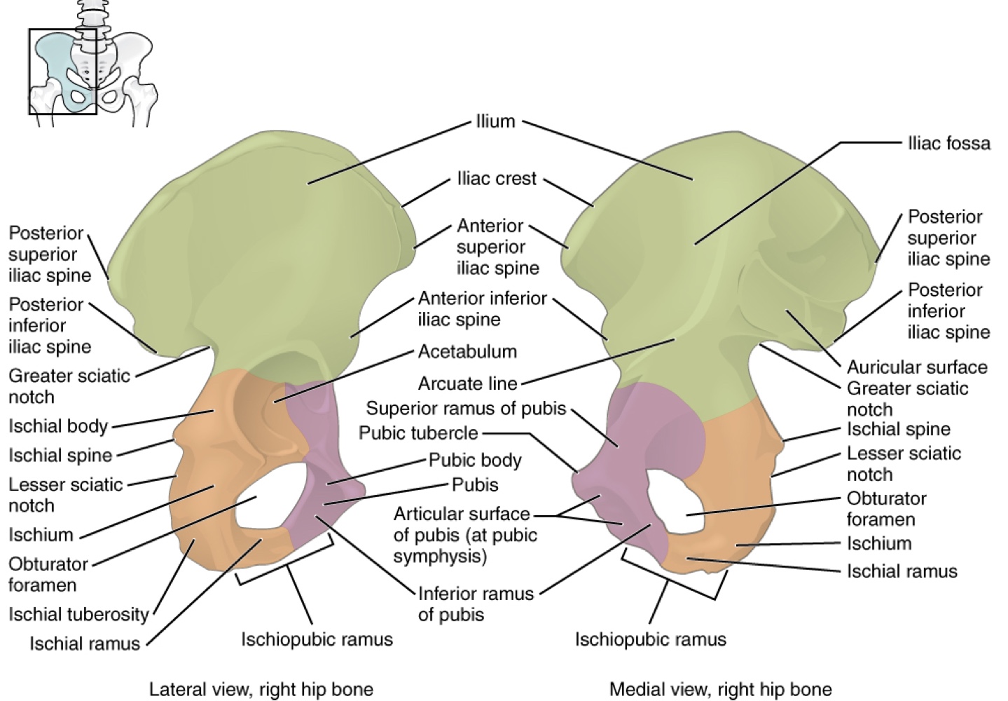
Ilium
the fan-like, superior region that forms the largest part of the hip bone. It is firmly united to the sacrum at the largely immobile sacroiliac joint.
When you place your hands on your waist, you can feel the arching, superior margin of the ilium along your waistline, which is called the iliac crest.
Anterior termination of the iliac crest: the anterior superior iliac spine (important bony landmark felt at anterolateral hip).
Inferior to the anterior superior iliac spine: the anterior inferior iliac spine (rounded protuberance).
These iliac spines serve as attachment points for muscles of the thigh.
Posteriorly, the iliac crest curves downward to terminate as the posterior superior iliac spine.
Muscles and ligaments surround but do not cover this bony landmark, sometimes producing a depression seen as a "dimple" located on the lower back.
More inferiorly: the posterior inferior iliac spine.
This is located at the inferior end of a large, roughened area called the auricular surface of the ilium.
The auricular surface articulates with the auricular surface of the sacrum to form the sacroiliac joint.
Both the posterior superior and posterior inferior iliac spines serve as attachment points for muscles and very strong ligaments that support the sacroiliac joint.
The shallow depression located on the anteromedial (internal) surface of the upper ilium is called the iliac fossa.
The inferior margin of this space is formed by the arcuate line of the ilium, a ridge formed by the pronounced change in curvature between the upper and lower portions of the ilium.
The large, inverted U-shaped indentation located on the posterior margin of the lower ilium is called the greater sciatic notch.
Ischium
The ischium forms the posterolateral portion of the hip bone. It supports the body when sitting.
The large, roughened area of the inferior ischium is the ischial tuberosity.
This serves as the attachment for the posterior thigh muscles and also carries the weight of the body when sitting.
You can feel the ischial tuberosity if you wiggle your pelvis against the seat of a chair.
Projecting superiorly and anteriorly from the ischial tuberosity is a narrow segment of bone called the ischial ramus.
The slightly curved posterior margin of the ischium above the ischial tuberosity is the lesser sciatic notch.
The bony projection separating the lesser sciatic notch and greater sciatic notch is the ischial spine.
Pubis
The pubis forms the anterior portion of the hip bone.
The enlarged medial portion of the pubis is the pubic body.
Located superiorly on the pubic body is a small bump called the pubic tubercle.
The narrow ridge running along the superior margin of the superior pubic ramus is the pectineal line of the pubis.
The pubic body is joined to the pubic body of the opposite hip bone by the pubic symphysis.
Extending downward and laterally from the body is the inferior pubic ramus.
The pubic arch is the bony structure formed by the pubic symphysis, and the bodies and inferior pubic rami of the adjacent pubic bones.
The inferior pubic ramus extends downward to join the ischial ramus.
Together, these form the single ischiopubic ramus, which extends from the pubic body to the ischial tuberosity.
The inverted V-shape formed as the ischiopubic rami from both sides come together at the pubic symphysis is called the subpubic angle.
Pelvis
The pelvis consists of four bones: the right and left hip bones, the sacrum, and the coccyx.
Its primary role is to support the weight of the upper body when sitting and to transfer this weight to the lower limbs when standing.
It serves as an attachment point for trunk and lower limb muscles, and also protects the internal pelvic organs.
When standing in the anatomical position, the pelvis is tilted anteriorly. In this position, the anterior superior iliac spines and the pubic tubercles lie in the same vertical plane, and the anterior (internal) surface of the sacrum faces forward and downward.
The three areas of each hip bone, the ilium, pubis, and ischium, converge centrally to form a deep, cup-shaped cavity called the acetabulum.
The large opening in the anteroinferior hip bone between the ischium and pubis is the obturator foramen. This space is largely filled in by a layer of connective tissue and serves for the attachment of muscles on both its internal and external surfaces.
Several ligaments unite the bones of the pelvis. The largely immobile sacroiliac joint is supported by a pair of strong ligaments:
anterior sacroiliac ligament on the anterior side of the joint.
posterior sacroiliac ligament on the posterior side.
Also spanning the sacrum and hip bone are two additional ligaments:
The sacrospinous ligament runs from the sacrum to the ischial spine.
The sacrotuberous ligament runs from the sacrum to the iscial spine, and the sacrotuberous ligament runs from the sacrum to the ischial tuberosity.
These ligaments help to support and immobilize the sacrum as it carries the weight of the body
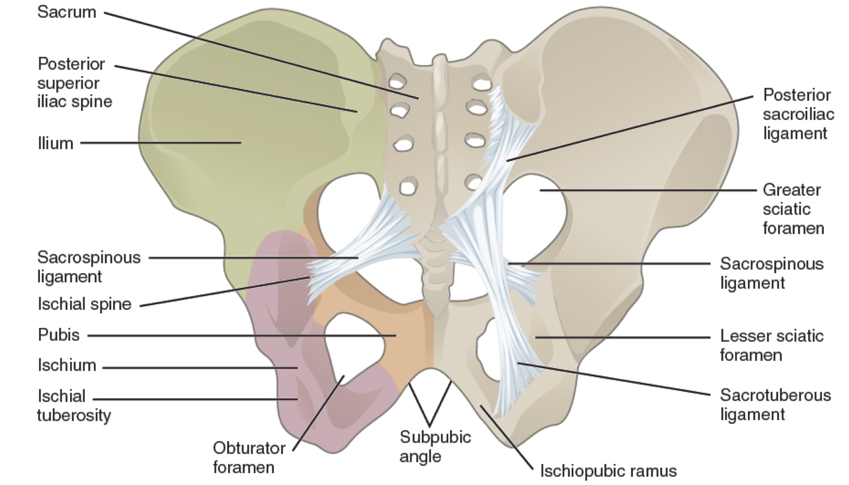
The sacrospinous and sacrotuberous ligaments also help to define two openings on the posterolateral sides of the pelvis through which muscles, nerves, and blood vessels for the lower limb exit:Greater sciatic foramen: The superior opening, formed by the greater sciatic notch of the hip bone, the sacrum, and the sacrospinous ligament.
Lesser sciatic foramen: The smaller, more inferior opening, formed by the lesser sciatic notch of the hip bone together with the sacrospinous and sacrotuberous ligaments.
Divisions of the Bony Pelvis
The space enclosed by the bony pelvis is divided into two regions:
Greater pelvis (greater pelvic cavity; false pelvis): Broad, superior region, defined laterally by the large, fan-like portion of the upper hip bone.
This area is occupied by portions of the small and large intestines. It is considered "false" because it is more closely associated with the abdominal cavity.
Lesser pelvis (lesser pelvic cavity; true pelvis): The narrow, rounded space inferior to the greater pelvis.
It contains the bladder and other pelvic organs and is also known as the true pelvis. Its superior margin is defined by the pelvic brim (pelvic inlet) and its inferior limit is defined by the pelvic outlet.
Pelvic brim (pelvic inlet): Forms the superior margin of the lesser pelvis. It is a line formed by the upper margin of the pubic symphysis anteriorly, the pectineal line of the pubis, the arcuate line of the ilium, and the sacral promontory (the anterior margin of the superior sacrum) posteriorly.
Pelvic outlet: Defines the inferior limit of the lesser pelvis. It is formed by the inferior margin of the pubic symphysis anteriorly, the ischiopubic ramus, the ischial tuberosity, the sacrotuberous ligament, and the inferior tip of the coccyx posteriorly.
Due to the anterior tilt of the pelvis, the lesser pelvis is also angled, giving it an anterosuperior (pelvic inlet) to posteroinferior (pelvic outlet) orientation.
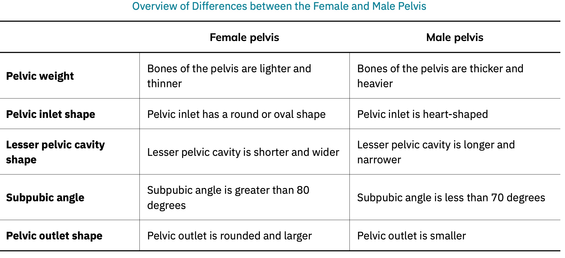
Comparison of the Female and Male Pelvis
The differences between the adult female and male pelvis relate to function and body size. In general, the bones of the male pelvis are thicker and heavier, adapted for support of the male's heavier physical build and stronger muscles. The female pelvis is adapted for childbirth
8.4 Bones of the Lower Limb
Like the upper limb, the lower limb is divided into three regions:
Thigh: portion between the hip joint and knee joint.
Leg: specifically the region between the knee joint and the ankle joint.
Foot: distal to the ankle.
The lower limb contains 3030 bones, including the femur, patella, tibia, fibula, tarsal bones, metatarsal bones, and phalanges.
The femur is the single bone of the thigh.
The patella is the kneecap and articulates with the distal femur.
The tibia is the larger, weight-bearing bone located on the medial side of the leg.
The fibula is the thin bone of the lateral leg.
The bones of the foot are divided into three groups:
Posterior portion: formed by a group of seven tarsal bones.
Mid-foot: contains five elongated metatarsal bones.
Toes: contain 1414 small bones, each a phalanx bone of the foot.
Femur
The femur, or thigh bone, is the single bone of the thigh region.
It is the longest and strongest bone of the body, accounting for approximately one-quarter of a person's total height.
The rounded, proximal end is the head of the femur, which articulates with the acetabulum of the hip bone to form the hip joint.
The fovea capitis is a minor indentation on the medial side of the femoral head.
It serves as the site of attachment for the ligament of the head of the femur.
This ligament spans the femur and acetabulum but is weak and provides little support for the hip joint.
It does, however, carry an important artery that supplies the head of the femur.
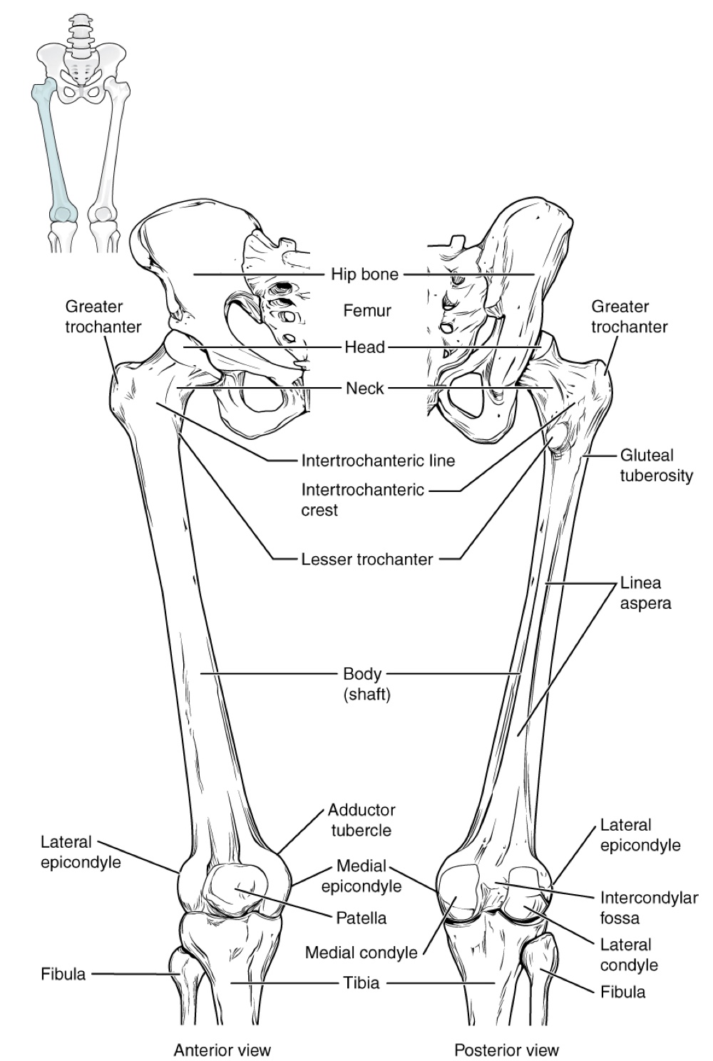
Neck of the femur: The narrowed region below the head, a common area for fractures.
Greater trochanter: A large, upward, bony projection above the base of the neck.
Serves as an attachment site for multiple hip joint muscles, providing additional leverage due to its projection.
Can be felt under the skin on the lateral side of the upper thigh.
Lesser trochanter: A small, bony prominence on the medial aspect of the femur, just below the neck.
Attachment site for a single, powerful muscle.
Intertrochanteric line: A roughened line on the anterior side of the femur, connecting the greater and lesser trochanters.
Intertrochanteric crest: A larger structure on the posterior side of the femur, connecting the trochanters.
Shaft of the femur: Elongated with a slight anterior bowing or curvature.
Gluteal tuberosity: A roughened area on the proximal posterior shaft, extending inferiorly from the greater trochanter.
Linea aspera ("rough line"): A roughened ridge continuous with the gluteal tuberosity, passing distally along the posterior mid-femur.
Provides long, thin attachment sites for multiple hip and thigh muscles.
Distal end of the femur: Features medial and lateral bony expansions.
Lateral condyle of the femur: smooth portion on the outer, lateral side of the condyle.
Medial condyle of the femur: smooth region on the distal and posterior medial femur.
Medial epicondyle of the femur: irregular outer, medial side.
The lateral and medial condyles articulate with the tibia to form the knee joint.
The epicondyles provide attachment for muscles and supporting ligaments of the knee.
Adductor tubercle: a small bump located at the superior margin of the medial epicondyle.
Intercondylar fossa: a deep depression posteriorly that separates the medial and lateral condyles.
Patellar surface: smooth surfaces of the condyles anteriorly that join to form a wide groove. This provides for articulation with the patella bone.
The combination of the medial and lateral condyles with the patellar surface gives the distal end of the femur a horseshoe (U) shape.
Patella
The patella (kneecap) is the largest sesamoid bone of the body.
A sesamoid bone is a bone that is incorporated into the tendon of a muscle where that tendon crosses a joint.
Sesamoid bones articulate with underlying bones to prevent damage to the muscle tendon due to rubbing against the bones during joint movements.
The patella is found in the tendon of the quadriceps femoris muscle, a large muscle of the anterior thigh that passes across the anterior knee to attach to the tibia.
It articulates with the patellar surface of the femur, preventing rubbing of the muscle tendon against the distal femur.
The patella also lifts the tendon away from the knee joint, which increases the leverage power of the quadriceps femoris muscle as it acts across the knee.
The patella does not articulate with the tibia.
Tibia
The tibia (shin bone) is the medial bone of the leg and is larger than the fibula, with which it is paired.
The tibia is the main weight-bearing bone of the lower leg and the second longest bone of the body, after the femur.
The medial side of the tibia is located immediately under the skin, allowing it to be easily palpated down the entire length of the medial leg
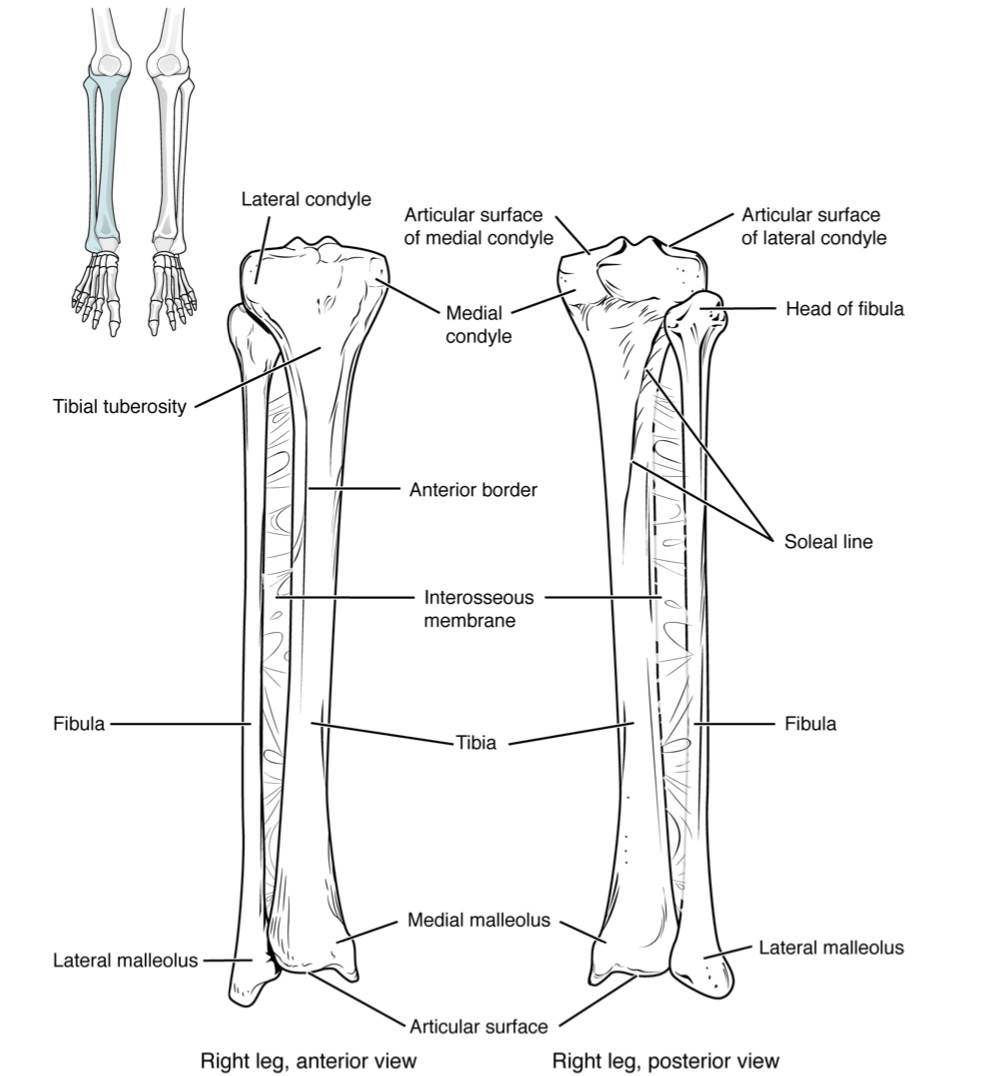
Proximal End of the Tibia:
Greatly expanded, forming two condyles:
Medial condyle of the tibia: Medial expansion, smooth and flattened surface.
Lateral condyle of the tibia: Lateral expansion, smooth and flattened surface.
These condyles articulate with the medial and lateral condyles of the femur to form the knee joint.
The tibia does not have epicondyles.
Between the articulating surfaces of the tibial condyles is the intercondylar eminence, an irregular, elevated area that serves as the inferior attachment point for two supporting ligaments of the knee.
Tibial Tuberosity:
An elevated area on the anterior side of the tibia, near its proximal end.
It is the final site of attachment for the muscle tendon associated with the patella.
Shaft of the Tibia:
Inferiorly, the shaft of the tibia becomes triangular in shape.
The anterior apex of this triangle forms the anterior border of the tibia, which begins at the tibial tuberosity and runs inferiorly along the length of the tibia.
Both the anterior border and the medial side of the triangular shaft are located immediately under the skin and can be easily palpated along the entire length of the tibia.
A small ridge running down the lateral side of the tibial shaft is the interosseous border of the tibia.
This is for the attachment of the interosseous membrane of the leg, a sheet of dense connective tissue that unites the tibia and fibula bones.
Located on the posterior side of the tibia is the soleal line, a diagonally running, roughened ridge.
This line begins below the base of the lateral condyle, and runs down and medially across the proximal third of the posterior tibia.
Muscles of the posterior leg attach to this line.
Distal End of the Tibia:
The large expansion found on the medial side is the medial malleolus ("little hammer").
This forms the large bony bump found on the medial side of the ankle region.
Both the smooth surface on the inside of the medial malleolus and the smooth area at the distal end of the tibia articulate with the talus bone of the foot as part of the ankle joint.
On the lateral side of the distal tibia is a wide groove called the fibular notch
This area articulates with the distal end of the fibula, forming the distal tibiofibular joint.
Fibula
The fibula is the slender bone located on the lateral side of the leg. It does not bear weight. It serves primarily for muscle attachments and thus is largely surrounded by muscles. Only the proximal and distal ends of the fibula can be palpated.
The head of the fibula is the small, knob-like, proximal end of the fibula. It articulates with the inferior aspect of the lateral tibial condyle, forming the proximal tibiofibular joint.
The thin shaft of the fibula has the interosseous border of the fibula, a narrow ridge running down its medial side for the attachment of the interosseous membrane that spans the fibula and tibia.
The distal end of the fibula forms the lateral malleolus, which forms the easily palpated bony bump on the lateral side of the ankle. The deep (medial) side of the lateral malleolus articulates with the talus bone of the foot as part of the ankle joint. The distal fibula also articulates with the fibular notch of the tibia.
Tarsal Bones
The posterior half of the foot is formed by seven tarsal bones. The most superior bone is the talus. This has a relatively square-shaped, upper surface that articulates with the tibia and fibula to form the ankle joint.
Three areas of articulation form the ankle joint:
The superomedial surface of the talus bone articulates with the medial malleolus of the tibia.
The top of the talus articulates with the distal end of the tibia.
The lateral side of the talus articulates with the lateral malleolus of the fibula.
Inferiorly, the talus articulates with the calcaneus (heel bone), the largest bone of the foot, which forms the heel.
Body weight is transferred from the tibia to the talus to the calcaneus, which rest on the ground.
The medial calcaneus has a prominent bony extension called the sustentaculum tail that supports the medial side of the talus bone.
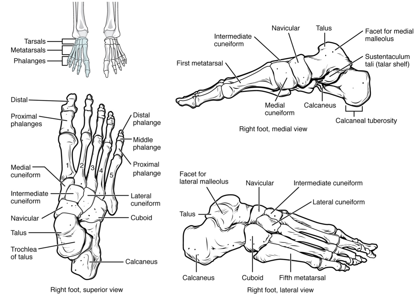
The cuboid bone articulates with the anterior end of the calcaneus bone.
It also has a deep grove running across its inferior surface, which provides passage for a muscle tendon.
The talus bone articulates anteriorly with the navicular bone, which in turn articulates anteriorly with the three cuneiform (wedge shaped) bones.
These bones are the medial, intermediate, and lateral cuneiform.
Each of these bones has a broad superior surface and a narrow inferior surface, which together produce the transverse curvature of the foot.
The navicular and lateral cuneiform bones also articulate with the medial side of the cuboid bone
Metatarsal Bones
The anterior half of the foot is formed by the five metatarsal bones, which are located between the tarsal bones of the posterior foot and the phalanges of the toes.
These elongated bones are numbered 1−51−5, starting with the medial side of the foot.
The first metatarsal bone is shorter and thicker than the others. The second metatarsal is the longest.
The base of the metatarsal bone is the proximal end of each metatarsal bone. These articulate with the cuboid or cuneiform bones.
The base of the fifth metatarsal has a large, lateral expansion that provides for muscle attachments.
The expanded distal end of each metatarsal is the head of the metatarsal bone. Each metatarsal bone articulates with the proximal phalanx of a toe to form a metatarsophalangeal joint.
The heads of the metatarsal bones also rest on the ground and form the ball (anterior end) of the foot.
Phalanges
The toes contain a total of 1414 phalanx bones (phalanges), arranged in a similar manner as the phalanges of the fingers.
The toes are numbered 1−51−5, starting with the big toe (hallux).
The big toe has two phalanx bones, the proximal and distal phalanges.
The remaining toes all have proximal, middle, and distal phalanges.
A joint between adjacent phalanx bones is called an interphalangeal joint.
Arches of the Foot
When the foot comes into contact with the ground during walking, running, or jumping activities, the impact of the body weight puts a tremendous amount of pressure and force on the foot.
During running, the force applied to each foot as it contacts the ground can be up to 2.5 times your body weight.
The bones, joints, ligaments, and muscles of the foot absorb this force, thus greatly reducing the amount of shock that is passed superiorly into the lower limb and body.
The arches of the foot play an important role in this shock-absorbing ability. When weight is applied to the foot, these arches will flatten somewhat, thus absorbing energy. When the weight is removed, the arch rebounds, giving "spring" to the step. The arches also serve to distribute body weight side to side and to either end of the foot.
Body weight is then conveyed from the talus to the ground by the anterior and posterior ends of these arches.
Strong ligaments unite the adjacent foot bones to prevent disruption of the arches during weight bearing.
On the bottom of the foot, additional ligaments tie together the anterior and posterior ends of the arches.
These ligaments have elasticity, which allows them to stretch somewhat during weight bearing, thus allowing the longitudinal arches to spread.
The stretching of these ligaments stores energy within the foot, rather than passing these forces into the leg.
Contraction of the foot muscles also plays an important role in this energy absorption.
When the weight is removed, the elastic ligaments recoil and pull the ends of the arches closer together. This recovery of the arches releases the stored energy and improves the energy efficiency of walking.
Stretching of the ligaments that support the longitudinal arches can lead to pain. This can occur in:
Overweight individuals.
People who have jobs that involve standing for long periods of time (such as a waitress).
Walking or running long distances.
If stretching of the ligaments is prolonged, excessive, or repeated, it can result in a gradual lengthening of the supporting ligaments, with subsequent depression or collapse of the longitudinal arches, particularly on the medial side of the foot. This condition is called pes planus ("flat foot" or "fallen arches").
The foot has a transverse arch, a medial longitudinal arch, and a lateral longitudinal arch.
The transverse arch forms the medial-lateral curvature of the mid-foot. It is formed by the wedge shapes of the cuneiform bones and bases (proximal ends) of the first to fourth metatarsal bones. This arch helps to distribute body weight from side to side within the foot, thus allowing the foot to accommodate uneven terrain.
The longitudinal arches run down the length of the foot.
The lateral longitudinal arch is relatively flat, whereas the medial longitudinal arch is larger (taller).
The longitudinal arches are formed by the tarsal bones posteriorly and the metatarsal bones anteriorly.
8.5 Development of the Appendicular Skeleton