Exhaustive Histology and Physiology of the Respiratory System
Functional Overview of the Respiratory System
Conducting Portion: Includes structures from the nasal cavity through the terminal bronchioles.
Respiring Portion: terminal bronchioles and alveoli where active gas exchange occurs.
Respiration Mechanics:
Inspiration of is an active process.
Expiration of is generally a passive process.
Central Nervous System Control: Respiratory centers are located in the CNS (brainstem). They send impulses to the diaphragm and intercostal muscles of the ribs to regulate the rate and depth of respiration.
Ventilation Process: Ventilation (drawing air in) involves the expansion of the chest wall to increase volume and create negative pressure. Negative pressure is the key driver for air entry. The diaphragm contracts to push the abdomen down, facilitating expansion.
Lung Anatomy and Physiology
Main Function: Gas exchange and ventilation.
Efficiency Factors: The amount of air reaching the lungs to carry in and out depends on:
Lung volume and capacity.
Lung compliance (the ability of the lungs to expand)
with fibrosis - stiff
with emphysema flabby - loss of elastin + collagen
Resistance to airflow = conductance
Gross Anatomy:
Right Lung: 3 lobes.
Left Lung: 2 lobes.
Fissures: Lobes are separated by fissures.
Ventilation-Perfusion (V/Q) Matching: The goal is to match Ventilation (V) with Perfusion (Q).
Ventilation (): The amount of air in the lungs/alveoli, typically .
Perfusion (): The amount of blood in pulmonary capillaries, typically .
Lung Volumes and Spirometry
Spirometry Procedure: Resulting data includes:
Tidal Volume ( or ): . Meaning: Amount of air entering lungs with each normal breath.
Residual Volume (): . Meaning: Amount of air remaining in the lungs after forced expiration. Measured via Helium () dilution or body plethysmograph.
Inspiratory Reserve Volume (): . Meaning: Maximal volume of air inspired following maximal expiration.
Expiratory Reserve Volume (): . Meaning: Maximal volume of air expired following a passive expiration (after tidal volume).
Vital Capacity (): . Meaning: Maximal amount of air expired following a maximal inspiration.
Total Lung Capacity (): . Meaning: Total volume of air in the lungs after maximal inspiration ( or ).
Embryological Development of the Respiratory System
Endoderm Derivatives: Gives rise to the epithelium and glands of the larynx, trachea, bronchi, and the pulmonary epithelium.
Mesoderm Derivatives: Gives rise to the connective tissue, cartilage, and smooth muscle of the respiratory tract.
Bronchial Branching: The lung bud divides into two bronchial buds, which further divide to form secondary and tertiary bronchi.
Mesenchymal Differentiation: As bronchi develop, the surrounding mesenchyme forms the associated cartilage, smooth muscle, connective tissue, and capillaries.
No respiration <24 weeks
respiratory distress syndrome: No surfactant often in premature infants
Surfactant Factors: Development requires the production of Surfactants (A, B, and C), which are critical for maintaining alveoli and preventing alveolar collapse.
Olfactory Tissue and Clinical Correlation
Olfactory Cells: These are bipolar neurons. They possess odorant-binding proteins on vesicles and transmit signals eventually to Cranial Nerve I (Olfactory Nerve).
Bowman’s Glands: They provide secretions to trap odorants for detection.
Sustentacular Cells: These serve as supporting cells for the neurons, provides nutrients
Basal Cells
Clinical Correlation: COVID-19 (SARS-CoV-2):
Symptoms: Loss of smell.
Mechanism: The virus enters oral and respiratory cells via the receptor. It targets the epithelium
Histology of the Larynx and Vocal Cords
Function: Passageway for air between the oropharynx and trachea; serves as the organ of speech (phonation).
Ventricular Folds: Also known as "false" vocal cords. They are lined with ciliated columnar epithelium.
Vocal Folds: Also known as "true" vocal cords. They are two folds of mucosa lined with squamous type epithelium.
Anatomy: The epiglottis is superior, followed by the false cords, the ventricle (recess), and then the true cords, leading into the trachea.
Clinical correlation:
bronchoscopy
Hyaline cartilage in conducting respiration
Cellular Composition of the Respiratory Epithelium
Mucosa:
epithelium + basement membrane
Conducting Epithelium: ciliated columnar
most vunerable to toxins (smoke) thus replaced by basal cells → stratified squamous (metaplasia)
Ciliated Columnar Cells: The most abundant cell type. Their cilia beat in unison to move mucus and trapped particles toward the oropharynx for swallowing or expectoration.
Goblet Cells: Produce mucus to trap debris
Basal Cells: Stem cells located at the basement membrane that replenish the epithelium.
Neuroendocrine Cells: Epithelial cells containing hormones.
respiratory epithelium - pseudostrat. ciliated columnar
3-4 goblet cells for every 10 cells: more mucus than necessary often in asthma
Clinical Correlation: Asthma:
Pathological changes include Basement Membrane () thickening, increased inflammation, and increased goblet cell.
Cartilage: chondrocytes, COL II, proteoglycans
submucosal glands & underlying tissue
mucous and serous glands
lymphoid tissue BALT (bronchial associated lymphoid tissue)
smooth muscle
cartilage - hyaline type
adventitia
Structural Anatomy: Trachea and Bronchi
Trachea Structure: Contains C-shaped hyaline cartilage rings. The posterior membranous portion contains smooth muscle.
Branching Hierarchy: Trachea → Primary Bronchi (2) → Secondary Bronchi (Lobar) → Tertiary Bronchi (Segmental) → Smaller Bronchi → Bronchioles.
Right Mainstem Bronchus: Lies more vertical and in direct line with the trachea, leading to an increased likelihood of aspiration compared to the left.
Histologic Layers of the Bronchus (Order from Lumen Outward):
Epithelium ()
Lamina Propria ()
Smooth Muscle ()
Cartilage ()
Progressive Changes during Branching (9-12 branches):
Connective tissue thickness decreases.
Relative amount of smooth muscle and elastic tissue increases.
Cartilage gradually disappears (it is completely gone by the time the airway reaches the bronchiole level).
The Bronchiolar Tree
Bronchioles: NO cartilage
Epithelium Transition: Larger bronchioles have respiratory epithelium; smaller bronchioles transition to low columnar epithelium.
in asthma the smooth muscles in bronchioles constricts → breathing difficulty
Clinical correlation:
bronchiectasis: clogged bronchioles, bluer image = big mucus plug, often seen in cystic fibrosis
Terminal Bronchioles:
Epithelium: Simple cuboidal with cilia.
Clara Cells : Non-ciliated epithelial cells with secretory granules. Functions include: producing surfactant components, breaking down mucus, detoxifying harmful substances, transferring , and fighting bacteria.
Rule of Loss: In the transition down the tract, goblet cells are lost before the cilia are lost.
Bronchi divide → bronchioles, once bronchi loses cartilage = bronchiole
bronchioles divide to become membranous than terminal bronchioles
distal to terminal bronchiole → acinus (terminal respiratory unit)
Acinus (Terminal Respiratory Unit): The unit of lung distal to the last terminal (membranous) bronchiole. It includes respiratory bronchioles, alveolar ducts, and alveolar sacs.
Alveolar duct (AD) → Alveolar saccules (AS)
separated by fibrous septa (contains veins and lymphatics) called a lobule
The Respiring Portion: Respiratory Bronchioles to Alveoli
Respiratory Bronchioles: Characterized by the appearance of alveoli along the wall. Cilia are lost by the end of the respiratory bronchiole.
Alveolar Ducts: Feature back-to-back alveolar openings along the wall. Smooth muscle between these openings appears as "knobs" in histologic sections.
Clinical correlation: Emphysema
large loose structure
commonly caused by cigarette smoke
Alveolar Sacs: Clusters of alveoli at the end of the ducts.
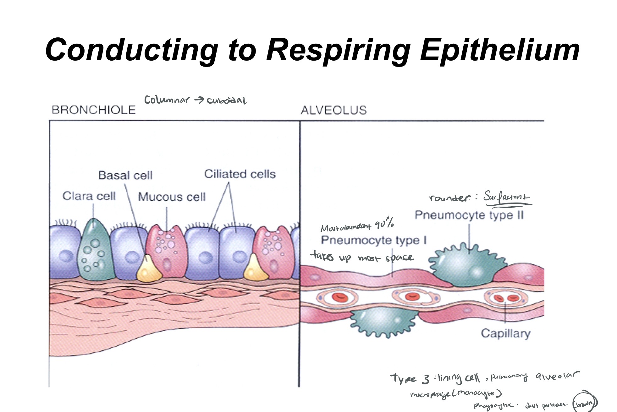
Alveolar Histology and the Blood-Air Barrier
Inter-alveolar Septae: Thin walls separating alveoli containing a dense capillary network, fibroblasts, and matrix components (collagen/elastic fibers).
Alveoli: sac like structures with thin walls so O2 and CO2 can diffuse between blood
Type I Pneumocytes:
Thin, flat squamous cells.
Cover of the alveolar surface.
Core component of the blood-air barrier.
Clinical correlation: edema
injury to type I pneumocytes occurring through infection + injury (fluid) epithelium and endothelium is broken → congested vessels
Type II Pneumocytes:
Large cuboidal cells with round nuclei, covering only of the surface.
Contain lamellar bodies in the cytoplasm which make and store surfactant.
Surfactant reduces surface tension to prevent alveolar collapse during expiration.
Blood-Air Barrier Layers (Air to Blood):
Type I Pneumocyte
Fused basal laminae (of Type I cell and capillary endothelial cell)
Capillary endothelial cell
Alveolar Macrophages (Dust Cells): Derived from blood monocytes; they phagocytose debris and particles that escape the conducting portion's defenses.
seen in smokers macrophages take up smoke looks like brown particles
LaPlace’s Law: Specifically applied to alveoli as , where is pressure, is surface tension, and is the radius. Lower surface tension (via surfactant) requires less pressure to keep the alveolus open.
Respiratory Blood Supply and Interstitium
Dual Arterial Supply:
Pulmonary Arteries (Low Pressure): Carry deoxygenated blood from the right heart. Branches run alongside bronchi and bronchioles, dividing into the capillary network in alveolar walls.
Bronchial Arteries (High Pressure): Systemic origin (aorta). Supply oxygenated blood to the tissues of the conducting airways down to the level of respiratory bronchioles.
Interstitium: The space between the basement membranes of the endothelial and epithelial cells. Contains collagen, elastic fibers, fibroblasts, and mononuclear cells.
Clinical Correlations Summary:
Pulmonary Edema: Injury to Type I pneumocytes leads to fluid accumulation and congested vessels.
Emphysema: Damage to alveolar walls; centrilobular emphysema is often visualized in the lobule center.
ARDS (Acute Respiratory Distress Syndrome): Results in thickened hyaline membranes, Type II pneumocyte hyperplasia, and interstitial fibrosis.
too pink - too much connective tissue
infection + thickening of membrane
The Pleura and Lymphatics
outer surface of lung and inner surface of thoracic cavity are covered by pleura, a serous membrane
Pleura: A serous membrane (serosa) consisting of simple squamous mesothelium and thin connective tissue.
Visceral Pleura: Covers and adheres directly to the lung surface.
Parietal Pleura: Lines and adheres to the walls of the thoracic cavity.
Pleural Cavity: The space between layers containing serous fluid.
Lymphatics:
Follow bronchovascular structures (arteries and airways).
Found in the pleura and interlobular septa.
Network is rich in the visceral pleura and extends along venules and veins in the periphery of lobules.
Clinical Correlation: Mesothelioma: A malignancy often associated with asbestos exposure (e.g., insulation workers), presenting with pleural effusion and chest pain.
Focus on:
Conducting vs. respiring portions of the respiratory system
Tissue layers and cellular/tissue components of the respiring and conducting structures of the respiratory tract, trachea, bronchi, bronchioles, respiratory and terminal bronchioles
Non-cellular components of the lung, collagen and elastin
The pulmonary acinus, its component structures on the alveolar side, interstitium and capillary side
Pulmonary circulation and bronchial circulation and lymphatics in the lung
Pleura- two components