Cardiovascular System
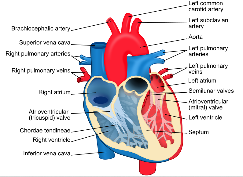
Pathway of Blood Flow through the Heart: Superior( deoxygenated blood from the upper body) & Inferior (deoxygenated blood from the lower body) Vena Cava → Right Atrium → Tricuspid Valve → Right Ventricle → Pulmonary Artery (only artery that carried deoxygenated blood) → Lungs ( gas changes of O2 and CO2 in the Alveoli ) → Pulmonary Vein (only vein that carries oxygenated blood) → Left Atrium → Biscupid/Mitral Valve → Left Ventricle → Aortic Valve → Aorta → Back to the rest of the body.
THE RIGHT SIDE OF THE HEART IS WHERE DEOXYGENATED BLOOD FLOWS
THE LEFT SIDE OF THE HEART IS WHERE OXYGENATED BLOOD FLOWS
From greatest to least in size:
Arteries: Oxygenated blood leaves the left side of the heart out the aorta, which branches into arteries, and then arterioles which carry blood and oxygen to the smallest blood vessels the capillaries.
Capillaries Function: Alveoli are tiny air sacs in the lungs—1.5 million per lung—encased within capillary networks located in their walls. Oxygen diffuses through the permeable walls of the alveoli into the capillaries (which are likewise permeable to oxygen and carbon dioxide) and carries the oxygen from the lungs into the pulmonary venules then to the pulmonary veins to the left atrium and eventually to the rest of the body; carbon dioxide diffuses into the alveoli and is then exhaled out of the body.
Veins: Deoxygenated blood leaves the capillary and enters the small venules. These venules become progressively larger vessels called veins. VEINS CONTAIN VALVES to keep the blood flow in one direction. The vena cava (s) are the two largest veins that carry blood into the right upper chamber of the heart (the right atrium).
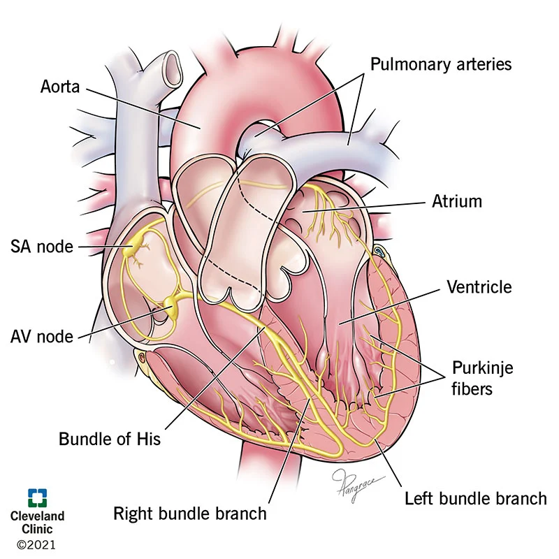
The Electrical Components of the Heart:
The heart is regulated by the Nervous and Intrinisic ( anatomy or use of certain nerves and muscles specific to an organ or body part) System.
SA node aka the “Pacemaker” (located in the right atrium) sends a signal to the AV (atrioventricular) node ( located at the base of the right atrium).
Bundle of His, Left/Right Bundle Branches, & Purkinje Fibers are located in the septum.
SA node → AV node → Bundle of His → Right/Left Bundle Branches → Purkinje Fibers.