exam II resp
key terms for this chapter:
ventilation: flow of air into alveoli
perfusion: blood flow through lungs that carries o2
flow: pressure of oxygen
fio2: how much oxygen is in the flow
respiratory failure vs ventilatory failure: ventilatory failure is the inability to remove co2, while respiratory failure is the inability to provide adequate oxygen
hypercapnia: paCO2 > 50
hypoxemia: paO2 < 60, normal or lowered paCO2
comorbidites: presence of 2+ diseases in a pt; ex: pt has diabetes and comorbidity of DVT
asterixis: involuntary jerking of the hand/wrist, usually upward or downward; a sign of hypercapnia
PaO2 to FiO2 ratio: partial pressure of arterial o2 to a fraction of inspired o2
acute respiratory failure (ARF)
pathophysiology
the inability of the body to sustain the respiratory drive, ↓ capacity to exchange o2 and co2. can be a result of a dysfunction in the structures responsible for ventilation, a condition that results in hypoperfusion, or a disease process that impairs the ventilatory capacity of the lungs like COPD. can be divided into 2 types— hypoxemic (PaO2 < 60mmHg and PaCO2 is norm/low) which occurs in many diseases where fluid is accumulating or collapse of an alveoli occurs or hypercapnic (PaCO2 >50mmHg) which results in hypoxemia for RA pts.
v/q mismatch is an imbalance between ventilation and perfusion, leading to reduced blood oxygenation and s/s like SOB and dyspnea. Low v/q is a result of decreased ventilation because of airway or interstitial lung disease, or from overperfusion. High v/q indicates decreased perfusion to the lungs. shunt occurs when hypoxemia is present even with 100% o2 is inhaled, and the o2 sat will be low, and doesn’t improve with supplemental oxygen. Deadspace is a physiological deadspace in the lungs where air doesn’t go through or reach, causing perfusion problems.
epidemiological / etiological risk fx
ventilatory failure = hypercapnia; results from sedatives, COPD, neuromuscular defects, CNS dysfunction, chest trauma
lung failure = hypoxemia; results from pulmonary edema, pulmonary emboli, hf, atelectasis, pneumonia, and emphysema
disorders that affect the CNS, obesity, and alveoli abnormalities may weaken the chest wall and respiratory muscles, or cause improper diffusion, causing susceptability to ARF.
affecting a client’s health
pts diagnosed with ARF are not similar to COPD patients, in which they can become accustomed to low o2 levels and hypoxemia. pts with ARF are considered to be going through a medical emergency, and must have the underlying cause treated. otherwise, risk of permanent damage raises, such as lung scarring, memory problems, brain damage, and muscle weakness.
clinical presentation of acute respiratory failure (arf)
if hypoxemic: dyspnea, irritability, confusion, somnolence, tachycardia, arrhythmias, tachypnea, cyanosis
if hypercapnic: headache, change of behavior, coma, warm extremities, asterixis
diagnostic and labs
ABGs are used to determine criteria for hypercapnic vs hypoxemic and may help more accurately monitor the values in relation to air exchange (o2, co2, pH). chest x-rays help identify lung abnormalities that can cause ARF (pneumothorax, infection/inflammation, pleural effusion, atelectasis). if the cause is unclear through chest x-ray, a physician may choose to use CBCs, ECGs, pulmonary and thyroid function tests, electrolytes, and sputum, blood, and urine cultures to identify roots.
clinical judgement and nursing management, care, and role
during assessment, it’s important to identify dyspnea, SOB, and other signs of lack of oxygenation (cyanosis, slower cap refill). asterixis may also be present. vital signs may reveal clinical manifestations like tachynea, tachycardia, and low o2 sat. in advanced respiratory failure, alterations in memory or cognitive processes may be identiiable.
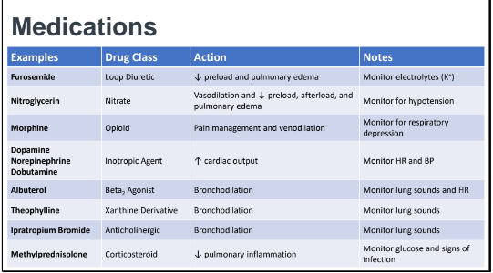
ABGs will be abnormal, and point to respiratory acidosis. biggest priority is restoring respiratory function through increasing o2 levels in the blood; least invasive measures first —> most invasive if necessary. address and find the underlying cause and reduce associated complications.
actions: elevate HOB, oral hygiene, suction prn, wean off ventilator, education, ROM exercises, prevent complications (DVT, peptic ulcers).
video references
ARF
acute respiratory distress syndrome (ARDs)
pathophysiology
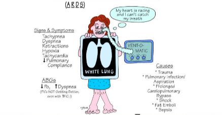
begins with a rapid onset (within 7 days) after lung injury. fluid builds up in the alveoli and surfactant in the lungs break down, causing stiffness that makes it hard to move oxygen throughout the body, leading to hypoxia. their hypoxic state may not respond to oxygen.
etiology / risk fxs
most cases are caused by sepsis, pneumonia, and aspiration. it is also often found in pts with mechanical ventilation.
risk fxs include being female, drowning, drug overdose, traumatic brain injury, recent cardiac-related surgery, smoking, and excessive alcohol use
.
impact on overall health
pts with ARDs are most likely going to be on mechanical ventilation; because of this, consistent airway management must be done. a g-tube may be put in place for nutritional support, muscle wasting from continuous dyspnea and an intolerance to exercise may occur, and lastly, cognitive loss from hypoxia.
clinical presentation
**dyspnea that worsens within 6-72 hrs, tachycardia, tachypnea, diffuse crackles in the lungs, respiratory distress, somnolence, cyanosis, diaphoresis.
labs and diagnostics
ABGs are used to manage and monitor the respiratory levels (PaO2 and PaCO2), chest x-rays help to find ‘white out lung’, where the lungs are filled with fluid. Condition of ARDs is determined through the Berlin criteria.
Berlin Criteria
Onset occur within 7 days
Noncardiac origin
Imaging indicating bilaterial lung infiltrates
Specific oxygenation measurements
ABGs report PaO2/FiO2 ratio of (x) < 300 mmHg
The true severity of ARDs is determined by the ratio of FiO2/PaO2. Normal PaO2 is 85 to 100 mmHg, and normal FiO2 is 0.21. Ratio is usually higher than 400 (e.g. 95/0.21 = 452)
Mild ARDs is less than 300, Moderate ARDs is less than 200, and Severe ARDs is less than 100.
clinical care, nursing role and actions, etc.
the nurse’s role in the care of ARDs is assessments— vitals, skin condition, progression, lab results, tubes and their placements, auscultation. care will be done based off ABCDEF (Assess, Both spontaneous awakening and breathing trials, Choice of analgesia/sedation, Delirium assessment, Early mobility/exercise, Family engagement and empowerment).
priorities are ventilatory assistance to increase assistance, decrease shunt, reduce o2 consumption, and prevent further lung injury. frequent monitoring of pain, respirations, and cardiac are necessary, and suction if needed. prevent VAD by performing oral hygiene, ROM, and prophylaxis.
prone positioning can be used to improve hypoxemia when other strategies don’t work in early stages of ARDs— pts can stay in prone for 16 hrs per day.
mechanical ventilation, ecmo, and chest tubes
necessity and usage, types
cpap: single pressure setting for whole breathing cycle— makes pt forcibly exhale against the pressure from CPAP. used for obstructive sleep apnea; not good for those who have large amounts of secretions or unable to maintain their own airway. cpaps increase oxygen, decreases the work of breathing and cardiac workload, preventing airway collapse.
bipap: separate pressure settings for inhalation and exhalation— 2 levels of positive pressure vs 1 lvl with cpap. often used for sleep apnea, copd, hf, and respiratory failure. not good for those who have decreased mental status, large amounts of secretions, or unable to spontaneously breathe. bipaps help remove co2 and make breathing easier.
endotracheal tube (ett): flexible tube inserted into the trachea from the mouth for breathing support. nurse should assist with intubation, inflating the cuff after placement. ventilate with an ambu-bag during the process as pt may be on a paralytic and sedation. placement should be confirmed with an end tidal co2 detector, noting the color change to indicate presence of co2. auscultate breath sounds for equal bilateral air movement in the chest, then observe the chest for wall movement that’s symmetrical. spo2 should rise, and a CXR will be last confirmation.
tracheotomy tube: tube placed into the trachea through an incision in the neck to maintain an airway. when assessing the et tube, check size and how far it is inserted. cuff should be inflated before auscultating breath sounds. use end tidal co2 capnography to ensure a color change, then pass a suction tube through the trach before removing. secure and obtain a chest x-ray.
vent modes + settings
full support/assist control a/c: assists with each breath by providing volume or pressure. vent takes over when RR falls below a predetermined setting.
partial support/synchronized intermittent mandatory ventilation SIMV: ventilates together with pt’s inspiratory efforts. pt can spontaneously breathe with the volume from effort, but otherwise preset rate ensures mandatory delivery.
pressure support ventilation or cpap (continuous positive airway pressure): respiratory rate, tidal volume, and inspiratory times are determined by the patient and not pre-set by the machine. pt must be awake and alert.
FiO2: fraction of inspired oxygen describes the percentage of oxygen present in the air
PEEP (positive end-expiratory pressure): amount of pressure remaining in lungs upon expiration. pressure is applied from the ventilator to the airways at the end of expiration, and prevents atelectasis by keeping the alveoli open.
pressure support (ps): preset pressure level that is delivered when the client breathes to make breathing easier
rate: a setting that shows lowest RR per min that the client can maintain before vent support begins.
tidal volume: amount of air moved into or out of lungs with each respiration
ventilator alarms
high pressure: caused by coughing, asynchrony, condensation, kinked tubing, high resistance, or low compliance
low pressure: disconnection in ventilation tubing or accidental extubation
apnea: oversedation, respiratory arrest
high v1 or rate: pain, anxiety, high metabolic demand, hypoxia, or hypercapnia
low v1: a disconnect, leak, or cuff leak
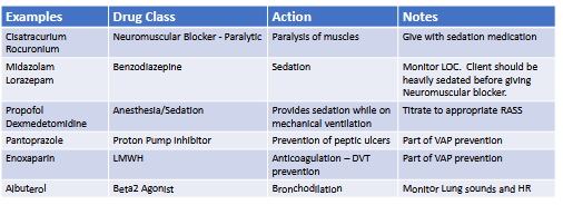
complications of mechanical ventilation include fluid retention and hemodynamic compromise (cardiovascular), and barotrauma, volutrauma, atelectasis, VAP, and oxygen toxicity (pulmonary). VAP (ventilator-associated pneumonia) may occur— if so, minimize sedation, HOB > 30, ROM, oral care, skin care, peptic ulcer prevention, nutritional care, and sedation vacation.
memory trick: 2 PB sandwiches can make you sick (high pressure)
2 P’s: pulmonary edema, pneumothorax
2 B’s: bronchospasm, biting
S-C-K: secretions, cough, kink in tubing
extracorporeal membrane oxygenation (ecmo)
blood is removed from the circulatory system and pulled outside of the body by a mechanical pump, and passed through an oxygenator and heat exchanger. this oxygenates hemoglobin and removes co2, then the blood is re-infused into the client’s circulation. bleeding is a major complication, and the nurse is responsible to administer heparin as ordered and monitor coagulation related lab values. platelet counts should remain above 150,000, fibrinogen > 200mg/L, < 1.5 prothrombin, clotting times between 180 - 240 seconds. be alert to signs of internal hemorrhaging like hypotension, tachycardia, reduced urine output and decreased central venous pressure.
complications include hypoxia, renal failure, sepsis, fluid imbalance, electrolyte imbalance, and malnutrition.
chest tubes
chest tubes are used to remove pleural fluid, for hemothoraxes and pneumothoraxes, and to drain fluid post-op
do NOT strip or milk chest tubes, as it can cause air to go back into the pleural cavity; notify provider if drainage > 1-1.5L in 1st hour, or >100 mL/hr after first hour, if there is subcutaneous emphysema or respiratory distress. do not elevate drainage system, do not clamp the tube. change the system when it is full, do not just empty it and measure the fluid levels. If tubing disconnects, reestablish the water seal.
IF the drainage unit is overturned, return the system upright, do NOT clamp, have the client take some deep breaths and replace the drainage unit.
pneumothorax
pathophysiology
latrogenic pneumothorax: occurs as a complication of a medical procedure
tension pneumothorax: air enters space but cannot escape
traumatic pneumothorax
spontaneous pneumothorax: who it occurs in (primary: healthy, secondary: Hx of pulmonary disease)
risk fxs
smoking, environment (low atmospheric pressure), genetics (tall/thin, pregnancy, marfan syndrome, family hx), medical procedures (cvc, transthoracic), and age
impact on overall health
pneumothorax may contribute to lower lung capacity and rib cage volume, leading to an overall lower o2 sat
clinical presentation (classic)
respiratory discomfort/distress, pleuritic chest pain, raised respiratory rate, asymmetrical lung expansion, decreased tactile fremitus (respiratory vibration on the chest wall, hyper resonance with percussion, lowered or absent breath sounds
tension pneumothorax clinical presentation
classic symptoms + tachycardia (>134 bpm), hypotension, tracheal deviation, JVD, cyanosis
hemothorax clinical presentation
similar to pneumothorax— blood in lungs, hypovolemia, no jvd
lab testing
chest x-ray, ultrasonography, blood work, ct scan
nurse role
prepare for chest tube to remove fluid/air, monitor respirations, pain relief, have pt splint and cough/deep breathe, give supplemental o2 and raise HOB, prepare for needle decompression or thoracentesis
expect hypotension, asymmetrical chest expansion, diminished/absent breath sounds, low spo2 and high paco2 on abgs
flail chest + embolism
pathophysiology
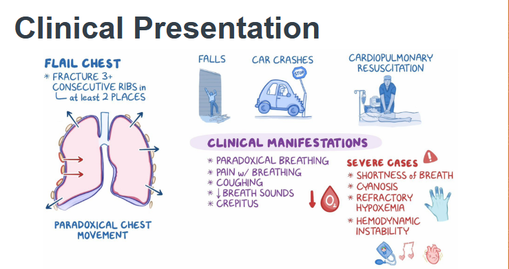
3 or more broken ribs 2 or more places, causing rib cage instability and paradoxical chest movement, causing broken ribs to move independently. eventual hypoventilation leads to atelectasis which leads to pulmonary contusion, then edema, hemorrhage, then decreased lung space.
epidemiology and risk fxs
blunt trauma (assault, fall, MVC)— 10-20% mortality rate and 7% of chest traumas
impact on overall health
causes altered gas exchange and may lead to a permanent chest wall deformity, making it hard to breathe for a lifetime. higher risk for respiratory distress in older adults
manifestations and presentation
paradoxical chest movement, decreased spo2, tachypnea, crepitus, decreased breathing sounds, pain w breathing
labs and diagnostics
chest x-ray, ct scan, abgs (High ph, everything else low)
role of the nurse
evaluate nutrition, cough/deep breathe, incentive spirometer, hob raised, ventilator maintenance (vap prevention, weaning), ambulation