Breathing
Ventilation and Control of Ventilation
The brain stem controls ventilations
There are various different respiratory centres:
Inspiratory area
Expiratory area
Medullary rhythmicity areas
Inspiratory area: made up of the dorsal respiratory group and cells from the solitary tract nucleus
Expiratory area: made up of the ventral respiratory group and cells from the ambiguous nucleus
→ The VRG contains the expiratory area but some is also a part of the Inspiratory area (e.g. pre-Bötzinger complex)
In the pons:
Apneuistic area
Pneumotaxic area
Pontine respiratory group
DRG: Inspiratory area
the first descending nerve through the lateral column synapse in C3, C4 and C5
The phrenic nerve projects from those vertebrae to inner cage the diaphragm
The descending nerve can also synapse in T1-T11
The intercostal nerves project from those vertebrae to unnerve the external intercostal muscles
→ It causes the diaphragm to contract so you can pull your ribcage up and out. Your lungs expand and move with your diaphragm
As your lungs expand, the pressure within the lungs goes down and inspiration occurs
Expiration = no signals from DRG Inspiratory area
In every 5 second period (roughly 12 breath a minute) for 3 seconds, the DRG will be sending out signals then for 2 seconds it stops.
Expiration is controlled by elastic recoil and the process of not inhaling.
Voluntary Control of Ventilation
The motor cortex in your brain sends out impulses and bypasses the Inspiratory area all together
→ The Inspiratory centre takes on inhalation responsibility so that we don’t have to consciously think about it
Ondine’s curse is another name for congenital central hypoventilation syndrome where when a person goes to sleep, their body forgets to breath.
The neuritis area projects down to the Inspiratory area (DRG) and activity in the apneuistic area triggers activity in the DRG.
If your DRG is not activating inspiration then the apneuistic area will.
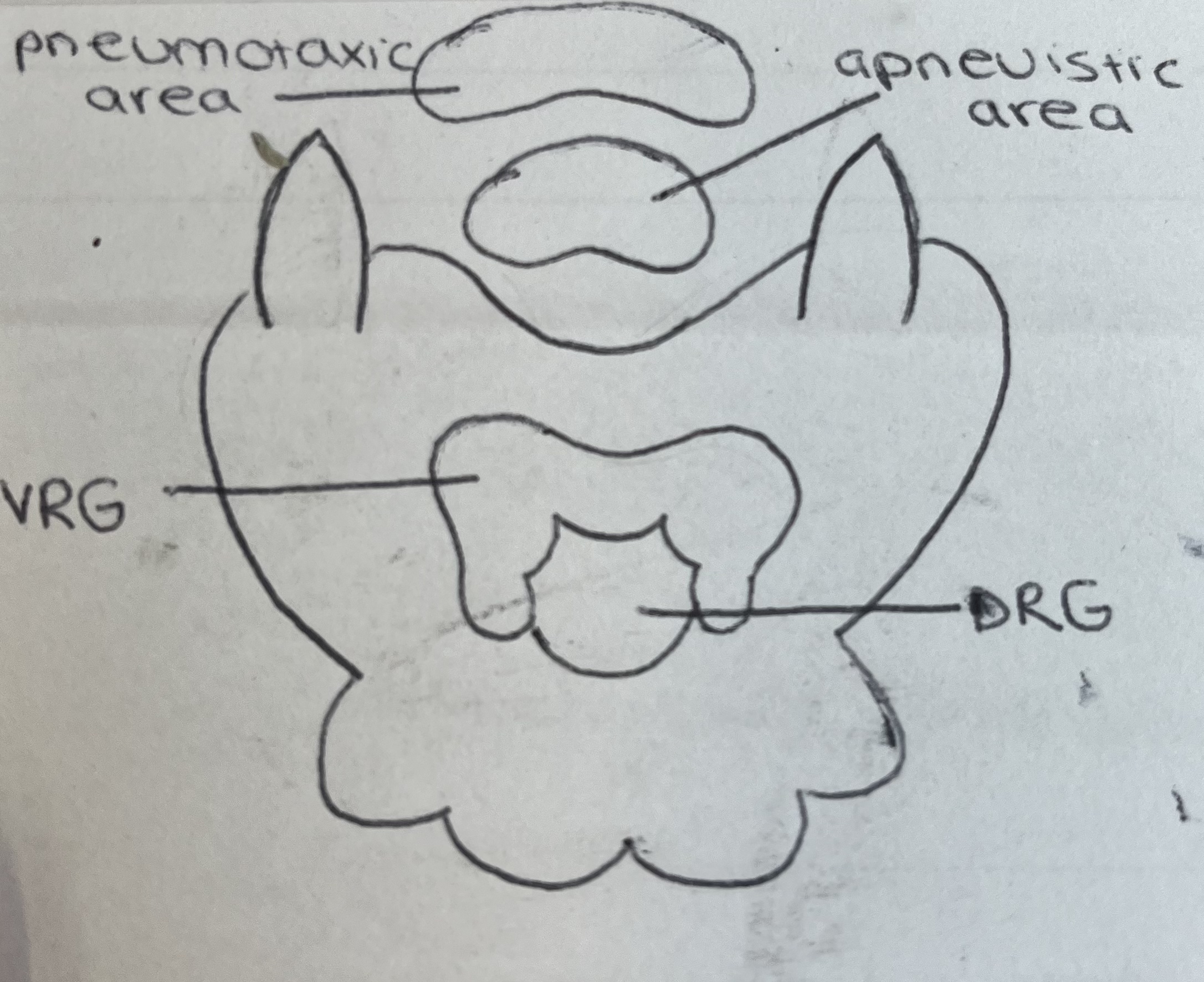
The pneumotaxic area is inhibitory. Inhibitory neurones can be sent to the apneuistic area and Inspiratory area to prevent them from activating.
This is a reinforcement of the rhythm
Pulmonary stretch receptors are found in smooth muscle in the airway. If there are excessively stretched, they trigger a reflex that would stop inhalation. This reflex is carried on the vagus nerve.
→ called the Hering-Breuer reflex
Increasing Inhalation
→ due to exercise and more CO2 produced
Central chemoreceptors in the medulla respond to protons (H+)
Henderson Hasselback Equation:
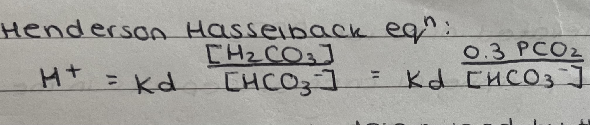 The concentration is determined by the partial pressure of CO2
In our circulating fluids, bicarbonate is the dominant buffer system
H+ is directly proportional to CO2
As CO2 diffuses into the brain, it activates the chemoreceptors by being converted into H+.
The increase in H+ increases activity in the Inspiratory centre causing more inhalation.
The increase in inhalation means increase in (more vigorous) exhalation which removes CO2 from the Boddy.
Peripheral chemoreceptors in your bloodstream respond to oxygen (in between internal and external carotid artery and in aortic arch)
They detect low partial pressure of O2.
They then send signals via the vagus and glossopharyngeal nerve to the Inspiratory area to increase inhalation.
Central chemoreceptors are responsible for normal breathing and peripheral chemoreceptors are if something is wrong.
Resting Ventilation
Inspiration: follows rhythmic activity in the DRG which leads to:
Activation of the phrenic nerve and contraction of the diaphragm
Activation of the intercostal nerves and contraction of the external intercostal muscles (raises and stiffens the ribcage)
Expiration: follows pauses in rhythmic activity in the DRF which leads to:
Relaxation of the diaphragm and the external intercostal muscles
Elastic recoil of the lungs
Exercise Ventilation
Inspiration: is deeper and more prolonged following:
Activation of the medullary central chemoreceptors by reduced CSF pH caused by elevated arterial PCO2
Activation of the peripheral chemoreceptors in the aortic and carotid bodies by hypoxia (reduced arterial PCO2)
Expiration: is more forceful because:
Inspiration is deeper and more prolonged
Expiratory muscles come into play
Control of Ventilation
The expiratory area (VRG) projects neurons from the ventral columns to the thoracic vertebrae (T1-11) which innervate the intercostal nerves and the internal intercostal muscles
The internal intercostal muscles lower the ribcage in and down
→ Important for forceful expiration
Other muscles for control of ventilation:
Internal intercostals, external oblique, internal oblique, transverse abdominus and rectus abdominus
Spirometry
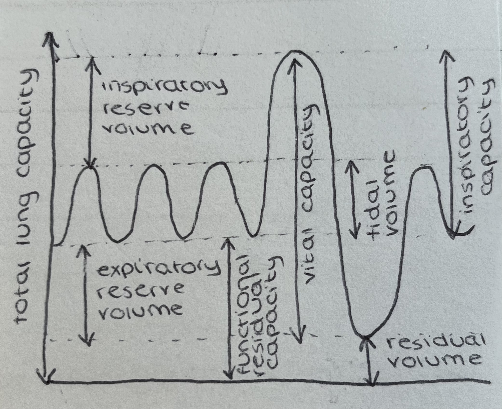
Measures the effects of ventilation
Average results:
TV = 500ml
FRC = 2400ml
ERV = 1200ml
RV = 1200ml
IRV = 3000ml
IC = 3500ml
VC = 4700ml
TLC = 5900ml
Bad = breathing in and out the same air depending on the volume that can be held in the tube
 Anatomical dead space (non alveolar airways) = 150ml
Spirometry
Carbon dioxide is the major regulator of body pH
Slow exchange is important to prevent wild swings in arterial PCO2 and pH
Hyperventilating will result in a respiratory alkalosis
→ which the kidney can compensate for by excreting HCO3-
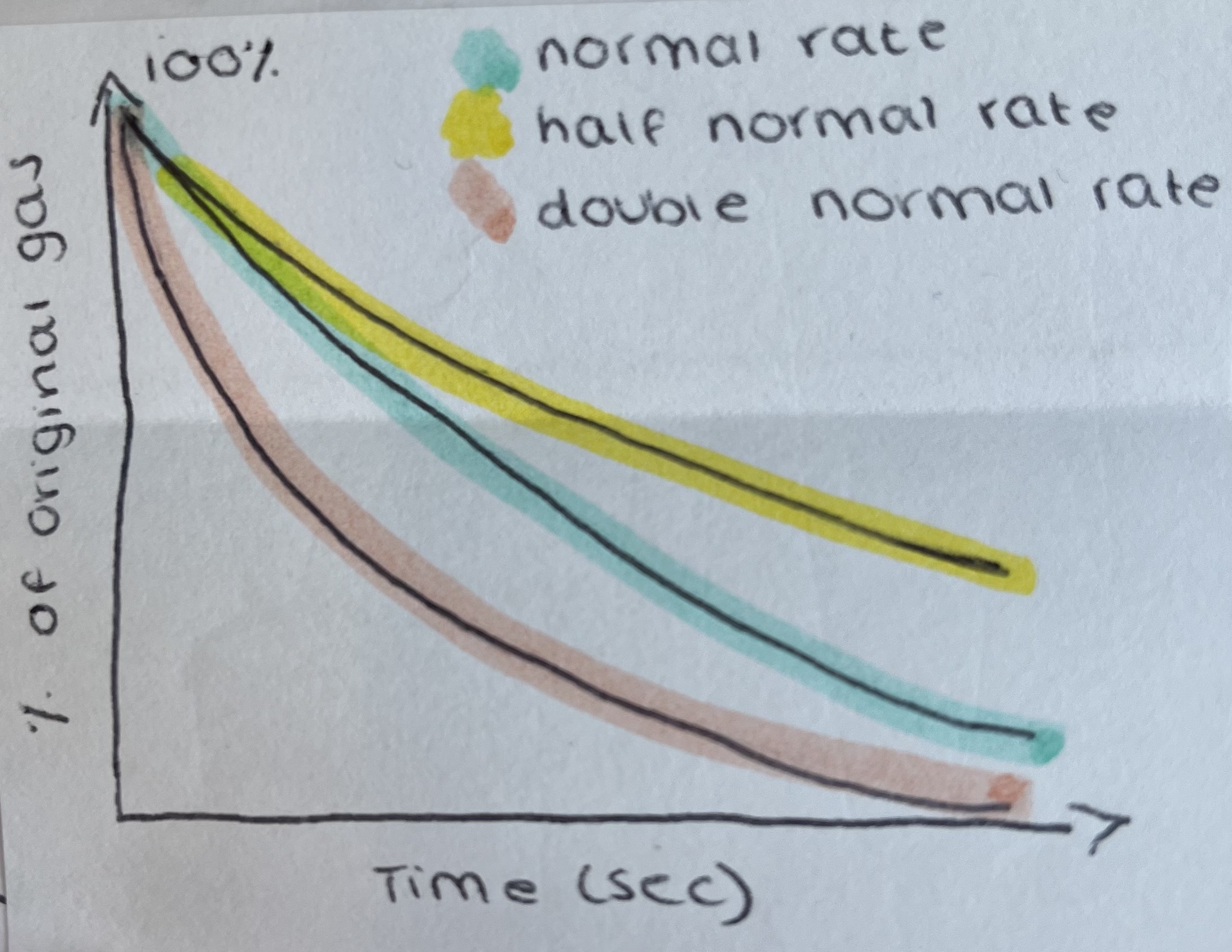
FEV1 = forced expiratory volume at 1s
FEV1/FVC x100
Healthy = around 80%
COPD = can be 60%
→ diagnostic indicator
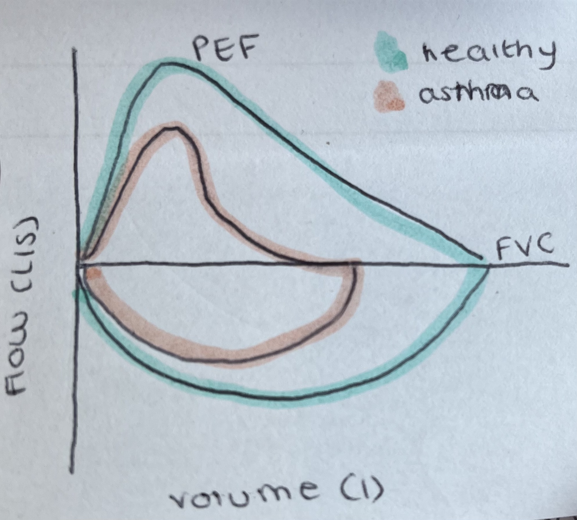
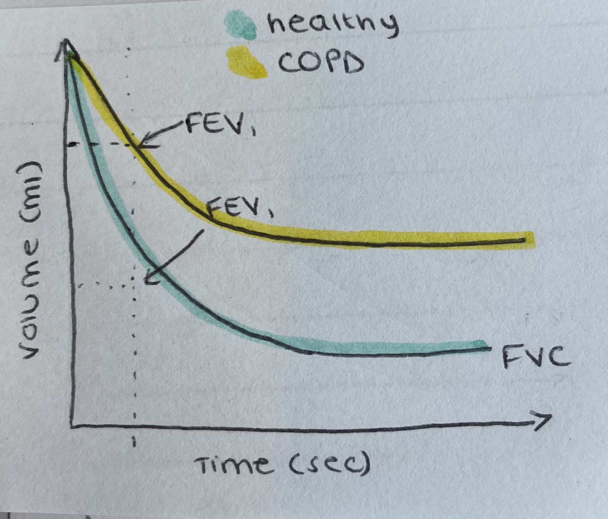 There are other condition that give curves like this however, they don’t change if you give them salbutamol
Diffusion
Gases diffuse, solutes in solution including gasses diffuse
Diffusion of gas’s in solution airways occurs from high partial pressure to low partial pressure e.g. PCO2, PO2, etc. (Fick’s first law of diffusion)
Partial pressure of a gas depends on both the amount dissolved and the solubility of the gas (P=[g]/gs) → CO2 is 20x more soluble than O2.
When equilibrium is reached, there is no further net movement
Diffusion of different solutes (including gases) may occur in opposite directions simultaneously
Ideally never reach equilibrium:
infinite source and infinite sink
Gas Exchange
Ventilation allows the alveolar air to represent an “infinite” oxygen supply at a PO2 of 100mmHg
→ Blood flow allows the alveolar capillaries to represent an “infinite” oxygen sink at PO2 of 40mmHg
Blood flow allows the alveolar capillaries to represent an “infinite” CO2 supply at PCO2 of 45mmHg
→ Ventilation allows the alveolar air to represent an “infinite” CO2 sink at PCO2 of 40mmHg
Fick’s first law
Flux = C(P1-P2)
Where: (P1-P2) = pressure gradient, C= conductance
If it is difficult to move down the partial pressure gradient then it is a small Flux and vice versa.
The lungs, through evolution, have a very low conductance
Specialised → thin so short diffusion distance
Internalised → increase SA
Alveolar layers
Fluid and surfactant
Alveolar epithelium
Epithelial basement membrane
Capillary layers:
Interstitial space
Capillary basement membrane
Capillary endothelium
The collection of the membrane is about 0.6uM and in some places as low as 0.2uM. (Red blood cell is 6uM)
→ this give high conductance and therefore high Flux
It is also kept moist so gases dissolve into fluids to diffuse across.
Around 70m2 of alveolar tissue with 60-140ml of blood
→ depth of blood coating is 1-2uM
Area of the lungs
All blood in the pulmonary capillaries is in very close contact with the air. Perfect for gas exchange
→ optimised for high Flux
Close by blood supply = erythrocytes
Haemoglobin
Contains a prosthetic group at its centre (Haem)
→ has iron wrapped around in porphyrin rings
Prosthetic group it’s into larger protein structure (globin)
→ 4 of these monomers make up a haemoglobin molecule (HbA)
2 beta chains and 2 alpha chains
Oxygen bind to the Haem group to form oxyhemoglobin.
CO2 binds to the protein to form carbaminohaemoglobin
CO binds to the Haem group to form carboxyhaemoglobin.
Oxygen binding to haemoglobin is cooperative which means that one the first oxygen is bound, it undergoes and conformational change
→ Therefore binding of the following oxygen is easier
The cooperative binding means higher haemoglobin saturation
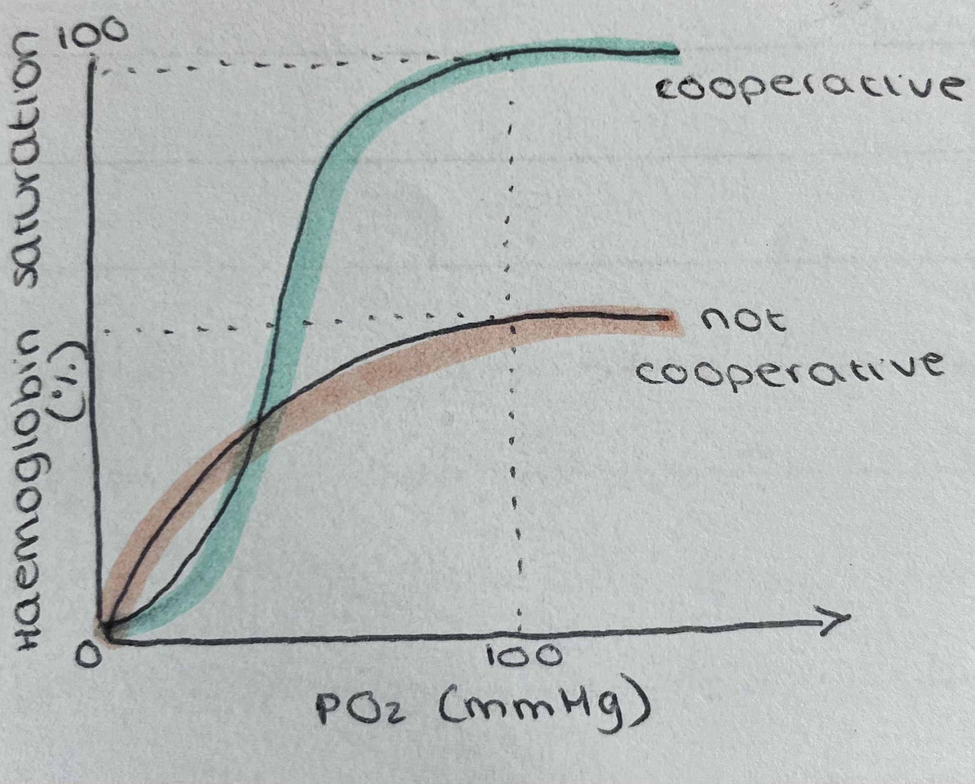 Between 20-40mmHg is where haemoglobin is most likely to give up its oxygen
Therefore, it is most saturated in the lungs where the PO2 is higher and least saturated in the tissues
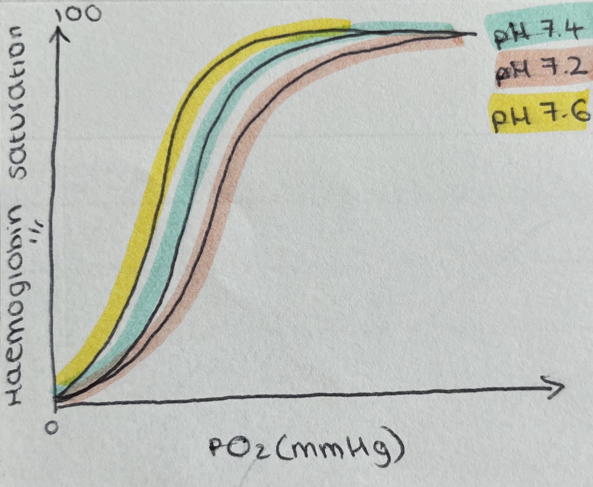 With lower pH, the curve shifts to the right. So with every PO2, haemoglobin will give up more oxygen.
Lower pH = raised [CO2]
This is the Bohr effect.
PCO2 will be lowest in the lungs so therefore haemoglobin will take up more oxygen and be less likely to give it up.
Raised temperature = more O2 released for any PO2
Raised metabolite [2,3-bisphosphoglycerate] = more O2 released for any PO2
→ shifts curve to the right (when exercising)
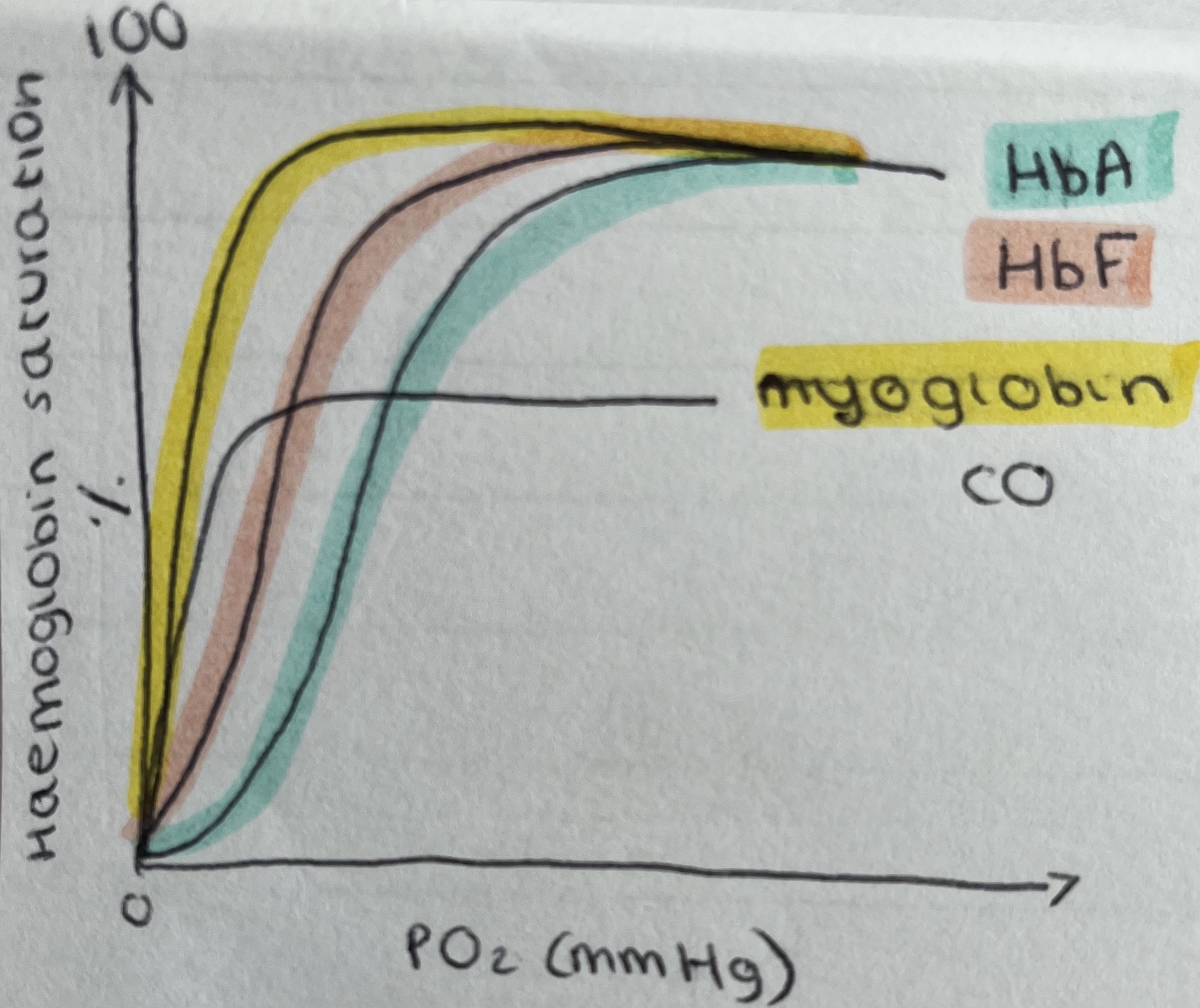 Adult haemoglobin = HbA = 2a and 2b
Foetal haemoglobin = HbF = 2a and 2y
At any PO2, foetal haemoglobin has a higher affinity for O2 than an adult
HbF doesn’t respond to 2,3-BOG.
HbF will be replaced with HbA between 6-12 months after birth
2,3-BPG is present in erythrocytes at 5mM
→ if this amount went up. The curve would shift to the right for HbA
Myoglobin is found in muscles and has a very high affinity for O2.
→ it will act as an O2 supply when PO2 gets very low
CO has a very high affinity (200x than O2) for Haem groups
→ affects haemoglobin cooperativity
CO is from incomplete combustion of carbon
Haldane Effect
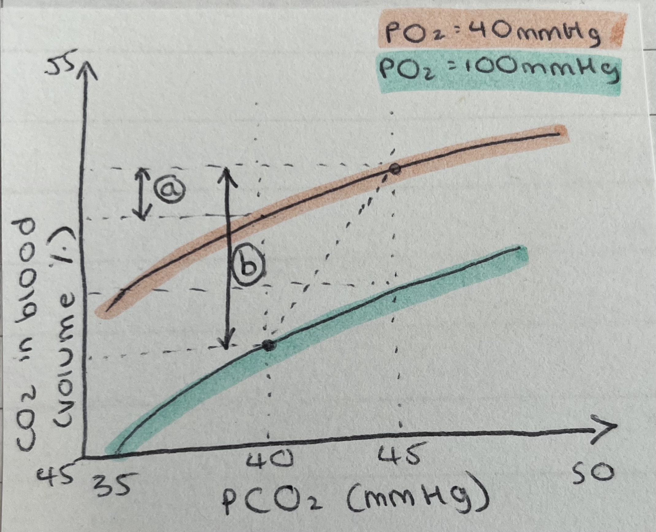 Lungs = 40 PCO2 mmHg
Tissue = 45 PCO2 mmHg
A) Amount of CO2 able to expire
When blood goes into the lungs, it gets oxygenated and the PO2 is higher. The oxygenated HbA is acidic which drives the bicarbonate buffering system. This makes the blood less able to hold CO2.
B) Increased amount of CO2 able to expire
→ The PCO2 is lower, the PO2 is higher and the haemoglobin is more acidic
Oxygenated haemoglobin is more acidic than deoxygenated haemoglobin
Drives the acid buffer system towards CO2 production (see Cl- shift)
Displaced CO2 from carbaminohaemoglobin
Summary
Gas exchange between the lungs and the tissues depends on the biochemical properties of haemoglobin
Haemoglobin is so effective because the anatomy of the lungs places the blood in close contact with alveolar air
Alveolar air is maintained as an infinite source of O2 and an infinite sink for CO2 by continual replenishment by the process of ventilation
Ventilation is automatically maintained by the medullary rhythmicity area